
Fluorescent microspheres for one-photon and two-photon imaging of mesenchymal stem cells†

Abstract
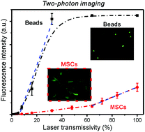
Stem cell-mediated therapy has emerged as a novel regenerative treatment for tissue repair in the last decade. However, noninvasive monitoring of stem cells remains a grand challenge for stem cell-based regenerative medicine for the complete understanding of the function and the behaviors of cells. Herein, acridine orange, a fluorescent dye was encapsulated into polymer nanospheres based on double emulsions. The fluorescent nanospheres with a diameter of around 200 nm were incubated with human mesenchymal stem cells (hMSCs) to produce nanosphere-endocytosed hMSCs. The fluorescent imaging of hMSCs can be directly and clearly captured using confocal microscopy in the one-photon and two-photon modes. The results indicated that the hMSCs adhered and spread well on the surface of the scaffolds with a high population and good distribution. Meanwhile, polystyrene microspheres were stained with dye complexes by using a gradual solvent evaporation method. The as-prepared fluorescent beads exhibited bright and multiple colors and a broad emission spectrum ranging from 500 to 800 nm, which was further used to quantitatively evaluate the one-photon and two-photon fluorescent signals of the hMSCs. Our study offers the possibility of direct monitoring of stem cells with high resolution, and encourages future quantitative clinical assessment in imaging-guided cell therapies.
 Please wait while we load your content...

