Mesoporous silica nanoparticles in injectable hydrogels: factors influencing cellular uptake and viability†
 ‡*b
and
Mika
Lindén
‡*a
‡*b
and
Mika
Lindén
‡*a
Abstract
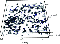
The incorporation of nanoparticles as drug vectors into 3D scaffolds has attracted a lot of recent interest. In particular, tissue engineering applications would benefit from a spatially and temporally regulated release of biological cues, which act on precursor/stem cells in a three-dimensional growth environment. Injectable cell- and nanoparticle-containing scaffolds are especially interesting in this respect, but require matrix self-assembly and coordinated interactions between cells, matrices, and nanoparticles, which are largely uncharacterized yet. In this proof of concept study we combined the matrix-forming self-assembling peptide RADA16-I, different mesoporous silica nanoparticles (MSN) as potential drug carriers, and MC3T3-E1 osteoblast precursor cells. When injected to physiological media, the mixtures rapidly formed hybrid peptide-silica hydrogels containing RADA16-I nanofiber scaffolds with uniform spatial distribution of viable cells and MSN. MSN surface chemistry was critical for interactions within the hydrogel and for RADA16-I adsorption, thereby dominantly influencing cellular uptake and cell viability, whereas the impact of serum protein was minor. Thus, important parameters which allow tuning of nanoparticulate drug vector interactions with cells in injectable 3D scaffolds are identified, which are of importance for the future design of smart scaffolds for advanced tissue engineering in vivo.
 Please wait while we load your content...

