Are different crystallinity-index-calculating methods of hydroxyapatite efficient and consistent?
Abstract
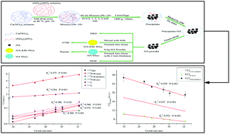
Crystallinity is related to the degree of order and the crystal size of a given crystalline substance. It can affect the quality of hydroxyapatite (HA) and its abnormality can lead to diseases of hard mineralized tissues. Crystallinity index (CI) is a quantitative indicator of crystallinity. Various techniques, such as X-ray diffraction (XRD), Fourier transform infrared spectroscopy (FTIR) and Raman spectroscopy, and many methods based on these techniques have been used to define the CI of HA. The present study compares these methods and summarizes their characters for hydrothermally synthesized HA crystals with different aging time. Additionally, correlation coefficients between CIs calculated by different methods and crystal size (R12) and correlation coefficients among various CIs (R22) were obtained from linear-regression analysis. Scanning electron microscopy (SEM) was utilized as a supplementary technique to observe the morphological changes during the HA aging process. The results showed larger crystal size, increased crystallinity and more regular morphology of HA with increased aging time. All the R12 and R22 were above 0.9 and there were no significant differences between R12. However, different CI-calculating methods showed individual characters or limitations during their application. Such results suggested XRD, FTIR and Raman techniques used in this study were generally consistent and efficient but their special characters should be considered during their application. Researchers should choose certain appropriate technique and method to obtain CI on the basis of sample characteristics and experimental conditions.


 Please wait while we load your content...