
A lysosome-locating and acidic pH-activatable fluorescent probe for visualizing endogenous H2O2 in lysosomes†
 *a
and
Yibing
Zhao
*a
*a
and
Yibing
Zhao
*a
Abstract
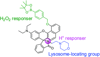
There is increasing evidence indicating that lysosomal H2O2 is closely related to autophagy and apoptotic pathways under both physiological and pathological conditions. Therefore, fluorescent probes that can be exploited to visualize H2O2 in lysosomes are potential tools for exploring diverse roles of H2O2 in cells. However, functional exploration of lysosomal H2O2 is limited by the lack of fluorescent probes capable of compatibly sensing H2O2 under weak acidic conditions (pH = 4.5) of lysosomes. Lower spatial resolution of the fluorescent visualization of lysosomal H2O2 might be caused by the interference of signals from cytosolic and mitochondrial H2O2, as well as the non-specific distribution of the probes in cells. In this work, we developed a lysosome-locating and acidic-pH-activatable fluorescent probe for the detection and visualization of H2O2 in lysosomes, which consists of a H2O2-responsive boronate unit, a lysosome-locating morpholine group, and a pH-activatable benzorhodol fluorophore. The response of the fluorescent probe to H2O2 is significantly more pronounced under acidic pH conditions than that under neutral pH conditions. Notably, the present probe enables the fluorescence sensing of endogenous lysosomal H2O2 in living cells without external stimulations, with signal interference from the cytoplasm and other intracellular organelles being negligible.
 Please wait while we load your content...

