Raman and infrared spectroscopy differentiate senescent from proliferating cells in a human dermal fibroblast 3D skin model†
 *ab
*ab
Abstract
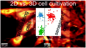
Senescent cells contribute to tissue aging and dysfunction. Therefore, detecting senescent cells in skin is of interest for skin tumor diagnostics and therapy. Here, we studied the transition into senescence of human dermal fibroblasts (HDFs) in a three-dimensional (3D) human fibroblast-derived matrix (FDM). Senescent and proliferating cells were imaged by Raman spectroscopy (RS) and Fourier transform infrared (FTIR) spectroscopy. The obtained averaged spectra were analyzed using PLS-LDA. For these 3D cultured cells, RS and FTIR could clearly distinguish senescent from proliferating cells. For both techniques, we detected senescence-associated alterations in almost all cellular macromolecules. Furthermore, we identified different biochemical properties of 3D compared to two-dimensional (2D) cultured cells, indicating that cells in their natural, skin-like 3D environment act differently than in (2D) cell cultivations in vitro. Compared to 2D cultured cells, cells grown in 3D models displayed a sharper contrast between the proliferating and senescent state, also affecting the abundance of biomolecules including nucleic acids. The training accuracies of both vibrational spectroscopic techniques were >96%, demonstrating the suitability of these label-free measurements for detecting these cellular states in 3D skin models.
 Please wait while we load your content...

