Retinal oxidative stress at the onset of diabetes determined by synchrotron FTIR widefield imaging: towards diabetes pathogenesis
Abstract
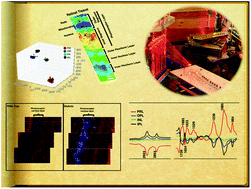
Diabetic retinopathy is a microvascular complication of diabetes that can lead to blindness. In the present study, we aimed to determine the nature of diabetes-induced, highly localized biochemical changes in the neuroretina at the onset of diabetes. High-resolution synchrotron Fourier transform infrared (s-FTIR) wide field microscopy coupled with multivariate analysis (PCA–LDA) was employed to identify biomarkers of diabetic retinopathy with spatial resolution at the cellular level. We compared the retinal tissue prepared from 6-week-old Ins2Akita/+ heterozygous (Akita/+, N = 6; a model of diabetes) male mice with the wild-type (control, N = 6) mice. Male Akita/+ mice become diabetic at 4-weeks of age. Significant differences (P < 0.001) in the presence of biomarkers associated with diabetes and segregation of spectra were achieved. Differentiating IR bands attributed to nucleic acids (964, 1051, 1087, 1226 and 1710 cm−1), proteins (1662 and 1608 cm−1) and fatty acids (2854, 2923, 2956 and 3012 cm−1) were observed between the Akita/+ and the WT samples. A comparison between distinctive layers of the retina, namely the photoreceptor retinal layer (PRL), outer plexiform layer (OPL), inner nucleus layer (INL) and inner plexiform layer (IPL) suggested that the photoreceptor layer is the most susceptible layer to oxidative stress in short-term diabetes. Spatially-resolved chemical images indicated heterogeneities and oxidative-stress induced alterations in the diabetic retina tissue morphology compared with the WT retina. In this study, the spectral biomarkers and the spatial biochemical alterations in the diabetic retina and in specific layers were identified for the first time. We believe that the conclusions drawn from these studies will help to bridge the gap in our understanding of the molecular and cellular mechanisms that contribute to the pathobiology of diabetic retinopathy.


 Please wait while we load your content...