
Cellular uptake, genotoxicity and cytotoxicity of cobalt ferrite magnetic nanoparticles in human breast cells
Abstract
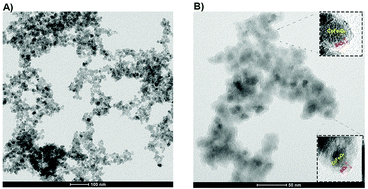
Magnetic nanoparticles (MNPs) have been increasingly used for many years as MRI agents and for gene delivery and hyperthermia therapy, although there have been conflicting results on their safety. In this study, cobalt ferrite magnetic nanoparticles (CoFe-MNPs) were prepared by the co-precipitation method and their surfaces were modified with silica by the sol–gel method. The particle and hydrodynamic sizes, morphology and crystal structure of the bare and silica-coated CoFe-MNPs were evaluated by transmission electron microscopy (TEM), dynamic light scattering (DLS), X-ray diffraction spectroscopy (XRD) and Fourier transform infrared spectroscopy (FTIR). The size of the bare CoFe-MNPs was in the range 8–20 nm and they were homogeneously coated with 3–4 nm silica shells. The bare and silica-coated CoFe-MNPs were agglomerated at physiological pH. However, the sizes of the agglomerates were below 200 nm both in water and complete medium. The cytotoxic and genotoxic potentials of the bare and silica-coated CoFe-MNPs were evaluated in a metastatic breast cancer cell line, MDA-MB-231, as well as a noncancerous mammary epithelial cell line, MCF-10A, by using XTT cytotoxicity, single-cell gel electrophoresis (comet), and cytokinesis-blocked (CB) micronucleus (CBMN) assays. Characterization studies with TEM, inductively coupled plasma optical emission spectroscopy (ICP-OES) and Prussian blue staining indicated that the CoFe-MNPs were internalized into the cells by energy-dependent endocytosis. The highest amount of uptake was observed in the cancer cells and the uptake of the silica-coated CoFe-MNPs was higher than that of the bare ones in both cell lines. The bare CoFe-MNPs showed higher levels of both cytotoxicity and genotoxicity than the silica-coated CoFe-MNPs. Moreover, the cancer cells seemed to be more susceptible to the CoFe-MNPs’ toxicity compared to the noncancerous cells. There was a concentration and time-dependent increase in DNA damage and the micronucleus (MN) frequency, which was statistically significant starting with the lowest concentration of bare CoFe-MNPs (p < 0.05), while no significance was observed below the concentration of 250 μg mL−1 for the silica-coated MNPs. Also, the extent of both DNA damage and MN frequency was much higher in the cancer cells compared to the noncancerous cells. According to our results, the silica coating ameliorated both the cytotoxicity and genotoxicity as well the internalization of the CoFe-MNPs.
 Please wait while we load your content...