Morphological single cell profiling of the epithelial–mesenchymal transition†
Abstract
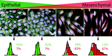
Single cells respond heterogeneously to biochemical treatments, which can complicate the analysis of in vitro and in vivo experiments. In particular, stressful perturbations may induce the epithelial–mesenchymal transition (EMT), a transformation through which compact, sensitive cells adopt an elongated, resistant phenotype. However, classical biochemical measurements based on population averages over large numbers cannot resolve single cell heterogeneity and plasticity. Here, we use high content imaging of single cell morphology to classify distinct phenotypic subpopulations after EMT. We first characterize a well-defined EMT induction through the master regulator Snail in mammary epithelial cells over 72 h. We find that EMT is associated with increased vimentin area as well as elongation of the nucleus and cytoplasm. These morphological features were integrated into a Gaussian mixture model that classified epithelial and mesenchymal phenotypes with >92% accuracy. We then applied this analysis to heterogeneous populations generated from less controlled EMT-inducing stimuli, including growth factors (TGF-β1), cell density, and chemotherapeutics (Taxol). Our quantitative, single cell approach has the potential to screen large heterogeneous cell populations for many types of phenotypic variability, and may thus provide a predictive assay for the preclinical assessment of targeted therapeutics.


 Please wait while we load your content...