Increased cell survival of cells exposed to superparamagnetic iron oxide nanoparticles through biomaterial substrate-induced autophagy†
Abstract
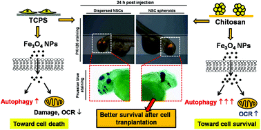
The cellular uptake of nanoparticles (NPs) can be promoted by NP surface modification but cell viability is often sacrificed. Our previous study has shown that intracellular uptake of iron oxide NPs was significantly increased for cells cultured on chitosan. However, the mechanism for having the higher cellular uptake as well as better cell survival on the chitosan surface remains unclear. In this study, we sought to clarify if the autophagic response may contribute to cell survival under excessive NP exposure conditions on chitosan. L929 fibroblasts and neural stem cells (NSCs) were challenged with different concentrations (0–300 μg ml−1) of superparamagnetic iron oxide NPs. The autophagic response as well as the metabolic activity of cells was evaluated. Results showed that culturing both types of cells on chitosan substrates significantly enhanced the cellular uptake of NPs. At higher NP concentrations, cells on chitosan showed a greater survival rate than those on TCPS. The expression levels of autophagy-related genes (Atg5 and Atg7 genes) and autophagy associated protein (LC3-II) on chitosan were higher than that on TCPS. The NP exposure further increased the expressions. We suggest that cells cultured on chitosan were more tolerant to NP cytotoxicity because of the increased autophagic response. Moreover, NP exposure increased the metabolic activity of cells grown on chitosan, while it decreased the metabolism of cells cultured on TCPS. In animal studies, iron oxide-labeled NSCs were injected in zebrafish embryos. Results also showed that cells grown on chitosan had better survival after transplantation than those grown on TCPS. Taken together, chitosan as a culture substrate can induce cell autophagy to increase cell survival in particular for NP-labeled cells. This will be valuable for the biomedical application of NPs in cell therapy.


 Please wait while we load your content...