
Toxicity mechanism of graphene oxide and nitrogen-doped graphene quantum dots in RBCs revealed by surface-enhanced infrared absorption spectroscopy†
Abstract
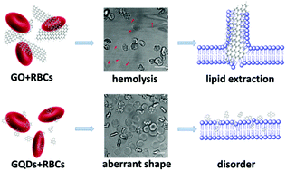
The study of the toxic effects of nanoparticles on biological systems at the molecular level is critical in order to gain a greater understanding of the origin of nanotoxicity. Recently, numerous forms of graphene materials have been synthesized and extensively applied in biosensors and biomedicine, but their toxicity has not yet been studied to the same extend, in particular the toxicity mechanism. In this work, we systematically studied the toxic effects of two typical graphene forms, graphene oxide (GO) and nitrogen-doped graphene quantum dots (N-GQDs), on red blood cells (RBCs) by testing their hemolytic activity, observing the morphological changes and detecting the ATP content of RBCs after being exposed to the two nanomaterials. The toxicity mechanism was further revealed by investigating the structural changes of RBCs lipid by surface-enhanced infrared absorption spectroscopy using model membranes. A detailed analysis of the infrared spectra revealed that the adsorption of GO destroys the integrity of a membrane by extracting the lipid bilayer, resulting in hemolysis and aberrant forms. In contrast, N-GQDs just disturb the structure and conformation of the lipid, resulting in only aberrant cells. To date, this is the first experimental study which has revealed the toxicity mechanism of graphene materials in RBCs at the molecular level.
- This article is part of the themed collection: Nanotoxicology
 Please wait while we load your content...