Issue 47, 2015

Formation of lipid/peptide tubules by IAPP and temporin B on supported lipidmembranes

Abstract

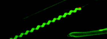
The conversion of various proteins and peptides to amyloid fibrils is accelerated by lipidmembranes, which are also postulated to represent targets mediating the cytotoxicity of amyloid protofibrils. Yet, our understanding of the molecular details governing membrane-catalyzed fibrillogenesis of amyloid precursors remains limited. To obtain insight into the intricate interplay of amyloidgrowth and membrane biophysics we have recently introduced supported lipid bilayers (SLBs) with fluorescent lipid analogs as model biomembranes, observed by time-lapse confocal microscopy. Here we demonstrate that human islet amyloidpolypeptide (IAPP) induces within minutes of its application on zwitterionic phosphatidylcholine bilayers the expulsion of numerous flexible lipid tubules from the SLB. Intriguingly, these flexible tubules gradually evolve into a network of straight tubes locally attached to the SLB substrate. Two-color imaging of the membrane and the fluorescently labeled peptide revealed IAPP to be distributed along the lipid tubes. Similar linear tubules were observed with the antimicrobial peptide temporin B and the non-amyloidogenic rat IAPP, revealing that the above mesoscopic membrane perturbations are not related to amyloid formation by the human IAPP. Micromanipulation experiments revealed that the linearity of the tubules was caused by tension, stretching the tubules between their points of attachment to the SLB substrate. After longer incubation times, for SLBs containing the oxidatively modified phospholipid 1-palmitoyl-2-azelaoyl-sn-glycero-3-phosphocholine (PazePC, bearing a terminal carboxyl group at the end of the sn-2azelaoyl chain) and human IAPP (but not the other peptides) some of the straight lipid tubes transformed into highly regular helices. This is likely to reflect tension originating from an efficient aggregation of the IAPP into parallelly aligned fibril bundles, associated with membrane tubes containing the oxidized phospholipid, possibly together with a concomitant flow of IAPPfibrils along the tubules to the immobile IAPP aggregates attaching the tubules to the SLB substrate, these two processes cause, upon shortening of the linear peptide scaffold, the attached excess lipid tubule to adopt a helical morphology, coiling around the peptidefibril core. The above fluorescence microscopy studies are in line with the multiphasic kinetics of IAPP fibrillation in the presence of oxidized lipid containing liposomes, assessed by thioflavin T fluorescence enhancement. In addition to demonstrating the feasibility of SLBs as biomimetic model system for studying lipid-assisted protein fibrillation, our results accentuate the role of membrane chemical composition in modulation of different stages of this process and the associated transformation of membrane architecture. Accordingly, changes in the chemical nature of cellular membranes arising from pathophysiological processes such as oxidative stress may participate in the triggering amyloidogenesis as well as amplification of its detrimental effects in vivo.

Graphical abstract: Formation of lipid/peptide tubules by IAPP and temporin B on supported lipidmembranes

Associated articles

Supplementary files

Article information

Article type
Paper
Submitted
01 Dec 2009
Accepted
15 Mar 2010
First published
17 Nov 2015

Soft Matter, 2015,11, 9188-9200

Formation of lipid/peptide tubules by IAPP and temporin B on supported lipidmembranes

P. K. J. Kinnunen, Y. A. Domanov, J. Mattila and T. Varis, Soft Matter, 2015, 11, 9188 DOI: 10.1039/B925228B

To request permission to reproduce material from this article, please go to the Copyright Clearance Center request page.

If you are an author contributing to an RSC publication, you do not need to request permission provided correct acknowledgement is given.

If you are the author of this article, you do not need to request permission to reproduce figures and diagrams provided correct acknowledgement is given. If you want to reproduce the whole article in a third-party publication (excluding your thesis/dissertation for which permission is not required) please go to the Copyright Clearance Center request page.

Read more about how to correctly acknowledge RSC content.

Social activity

Spotlight

Advertisements