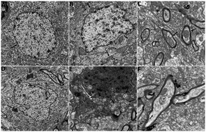
The discrepancy between the absence of copper deposition and the presence of neuronal damage in the brain of Atp7b−/− mice
Abstract
Wilson's disease (WD) is caused by mutations within the copper-transporting ATPase (ATP7B), characterized by copper deposition in various organs, principally the liver and the brain. With the availability of Atp7b−/− mice, the valid animal model of WD, the mechanism underlying copper-induced hepatocyte necrosis has been well understood. Nonetheless, little is known about the adverse impact of copper accumulation on the brain in WD. Therefore, the aim of this study was to identify copper disturbances according to various brain compartments and further dissect the causal relationship between copper storage and neuronal damage using Atp7b−/− mice. Copper levels in the liver, whole brain, brain compartments and basal ganglia mitochondria of Atp7b−/− mice and age-matched controls were measured by atomic absorption spectroscopy. Delicate electron microscopic studies on hepatocytes and neurons in the basal ganglia were performed. Here we further confirmed the remarkably elevated copper content and abnormal ultrastructure findings in livers of Atp7b−/− mice. Interestingly, we found the ultrastructure abnormalities in neurons of the basal ganglia of Atp7b−/− mice, whereas copper deposition was not detected in the whole brain, even within the basal ganglia and its mitochondria. The disparity provided a new understanding of neuronal dysfunction in WD, and strongly indicated that copper might not be the sole causative player and other unidentified pathogenic factors could enhance the toxic effects of copper on neurons in WD.


 Please wait while we load your content...