
A shotgun metalloproteomic approach enables identification of proteins involved in the speciation of a ruthenium anticancer drug in the cytosol of cancer cells†
Abstract
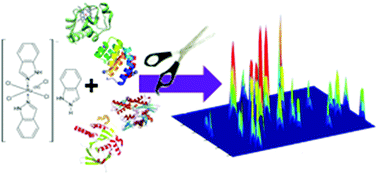
The study reported herein focused on the development and optimization of a versatile analytical methodology for characterization of intracellular distribution of protein-bound species of ruthenium originating from an anticancer Ru-based drug, indazolium trans-[tetrachloridobis(1H-indazole)ruthenate(III)]. A direct analysis of the drug-treated cytosol of cancer cells using size-exclusion chromatography (SEC) interfaced with inductively coupled plasma mass spectrometry (ICP-MS) revealed that over 85% of ruthenium is converted into a high molecular-mass fraction. To further determine the ruthenium binding pattern, a shotgun approach was used, with the entire proteome being digested and the resulting peptides being analyzed by capillary high-performance liquid chromatography (μHPLC) combined with electrospray ionization triple quadrupole MS. This allowed for identification of the ruthenated proteins on the basis of characteristic MS/MS spectra of the respective peptides. It was found that both Ru(III)- and Ru(II)-ligated functionalities participate in adduct formation, the hydrolyzed forms of the drug being attached to the majority of the binding proteins. Of an array of proteins responding to drug treatment, the most important – from the viewpoint of unveiling the exact mode of action – are inhibitor, pro-apoptotic, and DNA reparation proteins.
 Please wait while we load your content...

