
Label-free Raman imaging of the macrophage response to the malaria pigment hemozoin†
Abstract
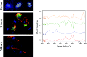
Hemozoin, the ‘malaria pigment’, is engulfed by phagocytic cells, such as macrophages, during malaria infection. This biocrystalline substance is difficult to degrade and often accumulates in phagocytes. The macrophage response to hemozoin relates to the severity of the disease and the potential for malaria-related disease complications. In this study we have used Raman spectroscopy as a label-free method to investigate the biochemical changes occurring in macrophages during the first few hours of hemozoin uptake. We found a number of distinct spectral groups, spectrally or spatially related to the presence of the hemozoin inside the cell. Intracellular hemozoin was spectrally identical to extracellular hemozoin, regardless of the location in the cell. A small proportion of hemozoin was found to be associated with lipid-based components, consistent with the uptake of hemozoin into vesicles such as phagosomes and lysosomes. The spatial distribution of the hemozoin was observed to be inhomogeneous, and its presence largely excluded that of proteins and lipids, demonstrating that cells were not able to break down the biocrystals on the time scales studied here. These results show that Raman imaging can be used to answer some of the open questions regarding the role of hemozoin in the immune response. How different combinations of hemozoin and other molecules are treated by macrophages, whether hemozoin can be broken down by the cell, and more importantly, which co-factors or products are involved in the subsequent cell reaction are the expected issues to be elucidated by this technique.
- This article is part of the themed collection: Optical Diagnosis (2014)
 Please wait while we load your content...

