
Imaging and quantification of trans-membrane protein diffusion in living bacteria†
Abstract
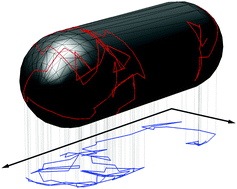
The cytoplasmic membrane forms the barrier between any cell's interior and the outside world. It contains many proteins that enable essential processes such as the transmission of signals, the uptake of nutrients, and cell division. In the case of prokaryotes, which do not contain intracellular membranes, the cytoplasmic membrane also contains proteins for respiration and protein folding. Mutual interactions and specific localization of these proteins depend on two-dimensional diffusion driven by thermal fluctuations. The experimental investigation of membrane–protein diffusion in bacteria is challenging due to their small size, only a few times larger than the resolution of an optical microscope. Here, we review fluorescence microscopy-based methods to study diffusion of membrane proteins in living bacteria. The main focus is on data-analysis tools to extract diffusion coefficients from single-particle tracking data obtained by single-molecule fluorescence microscopy. We introduce a novel approach, IPODD (inverse projection of displacement distributions), to obtain diffusion coefficients from the usually obtained 2-D projected diffusion trajectories of the highly 3-D curved bacterial membrane. This method provides, in contrast to traditional mean-squared-displacement methods, correct diffusion coefficients and allows unravelling of heterogeneously diffusing populations.
 Please wait while we load your content...

