
Small molecule phosphorescent probes for O2 imaging in 3D tissue models†
Abstract
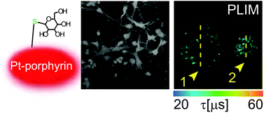
Monitoring of oxygenation is important for physiological experiments investigating the growth, differentiation and function of individual cells in 3D tissue models. Phosphorescence based O2 sensing and imaging potentially allow this task; however, current probes do not provide the desired bio-distribution and analytical performance. We present several new cell-penetrating phosphorescent conjugates of a Pt(II)-tetrakis(pentafluorophenyl)porphine (PtPFPP) dye produced by click-modification with thiols, and perform their evaluation as O2 imaging probes for 3D tissue models. The hydrophilic glucose (Pt-Glc) and galactose (Pt-Gal) conjugates demonstrated minimal aggregation and self-quenching in aqueous media, and efficient in-depth staining of different cell types and multi-cellular aggregates at working concentrations ≤10 μM. The Pt-Glc probe was applied in high-resolution phosphorescence lifetime based O2 imaging (PLIM) in multi-cellular spheroids of cancer cells (PC12), primary neural cells (neurospheres) and slices of brain tissue, where it showed good analytical performance, minimal effects on cell viability and appropriate responses to O2 with phosphorescence lifetimes changing from 20 μs in air-saturated to 57 μs under deoxygenated conditions. In contrast, mono- and tetra-substituted oligoarginine conjugates of PtPFPP showed marked aggregation and unstable photophysical properties precluding their use as O2 sensing probes.
 Please wait while we load your content...

