Investigating drug induced changes in single, living lymphocytes based on Raman micro-spectroscopy
Abstract
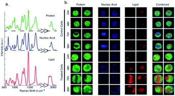
Raman spectroscopy is a powerful tool for label-free, single cell characterization. In many reported studies, a Raman spectrum is acquired from a fraction of the cell volume and used as a representative signature of the whole cell to identify and discriminate between cell populations. It has remained an open question whether this is the most suitable approach since the spectra may not truly represent the cell as a whole and critical biochemical information could therefore be lost. To address this question, we developed a line-scan Raman microscope to acquire Raman images of single lymphocytes exposed to the chemotherapeutic drug doxorubicin for 24 to 96 hours. Principal component analysis was able to separate cells based on their drug-exposure times. Difference spectra on the mean data for the different time-points revealed that changes are related to a decrease in mean nucleic acid content and an increase in mean protein and lipid content. Vertex component analysis was used to extract the pure component spectra of lipids, nucleic acids, and proteins. Quantitative analysis of the data revealed that biochemical changes occurred at both local subcellular (i.e. molecular density) and global cellular (i.e. total observable molecular content) levels. However, significant differences between the trends in the local and global changes were observed. While local nucleic acid content decreased with increasing drug exposure time, the total cellular nucleic acid content remained relatively constant. For protein, local content remained relatively constant for all exposure times while the total protein content in the cell increased ∼3 fold. Lipid content in the entire cell increased ∼5 fold, compared to a smaller increase in lipid at the local level. These results show that valuable information about the biochemical changes throughout the entire cell can be missed if only Raman spectra of localized cell regions are used. These findings are expected to have a major impact on the future development of Raman spectroscopy for cytometry applications.
 Please wait while we load your content...

