Issue 11, 2014

Single vs. two-photon microscopy for label free intrinsic tissue studies in the UV light region

Abstract

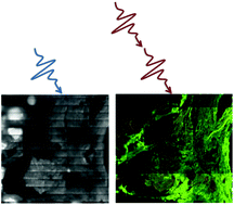
Fibrillar distribution in the rat tail tendon and mice liver can be measured using optical methods. Two-photon excitation provides easy assessment of fibrotic collagen types I and II. Single photon deep ultraviolet (DUV) excitation imaging highlights all collagen types without discrimination. Their combination on the same tissue area provides a better overview of collagens in fibrillar diseases.

Graphical abstract: Single vs. two-photon microscopy for label free intrinsic tissue studies in the UV light region

Article information

Article type
Communication
Submitted
27 Jan 2014
Accepted
20 Mar 2014
First published
21 Mar 2014

Analyst, 2014,139, 2663-2667

Author version available

Single vs. two-photon microscopy for label free intrinsic tissue studies in the UV light region

V. Zubkovs, F. Jamme, S. Kascakova, F. Chiappini, F. Le Naour and M. Réfrégiers, Analyst, 2014, 139, 2663 DOI: 10.1039/C4AN00203B

To request permission to reproduce material from this article, please go to the Copyright Clearance Center request page.

If you are an author contributing to an RSC publication, you do not need to request permission provided correct acknowledgement is given.

If you are the author of this article, you do not need to request permission to reproduce figures and diagrams provided correct acknowledgement is given. If you want to reproduce the whole article in a third-party publication (excluding your thesis/dissertation for which permission is not required) please go to the Copyright Clearance Center request page.

Read more about how to correctly acknowledge RSC content.

Social activity

Spotlight

Advertisements