pH-dependent adhesion of mycobacteria to surface-modified polymer nanofibers†
Abstract
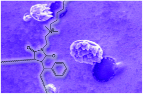
Children across the world are greatly affected by tuberculosis (TB) due to high morbidity and mortality. It is important to diagnose TB in children timeously and accurately in order to provide effective treatment. In this study we aimed to test the hypothesis that a modified polymer could be developed to capture Mycobacterium tuberculosis (M. tuberculosis), the pathogen that causes TB, under different pH conditions, mimicking different clinical specimens. The modified polymer was electrospun into nanofibrous mats thereby ensuring optimal surface areas. Affinity studies were conducted on these modified polymer nanofibers with Mycobacterium bovis bacillus Calmette–Guérin (BCG) and verified with M. tuberculosis to evaluate these nanofibrous surfaces as mycobacterium-capturing platforms. The results indicate that BCG and M. tuberculosis were successfully captured under different pH conditions depending on the affinity ligand. Concentration and time studies showed that binding efficiency is dependent on the incubation time and the concentration of mycobacteria. The affinity studies also reveal that the nanofibrous-capturing polymer should not be too hydrophobic in character as this causes poor wetting of the modified nanofibers, thus preventing close contact with the mycobacteria and a reduction in the capture effectivity of the polymer nanofibers. The detection of M. tuberculosis using microscopy is simplified by the tendency of the mycobacteria to aggregate on the hydrophobic surface of the modified nanofibers. As a result of this aggregation, fluorescence and light microscopy are regarded as feasible detection methods to image M. tuberculosis on the surface of the modified nanofibers, even at relatively low concentration. In this study a polymer was therefore successfully modified in such a way that it acquired an affinity for M. tuberculosis and enabled the capture of this organism onto the modified polymer nanofibrous surface.


 Please wait while we load your content...