Issue 37, 2013

Bismuth@US-tubes as a potential contrast agent for X-ray imaging applications

Abstract

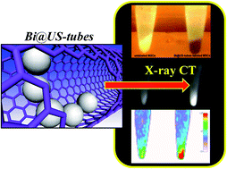
The encapsulation of bismuth as BiOCl/Bi2O3 within ultra-short (ca. 50 nm) single-walled carbon nanocapsules (US-tubes) has been achieved. The Bi@US-tubes have been characterized by high-resolution transmission electron microscopy (HR-TEM), energy-dispersive X-ray spectroscopy (EDS), thermogravimetric analysis (TGA), X-ray photoelectron spectroscopy (XPS), and Raman spectroscopy. Bi@US-tubes have been used for intracellular labeling of pig bone marrow-derived mesenchymal stem cells (MSCs) to show high X-ray contrast in computed tomography (CT) cellular imaging for the first time. The relatively high contrast is achieved with low bismuth loading (2.66% by weight) within the US-tubes and without compromising cell viability. X-ray CT imaging of Bi@US-tubes-labeled MSCs showed a nearly two-fold increase in contrast enhancement when compared to unlabeled MSCs in a 100 kV CT clinical scanner. The CT signal enhancement from the Bi@US-tubes is 500 times greater than polymer-coated Bi2S3 nanoparticles and several-fold that of any clinical iodinated contrast agent (CA) at the same concentration. Our findings suggest that the Bi@US-tubes can be used as a potential new class of X-ray CT agent for stem cell labeling and possibly in vivo tracking.

Graphical abstract: Bismuth@US-tubes as a potential contrast agent for X-ray imaging applications

Supplementary files

Article information

Article type
Paper
Submitted
25 May 2013
Accepted
29 Jul 2013
First published
30 Jul 2013

J. Mater. Chem. B, 2013,1, 4792-4800

Bismuth@US-tubes as a potential contrast agent for X-ray imaging applications

E. J. Rivera, L. A. Tran, M. Hernández-Rivera, D. Yoon, A. G. Mikos, I. A. Rusakova, B. Y. Cheong, M. D. G. Cabreira-Hansen, J. T. Willerson, E. C. Perin and L. J. Wilson, J. Mater. Chem. B, 2013, 1, 4792 DOI: 10.1039/C3TB20742K

To request permission to reproduce material from this article, please go to the Copyright Clearance Center request page.

If you are an author contributing to an RSC publication, you do not need to request permission provided correct acknowledgement is given.

If you are the author of this article, you do not need to request permission to reproduce figures and diagrams provided correct acknowledgement is given. If you want to reproduce the whole article in a third-party publication (excluding your thesis/dissertation for which permission is not required) please go to the Copyright Clearance Center request page.

Read more about how to correctly acknowledge RSC content.

Social activity

Spotlight

Advertisements