Proteomic signatures differentiating Bacillus anthracis Sterne sporulation on soil relative to laboratory media†
D. S.
Wunschel
 *,
J. R.
Hutchison
,
B. L.
Deatherage Kaiser
,
E. D.
Merkley
,
B. M.
Hess
,
A.
Lin
 and
M. G.
Warner

Chemical and Biological Signature Sciences, Pacific Northwest National Laboratory, Richland, WA 99354, USA. E-mail: David.Wunschel@pnnl.gov; Tel: (509) 371-6852
First published on 14th November 2017
Abstract
The process of sporulation is vital for the stability and infectious cycle of Bacillus anthracis. The spore is the infectious form of the organism and therefore relevant to biodefense. While the morphological and molecular events occurring during sporulation have been well studied, the influence of growth medium and temperature on the proteins expressed in sporulated cultures is not well understood. Understanding the features of B. anthracis sporulation specific to natural vs. laboratory production will address an important question in microbial forensics. In an effort to bridge this knowledge gap, a system for sporulation on two types of agar-immobilized soils was used for comparison to cultures sporulated on two common types of solid laboratory media, and one liquid sporulation medium. The total number of proteins identified as well as their identity differed between samples generated in each medium and growth temperature, demonstrating that sporulation environment significantly impacts the protein content of the spore. In addition, a subset of proteins common in all of the soil-cultivated samples was distinct from the expression profiles in laboratory medium (and vice versa). These differences included proteins involved in thiamine and phosphate metabolism in the sporulated cultures produced on soils with a notable increase in expression of ATP binding cassette (ABC) transporters annotated to be for phosphate and antimicrobial peptides. A distinct set of ABC transporters for amino acids, sugars and oligopeptides were found in cultures produced on laboratory media as well as increases in carbon and amino acid metabolism-related proteins. These protein expression changes indicate that the sporulation environment impacts the protein profiles in specific ways that are reflected in the metabolic and membrane transporter proteins present in sporulated cultures.
Introduction
The spores of Bacillus spp. have been widely studied and are known to be stable structures which are resistant to heat, pressure, and desiccation.1 The B. anthracis spore has been particularly well studied as the infective form and causative agent of anthrax, a serious animal and human disease. Human exposure typically occurs through contact with infected animals and animal products.2 The inhalation form of the disease, traditionally called “wool sorter's disease”, is typically fatal if not treated. This form of the disease re-emerged unexpectedly in 2001 during the anthrax letter attacks where a common laboratory strain, Ames, was used for intentional exposure.3 Within the last decade, several cases have appeared due to human activity with untreated animal skins used for traditional drum making and playing.4 These examples also serve as a backdrop to a study conducted by the U.S. National Academy of Science in cooperation with The Croatian Academy of Science and Arts, The U.K. Royal Society and The International Union of Microbiological Societies that outlined the need for more fundamental research in microbial forensics.5 The differentiation between naturally occurring and laboratory cultivated pathogens was found to represent a potential forensic challenge for responding to a disease outbreak. Furthermore, much is still unknown about the fundamental biology of B. anthracis in the soil environment.The composition and dynamics of the B. anthracis sporulation proteome has been studied by Liu et al. during different stages of sporulation. A number of distinct changes were observed in addition to sporulation proteins, including hydrolytic and exosporium specific proteins.6 The protein content of specific B. anthracis spore structures, such as the exosporium and coat, have also been studied in detail.7,8 As a result, proteins considered to be specific to spore formation and mature spores have been identified.6 Liu et al. also hypothesized that the proteins present in the spore are likely to be a reflection of the accumulated proteome from vegetative growth in addition to proteins produced in the mother cell and actively recruited into the mature spore, such as the coat and exosporium proteins. If true, it would be extremely useful to understand how different environmental conditions impact the protein composition of a mature spore. Differences in the B. anthracis sporulation proteome related to culture conditions have not been previously examined, nor has the proteome of spores produced on soil.
The proteomic changes in B. cereus and B. subtilis strains have been examined following broth culture to exponential phase in a medium containing soil extracted soluble organic matter (SESOM) and Luria Bertani broth.9,10 The ability of each species to germinate and form spores in each culture system is well documented.9 A comparison of the proteomes following growth in each medium showed that cultivation on soil extract stimulated an increase in the expression of ABC transporter proteins involved in importing peptides and amino acids into the cell. Expression of proteins involved with protein expression, polyamine, fatty acid, and amino acid biosynthesis was also increased in the soil medium samples. While these are important observations, not all members of genus Bacillus, or even the B. cereus group of species, have adopted similar survival strategies.
The B. cereus group of organisms includes strains belonging to the species B. cereus, B. thuringiensis, B. anthracis, as well as the lesser studied B. mycoides, B. pseudomycoides and B. weihenstephanensis. Members of genus Bacillus are abundant members of soil communities and B. cereus strains are saprophytic soil organisms that are able to cycle between germination, active vegetative growth, and sporulation in soils.9 By contrast, the relative genetic homogeneity of B. anthracis strains isolated from soils suggests that this species is largely dependent on a mammalian host to support germination and vegetative growth with relatively little vegetative growth in soils.11 Germination and vegetative growth has been suggested for B. anthracis in some soil environments,12,13 however different survival strategies are employed by B. anthracis relative to B. cereus. This suggests that the response of B. anthracis to the soil environment needs to be investigated.
Previous reports on B. anthracis spore proteomics have generated samples grown in laboratory media, while the proteomics of B. cereus cells cultured on a soil extract were performed on vegetative cells. A significant gap remains in our knowledge regarding the influence of soils on B. anthracis proteomic composition during the terminal stages of sporulation. This is particularly important for understanding sporulation events in the natural cycle of sporulation following release from an infected animal relative to intentional production on a laboratory medium. In order to address this gap, we have used a bottom-up proteomic analysis to examine the proteome of B. anthracis spores produced on three different laboratory culture media in comparison to spores produced on soil-agar systems. By comparing the proteomes of each type of sporulated culture, we sought to determine which proteins might be present in soil-produced spores that are missing or underrepresented in spores produced on laboratory media. We have identified such differences, leading to hypotheses about the environmental influences on soil-produced spores that may be absent when spores are produced on laboratory media.
Materials and methods
Chemicals
The following chemicals were obtained in ACS grade or higher from Sigma (St Louis MO): trichloroacetic acid (TCA), urea, ammonium bicarbonate, β-mercaptoethanol, methanol, acetonitrile, HPLC grade H2O, and trifluoroacetic acid. A Tris-HCl solution (50 mM) was made from purchased Tris-HCL (Life Tech. Grand Is. NY). Trypsin gold was purchased from Promega (Madison WI). ACS grade formic acid was purchased from EMD Millipore (Billerica MA).Bacterial culture and quantitation
Bacillus anthracis Sterne 34F2 seed stock was a gift from Robert Bull (Naval Medical Research Center), and was used to start all cultures. Primary spore stocks were generated by growing an overnight culture in Tryptic Soy Broth (TSB – Becton-Dickinson, Sparks, MD) in triplicate to generate three biological replicates. Each culture was diluted 1![[thin space (1/6-em)]](https://www.rsc.org/images/entities/char_2009.gif) :
:![[thin space (1/6-em)]](https://www.rsc.org/images/entities/char_2009.gif) 10 and 0.1 mL of this dilution was plated onto bovine blood agar (Hardy Diagnostics A188, Santa Maria, CA). Spores were visually inspected by phase contrast microscopy and harvested on day seven by flooding the plates with water, scraping with a spreader, and aspirating into a conical tube. Spores were washed three times with sterile water, enumerated on Tryptic Soy Agar (TSA – Becton-Dickinson, Sparks MD) plates, and diluted to 1 × 10⁸ spores per mL. Each of the three spore stocks were diluted in 0.1% w/v peptone such that ∼150 CFU were used to inoculate liquid cultures. CFU of inocula were verified with plate counts. Liquid cultures for vegetative growth were 10 mL of a 3
10 and 0.1 mL of this dilution was plated onto bovine blood agar (Hardy Diagnostics A188, Santa Maria, CA). Spores were visually inspected by phase contrast microscopy and harvested on day seven by flooding the plates with water, scraping with a spreader, and aspirating into a conical tube. Spores were washed three times with sterile water, enumerated on Tryptic Soy Agar (TSA – Becton-Dickinson, Sparks MD) plates, and diluted to 1 × 10⁸ spores per mL. Each of the three spore stocks were diluted in 0.1% w/v peptone such that ∼150 CFU were used to inoculate liquid cultures. CFU of inocula were verified with plate counts. Liquid cultures for vegetative growth were 10 mL of a 3![[thin space (1/6-em)]](https://www.rsc.org/images/entities/char_2009.gif) :
:![[thin space (1/6-em)]](https://www.rsc.org/images/entities/char_2009.gif) 1 mixture of bovine calf blood (Becton-Dickinson L12379, Sparks, MD) and Brain Heart Infusion broth (Becton-Dickinson, Sparks, MD). Following overnight growth in liquid bovine blood-BHI medium at 37 °C with gentle shaking, the number of vegetative cells was enumerated on TSA plates. Heat shock treatment of a 0.1 mL aliquot of each vegetative culture was performed at 70 °C for 30 minutes and enumerated on TSA plates. The heat-shocked cultures showed no growth, indicating a purely vegetative culture. A 0.1 mL portion of the vegetative culture containing an estimated 4.6 × 10⁶ to 4.8 × 10⁶ cells was used to inoculate each of the media for sporulation. Three biological replicates were created for each medium. Each replicate of solid medium consisted of three agar plates. For liquid medium, each of the replicates consisted of a single flask containing 100 mL of broth. For soil agars, an additional 0.5 mL of bovine calf blood was added to the surface of the agar plates to approximate the natural environment during animal death and to improve spore yields (data not shown).
1 mixture of bovine calf blood (Becton-Dickinson L12379, Sparks, MD) and Brain Heart Infusion broth (Becton-Dickinson, Sparks, MD). Following overnight growth in liquid bovine blood-BHI medium at 37 °C with gentle shaking, the number of vegetative cells was enumerated on TSA plates. Heat shock treatment of a 0.1 mL aliquot of each vegetative culture was performed at 70 °C for 30 minutes and enumerated on TSA plates. The heat-shocked cultures showed no growth, indicating a purely vegetative culture. A 0.1 mL portion of the vegetative culture containing an estimated 4.6 × 10⁶ to 4.8 × 10⁶ cells was used to inoculate each of the media for sporulation. Three biological replicates were created for each medium. Each replicate of solid medium consisted of three agar plates. For liquid medium, each of the replicates consisted of a single flask containing 100 mL of broth. For soil agars, an additional 0.5 mL of bovine calf blood was added to the surface of the agar plates to approximate the natural environment during animal death and to improve spore yields (data not shown).
Four types of solid media and one type of liquid medium were used for sporulation. The culture media used included: Nutrient Broth (NB) with CCY salts prepared as reported Buhr et al.14 Blood agar plates prepared as Tryptic Soy Agar (TSA) plates with 5% bovine blood (Hardy Diagnostics A188, Santa Maria, CA), Brain Heart Infusion (BHI) agar (BD Diagnostics, Sparks MD), and two immobilized soil agars. Soil agars were prepared by suspending 200 g of soil and 15 g of agar (BD 214530) in 1 L of water prior to autoclaving and dispensing into petri dishes. The soils used to create the soil agars were: Acid soil (WARD Scientific 470021-070, Rochester, NY) a commercially available potting soil, and Horizon C soil (WARD Scientific 470026-312) a subsurface soil. Three sets of replicate plates were inoculated for each medium and condition. In the case of blood agar, three sets of replicate plates were inoculated for culture at 25 °C for 7 days, three sets inoculated for culture at 37 °C for 7 days, and three sets inoculated for culture at 37 °C for 3 days. The combinations of experimental factors used in the study are outlined in Table 1.
| Culture variables | Laboratory sporulation media | Soil sporulation media | ||||
|---|---|---|---|---|---|---|
| Sporulation time | Sporulation temperature | TSA with 5% blood agar | Brain heart infusion agar (BHI) | Nutrient broth-CCY salts (NB-CCY) | Acid soil agar (+ blood amendment) | Horizon soil agar (+ blood amendment) |
| 3 day | 37 °C | 3 replicates | 3 replicates | |||
| 7 day | 25 °C | 3 replicates | 3 replicates | 3 replicates | ||
| 7 day | 37 °C | 3 replicates | 3 replicates | 3 replicates | 3 replicates | |
Cultures were incubated for 3 to 7 days at either 37 °C or 25 °C. Spores were harvested by centrifugation (liquid cultures) or by flooding the plates with 10 mL of sterile water and scraping from agar plates. Each of three replicate sets of plates were combined and transferred to a conical tube and centrifuged for 20 minutes at 5000g. The supernatant was discarded and spores were resuspended in 30 mL sterile water. The spore suspensions were incubated at 4 °C for 7 days to promote vegetative cell lysis. Suspensions were then washed three additional times in the same manner in sterile water. Differential plate counts (with and without heating) as described above were done to enumerate the total number of cells and the total number of spores within a spore suspension and the percent spore values are provided in Table 2. Samples were inspected using phase contrast microscopy to confirm the presence of spores with less than 5% vegetative cells or debris. The total spore yield was calculated from the harvested sample and the fold increase calculated by dividing the yield by the inocula of vegetative cells in blood deposited on the plate. All samples were stored at 4 °C until protein sample preparation.
| Medium, temperature, time | Ave spore yield | Yield % RSD | Ave % spore | Spore % RSD | Ave # proteins | Protein # % RSD |
|---|---|---|---|---|---|---|
| Blood agar, 37 °C, 3 day | 3.7 × 10¹⁰ | 11 | 86.5 | 5.5 | 367 | 6 |
| Blood agar, 37 °C, 7 day | 3.9 × 10¹⁰ | 11 | 85.7 | 7.7 | 353 | 6 |
| Blood agar, 25 °C, 7 day | 1.8 × 10¹⁰ | 15 | 83.2 | 5.3 | 350 | 19 |
| BHI agar, 37 °C, 7 day | 4.1 × 10¹⁰ | 9 | 97.9 | 1.3 | 363 | 5 |
| NB-CCY salts, 37 °C, 3 day | 2.7 × 10¹⁰ | 50 | 120.5 | 25.0 | 346 | 8 |
| Horizon soil, 25 °C, 7 day | 4.8 × 10⁹ | 5 | 97.8 | 9.2 | 285 | 5 |
| Horizon soil, 37 °C, 7 day | 2.2 × 10⁸ | 79 | 120.8 | 22.4 | 293 | 14 |
| Acid soil 25 °C, 7 day | 4.0 × 10⁹ | 25 | 108.4 | 11.0 | 284 | 10 |
| Acid soil 37° C, 7 day | 2.2 × 10⁸ | 79 | 42.9 | 7.8 | 315 | 11 |
The spore purification method used in this study did not employ a gradient purification, instead a cold shock and water washing method was used. This method is similar to the spore preparation method used in previous proteomic studies.6,7 Spore preparations were visually inspected to ensure that less than 5% non-spore material was visible. This is consistent with the criteria used by Redmond et al.7 In the first comprehensive study on B. anthracis spore proteomics, Liu et al. previously recognized that some binding of residual debris or proteins may occur, but postulated that water washing was preferable to harsher methods because these proteins (1) likely bind in nature and (2) these protein may play unanticipated roles in B. anthracis biology.6
Protein sample preparation for liquid chromatography-mass spectrometry analysis
Three replicate sample volumes containing approximately 1 × 10⁸ spores were analyzed for each culture replicate. The proteins were extracted from spores and prepared for analysis using a method previously described by Deatherage Kaiser et al.15 Briefly, following removal of sample supernatant, the cell pellet was resuspended in 0.5 mL of 20% w/v TCA and incubated for 24 hours at −20 °C. Following thawing, the spores were centrifuged, supernatant removed and washed two times with 0.2 mL of cold acetone. The pellet was dried and then resuspended in 0.1 mL of a denaturation solution containing 6 M urea and 14.3 mM β-mercaptoethanol for 60 min at 60 °C with vigorous shaking in a Thermomixer (Thermo scientific). Following denaturation, samples were diluted in 0.9 mL of 50 mM NH4CO3 and 0.1% v/v TFA in water to reduce the urea concentration below 1 M. After samples cooled to room temperature, 2.5 μg of trypsin (Promega V5280) was added to each sample and incubated 16 hours at 37 °C. Following filtration with a 0.2 μM pore filter to remove viable cells, the digested peptides purified using 1 mL C-18-T solid phase extraction (SPE) cartridges (Phenomenex, Torrence CA) following the manufacturers recommended protocol. Peptides were eluted into clean, low-protein binding 1.5 mL tubes with 1 mL of 80% v/v acetonitrile and 0.1% v/vTFA in water. The sample volume was decreased to <0.05 mL by Speedvac centrifugal vacuum drying (Thermo Scientific, Thousand Oaks, CA) and resuspended in 0.1% v/v formic acid in water. A protein quantitation using the BCA assay was performed to estimate the protein quantity and the sample volume adjusted to reach a final concentration of 1 mg mL−1 in an HPLC sample vial. The samples were stored at −20 °C until LC-MS/MS analysis.Liquid chromatography-tandem mass spectrometry analysis
Protein extracts from spores formed on each medium were analyzed in triplicate by LC-MS/MS (n = 9 per culture condition). A 1 μl volume of each sample containing 1 μg peptides was injected into an Agilent 1200 HPLC system. The peptides were separated using with a 40 cm long 0.15 mm ID fused silica capillary packed with Jupiter 5 μm C-18 reversed-phase resin (Phenomenex, Torrance, CA). The peptides were separated using a 2 μL min−1 flow rate was and a gradient established by adjusting the relative concentrations of Solvent A (5% v/v acetonitrile, 0.1% v/v formic acid in water) and Solvent B (95% v/v acetonitrile, 0.1% v/v formic acid in water) using the following program: 0% Solvent B (100% Solvent A) for 10 min; 0% to 7.5% B over 1 min; 7.5% B to 45% B over 110 min; 45% B to 95% B over 2 min; followed by regeneration at 95% B for 10 min equilibration with 0% B for 20 minPeptide parent and fragment mass information was captured using an Orbitrap XL system (Thermo Electron, Thousand Oaks, CA). Eluate from the HPLC was ionized using a 75 μm ID fused electrospray emitter etched to a tip and operated at 2.25 kV relative to the mass spectrometer interface. The ion transfer tube was maintained at 200 °C and 200 V. The ion injection time was set for automatic gain control with a maximum injection time of 200 ms for 5 × 10⁷ charges in the linear ion trap. Dynamic, data-dependent parent ion selection was performed where the top eight most abundant ions detected from the parent mass measurement in the Orbitrap were selected for MS-MS in the linear ion-trap analyzer using a 3 m/z window and relative collision energy of 35%.
Data processing and analysis
LC-MS/MS data processing and analysis was performed as previously described in Lin et al.16 The B. anthracis Sterne proteome used in the database search was downloaded from PATRIC.17 Parameters used in the database search are also specified in Lin et al.16 The MS-GF+ output of peptide-spectrum matches were filtered to a ≤0.1% False Discovery Rate (FDR) using q value (≤0.001) in Microsoft Access.18 The number of peptide observations were calculated for each protein in each dataset and combined for all peptides matching each protein accession number (spectral counts). A log2 transformation of the raw peptide spectral count data values was used for semi-quantitative comparison of data sets. Additional data analysis was conducted using Inferno, formerly known as DAnTE19 where Pearson's correlation analysis for whole dataset similarity comparison and hierarchical clustering using K-means testing were performed based on the experimental variables. Protein lists were further filtered based on the number of peptide spectral counts per dataset (at least 2) and number of total datasets containing peptides for each protein (at least 9). Statistical hypothesis testing for differential protein abundance was done using Quasitel.20 In each pairwise comparison of conditions, proteins considered to be significantly changing had to meet three criteria: (1) the q-value (the “quasi.fdr” value in the Quasitel output) was less than 0.05; (2) log2 fold change ratio of average spectral counts per LC-MS/MS run was at least 5; (3) average number of spectral counts per analysis was great enough to confidently detect a significant change, following the results of Old et al.21 A log2 fold change ratio cut-off used to down select proteins was very stringent and represented a presence/absence difference between conditions in many cases. The proteins identified as being significant based on spectral count differences were categorized by cellular function using the online KEGG metabolic pathway map (http://www.genome.jp/kegg-bin/) for Bacillus anthracis Sterne.Elemental analysis
An analysis of the media was performed using inductively coupled plasma optical emission spectroscopy (ICP-OES). One to three grams of spore sample was mixed with 10 mL of concentrated nitric acid, and heated in a ModBlock digestion block (CPI International) at 105 °C for 2 hours. After samples cooled, samples were weighed to determine the density in order to obtain a more accurate final volume. The samples were filtered using a 0.45 μm pore size syringe filter prior to submittal for ICP-OES.Results and discussion
Spore yields
Natural infection by B. anthracis is thought to begin with ingestion or inhalation of spores from soil or vegetation by herbivores followed by spore germination into vegetative cells within the host. Following vegetative cell replication, systemic disease including bacteremia with up to 108 vegetative cells per mL can be present in the blood preceding mortality.2 Exposure of the vegetative cells in the blood to the air and soil provide the triggers for sporulation resulting in eventual spore deposition in soil. Soil samples adjacent to zebra carcasses in Etosha national park were found to have soil burdens as between 103 and 108 spores per gram of soil.22,23The goal of this study was to characterize the proteomic differences induced by sporulation in natural vs. laboratory environments. Vegetative cells were propagated in bovine blood augmented with Brain Heart Infusion broth and used as an inoculum of approximately 4 × 10⁶ cells per plate for each of the nine sporulation conditions listed in Table 1. The highest spore yields were obtained from the blood agar, BHI agar, and NB-CCY liquid sporulation media above 2.0 × 10¹⁰ spores per replicate. Horizon soil and Acid soil at 37 °C had the lowest spore yields at 2.2 × 10⁸ spores per replicate (Table 2). By contrast, the media showing the highest percentage of spores were the soil, NB-CCY medium ore and BHI medium. The exception was the Acid soil cultures at 37 °C which had the lowest percentage of spores and lowest yield.
Comparison of datasets for estimates of dataset similarity
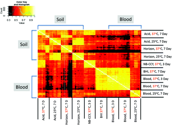
Proteins that met the filtering criteria within the 81 datasets were retained for data comparisons. A total of 672 unique proteins were observed across the various spore preparations. The average number of proteins identified in each type of spore sample using these criteria was between 284 and 367 (Table 1). Despite the similar spore mass used (1 × 10⁸ spores) and the amount of protein injected onto the LC-MS/MS system normalized to 1 mg mL−1, more proteins were generally identified in the spores cultured on laboratory media than the soil agars.A Pearson correlation analysis was performed on all 81 datasets to measure the similarity of each dataset to each of the others (Fig. 1). The correlation analysis provided a measure of relative similarity from negatively correlated (−1) to uncorrelated (0) to positively correlated (+1), represented by a color scheme in the plot. The datasets from spores produced on Horizon soil at 25 °C appeared to have the best correlation to other Horizon 25 °C datasets and, to a lesser degree, the 25 °C Acid soil samples. However the trends were less clear for the remainder of the datasets from soil. The NB-CCY 37 °C datasets appeared to be most similar within the replicates of that medium type. The BHI 37 °C and blood agar 37 °C datasets appear to have better correlation to one another than to the blood agar 25 °C datasets and those from sporulated on other media.
While some general trends appear in the correlation analysis data, the specific proteins that differ between sporulated samples needed to be identified to help elucidate differences between natural vs. laboratory environments. To identify these protein profiles, three comparisons were done. The first comparison was performed to find the proteins that distinguish between laboratory and soil media conditions. The second comparison was performed to identify proteins that distinguish between cultures sporulated at 25 °C and 37 °C within the blood and soil media datasets. Finally, comparisons between specific media to find proteins that distinguished Horizon soil from NB-CCY as well as Horizon soil from blood agar media were completed.
Protein profiles in soil vs. laboratory media
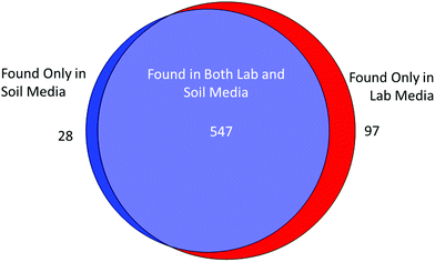
Of the 672 spore proteins, 547 were shared between one or more of the spore samples produced on soil as well as one or more spore samples produced on laboratory media (Fig. 2). Of the remaining proteins, 97 proteins were found only following sporulation in at least one type of laboratory media. By contrast, 28 proteins were found only in soil media data sets and were absent in all laboratory media datasets. | ||
| Fig. 2 Venn diagram of proteins found in soil or laboratory media using spectral count values used for 672 proteins found with at least 2 counts and at least 10% of data sets. | ||
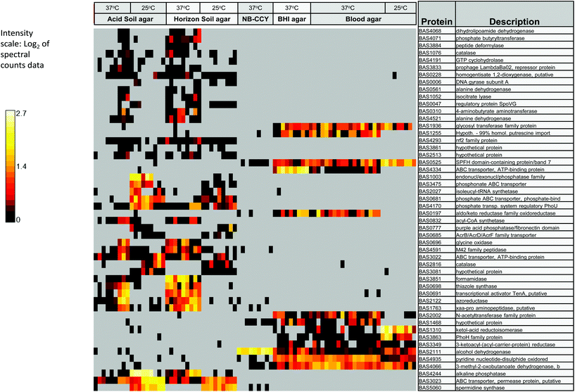
To identify proteins that were differentially expressed in sporulated cultures cultivated on soil vs. laboratory media, significance testing on relative protein abundances was performed. Proteins meeting the statistical significance (<0.05) and minimum log2 abundance difference (5.0) criteria were considered differentially expressed in datasets from soil agars (Acid or Horizon) and laboratory media (BHI agar, blood agar, or NB-CCY). A total of 154 proteins met this criteria and included proteins that were both unique to one group and those that were significantly increased in either soil or lab media; 37 proteins were enriched in samples sporulated on soil, and 117 proteins were enriched following sporulation on laboratory media (ESI Table 1†). Fig. 3 contains a heat map of 50 of the 154 proteins observed to be differentially present in spores generated on laboratory and soil media, including all of the proteins increased in or found only in spores from soil agars. The additional 104 proteins not included in Fig. 3 were only found in one or more types of laboratory media. Within these data, distinct clusters of proteins were present in BHI and blood media datasets but largely absent in the NB-CCY broth, Acid and Horizon soil agar datasets. Likewise, there were proteins present in the two types of immobilized soils that were largely absent in the NB-CCY broth, BHI and blood media datasets.
 | ||
| Fig. 3 Heat map showing the distribution of differentially expressed proteins in soil vs. lab media for 50 of the 154 proteins differentially regulated between the two growth conditions. | ||
The proteins enriched in laboratory media samples generally fell into the following predicted functional categories: nucleotide triphosphate metabolism, cysteine to thiamine metabolism, and general sugar and energy metabolism (specifically glycolysis, pyruvate metabolism and the TCA cycle) (Table 3). Additional proteins that showed increased abundance in laboratory media are annotated to be involved in fatty acid biosynthesis and degradation as well as valine, leucine, isoleucine amino acid metabolism. A number of ABC transporters annotated to be specific for amino acids, oligopeptides, lipoproteins, heme, sodium, and phosphonate were more abundant on laboratory media. Finally, the rod-shape determining protein Mbl and those involved in cell wall biosynthesis were also generally enriched in these samples.
| Soil vs. laboratory media | 25 °C vs. 37 °C | ||
|---|---|---|---|
| Increased in soil | Increased in laboratory media | Increased in 25 °C | Increased in 37 °C |
| Phosphate transport and metabolism | Glycolysis, pyruvate metabolism, TCA cycle | Cell envelope structural proteins | TCA cycle proteins |
| Catalases | Fatty acid biosynthesis/degradation | Cell wall lytic proteins | Thiamine and biotin metabolism |
| Antimicrobial peptide transport | Pyrimidine metabolism | Peptidases and Proteases | |
| Sugar, amino acid and peptide transport | |||
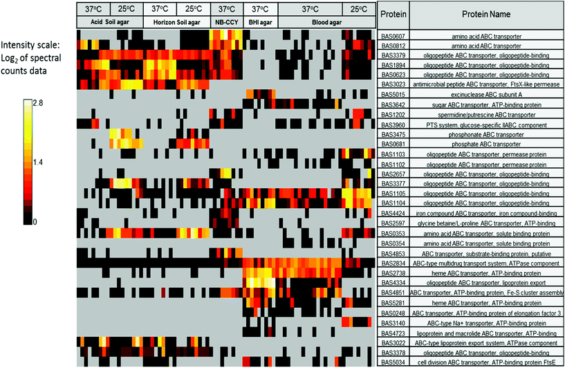
Proteins with increased abundance in sporulated cultures produced on soil agars are annotated to function in phosphate metabolism and vitamin biosynthesis. The proteins annotated to function in glycine to thiamine interconversion and as catalases were also more abundant in spores produced on one or both soil agars relative to those from laboratory medium spores (Table 3) Finally, proteins annotated as ABC transporter proteins for phosphate and antimicrobial peptide transport efflux were also detected in spores generated on the soil media (BAS3022 and BAS3023) (Fig. 4).
 | ||
| Fig. 4 Heat map showing the distribution of the detected ABC transporters based on their spectral count values across all medium types and temperatures. | ||
Specific protein profiles in Horizon soil, blood and NB-CCY medium
The correlation analysis in Fig. 1 illustrated that specific media also formed distinct clusters. Horizon, blood and NB-CCY in particular had the best correlation with themselves. Specific comparisons of these sporulation samples were performed and the results are found in ESI Tables 2 and 3.† The samples produced in blood agar and NB-CCY generally contained more abundant proteins predicted to be involved in carbon and energy metabolism. Specifically proteins predicted to be involved in phosphoenolpyruvate, ethanol and lactic acid production. The proteins annotated to be involved in nucleotide metabolism were increased in blood agar and NB-CCY cultures. By contrast, Horizon soil samples were enriched in proteins predicted to be involved in amino acid metabolism, fatty acid degradation and vitamin metabolism. Specifically, the Horizon soil samples contained more proteins annotated to function in thiamine metabolism. ABC transporter proteins annotated to function in amino acid and oligopeptide transport were increased in both blood and NB-CCY relative to Horizon soil (Fig. 4). The ABC transporters found to be increased in the Horizon soil sporulated cultures over blood and NB-CCY produced samples are annotated to be involved with phosphate, phosphonate and antimicrobial peptide transport (Fig. 4). Finally, the catalase proteins were increased in the Horizon samples as well. These were trends similar to, but more clearly defined than, the comparison of all soil agar vs. laboratory media produced samples.The media samples were analyzed for their elemental content using ICP-OES (Table 4). The amount of potassium, phosphorus and sodium in the soil media was lower or undetectable in the soil media using optical emission. By contrast, the amount of aluminum, manganese and barium was higher in the soil media. The decrease in phosphorus in the two soil media correlates with the increase in ABC phosphate transporters observed in samples from cultures sporulated on soil (Fig. 4).
| Analyte | Media type | |||||
|---|---|---|---|---|---|---|
| Blood agar | BHI | Horizon soil | Acid soil | NB-CCY salts | Bovine blood | |
| Aluminum | ND | ND | 954 | 248 | ND | ND |
| Barium | ND | ND | 6.4 | 5.27 | ND | ND |
| Calcium | ND | ND | 2550 | 1330 | 86![[thin space (1/6-em)]](https://www.rsc.org/images/entities/char_2009.gif) 300 300 |
203![[thin space (1/6-em)]](https://www.rsc.org/images/entities/char_2009.gif) 000 000 |
| Iron | ND | ND | 1590 | 486 | ND | 136![[thin space (1/6-em)]](https://www.rsc.org/images/entities/char_2009.gif) 000 000 |
| Magnesium | 69.7 | ND | 1200 | 133 | 42![[thin space (1/6-em)]](https://www.rsc.org/images/entities/char_2009.gif) 200 200 |
8890 |
| Manganese | ND | ND | 43.4 | 14.2 | ND | ND |
| Phosphorus | 599 | 971 | 64.9 | ND | 1![[thin space (1/6-em)]](https://www.rsc.org/images/entities/char_2009.gif) 380 380![[thin space (1/6-em)]](https://www.rsc.org/images/entities/char_2009.gif) 000 000 |
83![[thin space (1/6-em)]](https://www.rsc.org/images/entities/char_2009.gif) 800 800 |
| Potassium | 419 | 890 | ND | ND | 3![[thin space (1/6-em)]](https://www.rsc.org/images/entities/char_2009.gif) 010 010![[thin space (1/6-em)]](https://www.rsc.org/images/entities/char_2009.gif) 000 000 |
438![[thin space (1/6-em)]](https://www.rsc.org/images/entities/char_2009.gif) 000 000 |
| Sodium | 3200 | 3350 | ND | ND | 383![[thin space (1/6-em)]](https://www.rsc.org/images/entities/char_2009.gif) 000 000 |
2![[thin space (1/6-em)]](https://www.rsc.org/images/entities/char_2009.gif) 710 710![[thin space (1/6-em)]](https://www.rsc.org/images/entities/char_2009.gif) 000 000 |
| Strontium | ND | ND | 3.64 | ND | ND | ND |
| Zinc | ND | ND | 6.78 | ND | ND | ND |
Protein profiles in relation to temperature
The correlation analysis also indicated differences between the datasets related to growth temperature. The impact of sporulation at 25 °C and 37 °C was examined in both the soil and blood media spore datasets using proteins selected based on log2 spectral count abundance criteria as described above. For spores produced on soil media, a total of 144 proteins met these criteria, with 94 being differentially present in the 37 °C soil samples while 50 were differentially found in the 25 °C soil samples (ESI Table 4†). For spores produced on laboratory media containing blood, a total of 162 proteins met the filtering criteria. A nearly equal number of proteins (82) were present in the 37 °C blood samples relative to the 25 °C blood samples (80) (ESI Table 5†).While there are many differences between sporulated cultures produced at each temperature, a few consistent themes were observed independent of whether the sporulated cultures were produced on blood or the two soils. The proteins annotated to be involved in vitamin metabolism, glycolysis, and the TCA cycle were increased at 37 °C relative to 25 °C. By contrast, more proteins thought to be present on or in the vegetative cell and spore surface, such as S-layer proteins, spore coat, cell wall lytic enzymes (e.g. muramyl-alanine amidase), were found in 25 °C samples. The effect was more pronounced in blood media where an increased amount of immune inhibitors (BAS0638 and BAS1197) and cell wall enzymes were observed at the lower temperature. A similar increase in spore surface and spore coat proteins was observed during sporulation of B. cereus at 20 °C by Bressuire-Isoard et al.24
There were cases where temperature had different impacts depending on which media the spores were produced on. The proteins annotated to be involved in pyrimidine and purine metabolism were increased at 37 °C in the cultures produced on soils while these proteins were more abundant in cultures sporulated at 25 °C in blood medium. This reinforces the complexity of environmental impacts on protein expression.
ABC transporters as signatures of sporulation on different media
The cultures produced on immobilized soils showed differences in the abundance of putative ABC transporters for export of antimicrobial peptides (e.g. FtsX-like permease). Similar transporters have previously been observed to respond to the presence of antimicrobial peptides produced by other microbes.25 The spores produced on immobilized soil also had more abundant ABC transporters annotated for phosphate and phosphonate transport. This observation parallels a previous report that examined the extracellular metaproteome of chitin-rich soil dominated by Proteobacteria, Actinobacteria and Bacteroidetes where ABC transporter proteins specific for phosphate and phosphonate transport were observed.26 While these proteins have complete homology with ABC transporters described in other B. cereus group species, it is important to note that the annotation is based on homology and not demonstrated substrate specificity.Different sets of ABC transporter proteins were found to be more abundant in sporulated cultures produced on laboratory media. ABC transporters annotated to be specific for sugars, amino acids, peptides and lipoproteins were more abundant in cultures produced on laboratory media. One class of ABC transporters annotated to be specific for bacteriocin (BAS3140) and bacitracin (BAS4334) were found to be increased in cultures sporulated on blood over those produced on soil. Based on the gene annotation, these ABC transporters are predicted to have a similar function as the FtsX-like permease transporter for antimicrobial peptides found in sporulated cultures produced on soil. This observation was largely driven by the blood 37 °C datasets and the same transporters were also found in the BHI, but not NB-CCY, samples (Fig. 4).
Conclusions
The observations described here indicate that the sporulated samples from different media and temperatures contain distinct protein profiles that represent distinct functional classes. Although sporulated cultures were washed twice to remove vegetative debris, we recognize that the detected proteins likely represent proteins present within the spore, attached to spores surfaces as well as recalcitrant vegetative debris present in the samples. This possibility was previously noted by Liu et al.6 However the same report stated that water washing of B. anthracis spores would more closely mimic natural conditions than more stringent purification methods.Generalized differences between laboratory media and soil systems were observed. These observations need to be expanded to other strains and extended to B. anthracis spores found naturally in soils, however ABC transporters and proteins with specific predicted functions appear to reflect the nutritional environment experienced during sporulation. Additional patterns of protein expression related to sporulation on soil that have not been described here may also exist. These may be present within the set of identified proteins whose functions are not all fully understood as well as those proteins currently identified only as hypothetical.
These observations aid our understanding of B. anthracis sporulation in a soil environment and may be useful in discrimination of laboratory-produced spores from those formed in soil. In turn, these observations help to addresses a basic research need in microbial forensics outlined in the U.S. National Academy of Science report on science needs in microbial forensics for, “⋯development of high-confidence methodologies to distinguish among natural, accidental, and deliberate outbreaks of infectious disease”.5 Additional research on the phenotypic state of B. anthracis and other pathogens in their natural state will be needed to address this research challenge in conjunction with the genomic methods discussed in the report. Furthermore, targeted studies on specific proteins that distinguish between growth environments need to be performed. These studies are necessary to establish their detectability following varied methods of sample production, preparation, storage and environmental aging.
Conflicts of interest
There are no conflicts to declare.Acknowledgements
The authors would like to thank Charlie Doll for his instrumentation expertise and Michael Sydor for his laboratory assistance and Michelle Snyder for elemental analysis of media samples. The research described in this paper was conducted under the Laboratory Directed Research and Development Program at Pacific Northwest National Laboratory, a multiprogram national laboratory operated by Battelle for the U.S. Department of Energy. Battelle Memorial Institute operates Pacific Northwest National Laboratory for the U.S. DOE under Contract DE-AC06-76RLO.References
- A. Driks, Proc. Natl. Acad. Sci. U. S. A., 2003, 100, 3007 CrossRef CAS PubMed.
- P. Turnbull, in Anthrax in humans and animals, World Health Organization, Geneva, Switzerland, 4th edn, 2008, ISBN: 978-92-4-154753-6 Search PubMed.
- A. Hoffmaster, C. Fitzgerald, E. Ribot, L. Mayer and T. Popovic, Emerging Infect. Dis., 2002, 8, 111 Search PubMed.
- S. Anaraki, S. Addiman, G. Nixon, D. Krahé, R. Ghosh, T. Brooks, G. Lloyd, R. Spencer, A. Walsh, B. McCloskey and N. Lightfoot, Eurosurveillance, 2008, 13, 18 Search PubMed.
- National Research Council, in: Science needs for microbial forensics: developing initial international research priorities, National Academy of Sciences Press, Washington D.C. U. S. A., 2014, 252 pages, ISBN: 978-0-309-30245-6 Search PubMed.
- H. Liu, N. Bergman, B. Thomason, S. Shallom, A. Hazen and J. Crossno, et al. , J. Bacteriol., 2004, 186, 164 CrossRef CAS PubMed.
- C. Redmond, L. Baillie, S. Hibbs, A. Moir and A. Moir, Microbiol., 2004, 150, 355 CrossRef CAS PubMed.
- E.-M. Lai, N. Phadke, M. Kachman, R. Giorno, S. Vazquez and J. Vazquez, et al. , J. Bacteriol., 2003, 185, 1443 CrossRef CAS PubMed.
- S. Vilain, Y. Luo, M. Hildreth and V. Brözel, Appl. Environ. Microbiol., 2006, 72, 4970 CrossRef CAS PubMed.
- Y. Luo, S. Vilain, B. Voigt, D. Albrecht, M. Hecker and V. Brozel, FEMS Mirobiol. Lett., 2007, 271, 40 CrossRef CAS PubMed.
- M. Van Ert, W. Easterday, L. Huynh, R. Okinaka, M. Hugh-Jones, J. Ravel, S. Zanecki, T. Pearson and T. Simonson, PLoS One, 2007, 2, e461 Search PubMed.
- G. Van Ness, Science, 1971, 172, 1303 CAS.
- E. Saile and T. Koehler, Appl. Environ. Microbiol., 2006, 72, 3168 CrossRef CAS PubMed.
- T. Buhr, D. McPherson and B. W. Gutting, J. Appl. Microbiol., 2008, 105, 1604 CrossRef CAS PubMed.
- B. Deathridge-Kaiser, D. Wunschel, M. Sydor, M. Warner, K. Wahl and J. Hutchison, J. Microbiol. Methods, 2015, 118, 18 CrossRef PubMed.
- A. Lin, E. Merkley, B. Clowers, J. Hutchison and H. Kreuzer, J. Microbiol. Methods, 2015, 112, 3 CrossRef CAS PubMed.
- A. Wattam, D. Abraham, O. Dalay, T. Disz, T. Driscoll, J. Gabbard, J. Gillespie, R. Gough and D. Hix, et al. , Nucleic Acids Res., 2014, 42, D581 CrossRef CAS PubMed.
- S. Kim, N. Gupta and P. Pevzner, J. Proteome Res., 2008, 7, 3354 CrossRef CAS PubMed.
- A. Polpitiya, W.-J. Qian, N. Jaitly, V. Petyuk, J. Adkins, D. Camp and G. Anderson, et al. , Bioinformatics, 2008, 24, 1556 CrossRef CAS PubMed.
- M. Li, W. Gray, H. Zhang, C. Chung, D. Billheimer, W. Yarbrough, D. Liebler, Y. Shyr and R. Slebos, J. Proteome Res., 2010, 9, 4295 CrossRef CAS PubMed.
- W. Old, K. Meyer-Arendt, L. Aveline-Wolf, K. Pierce, A. Mendoza, J. Sevinsky, K. Resing and N. Ahn, Mol. Cell. Proteomics, 2005, 4, 1487 CAS.
- P. Turnbull, P. Lindeque, J. Le Roux, A. Bennett and S. Parks, J. Appl. Microbiol., 1998, 84, 667 CAS.
- S. Bellan, P. Turnbull, W. Beyer and W. Getz, Appl. Environ. Microbiol., 2013, 79, 3756 CrossRef CAS PubMed.
- C. Bressuire-Isoard, I. Bornard, A. Henriques, F. Carlin and V. Broussolle, Appl. Environ. Microbiol., 2016, 82, 232 CrossRef CAS PubMed.
- S. Dintner, R. Heermann, C. Fang, K. Jung and S. Gebhard, J. Biol. Chem., 2014, 289, 27899 CrossRef CAS PubMed.
- A. Johnson-Rollings, H. Wright, G. Masciandaro, C. Macci, S. Doni, L. Calvo-Bado, S. Slade, C. Plou and E. Wellington, ISME J., 2014, 6, 2148 CrossRef PubMed.
Footnote |
| † Electronic supplementary information (ESI) available. See DOI: 10.1039/c7an01412k |
| This journal is © The Royal Society of Chemistry 2018 |

