Hakim Rahmaa,
Rachel Nickelb,
Elizabeth Skoropatab,
Yaroslav Wroczynskyjb,
Christopher Rutleyb,
Palash K. Mannab,
Ching Hung Hsiaoc,
Hao Ouyangc,
Johan van Lierop*b and
Song Liu*a
aDepartment of Biosystems Engineering, Faculty of Agricultural and Food Sciences, University of Manitoba, Winnipeg, Canada R3T 2N2. E-mail: Song.Liu@umanitoba.ca; Fax: +1-204-474-7512; Tel: +1-204-474-9616
bDepartment of Physics and Astronomy, University of Manitoba, Winnipeg, MB R3T 2N2, Canada. E-mail: Johan.van.Lierop@umanitoba.ca
cMaterials Science and Engineering, National Tsing Hua University, Hsinchu, Taiwan
First published on 5th July 2016
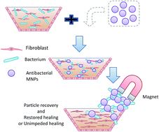
We propose a new and highly effective tool to add to the ever shrinking toolbox for combating infections caused by antibiotic resistant bacteria; N-chloramine and quaternized N-chloramine were coated onto iron-oxide magnetic nanoparticles to generate antibacterial MNPs. Two differently-sized primary iron-oxide nanoparticles (3 nm and 10 nm) were synthesized and coated with silica and (3-chloropropyl)triethoxysilane, allowing subsequent introduction of N-chloramine precursors – dimethyl hydantoin (DMH) and quaternized dimethyl hydantoin (QDMH). The functionalized MNPs (MNP@DMH and MNP@QDMH) have a clear core–shell structure as evidenced by TEM images. Fe3O4 was identified (by combining X-ray diffraction with Mössbauer spectroscopy) to be the iron oxide in the 10 nm MNPs, while γ-Fe2O3 and Fe3O4 were the 3 nm MNP's oxide phases. Both MNPs (3 nm and 10 nm) have good magnetic responses, with saturation magnetizations of 40 ± 4 emu g−1 and 65 ± 2 emu g−1, respectively. Chlorination activated the antibacterial function and yielded two antibacterial MNPs: MNP@DMCl and MNP@QASCl. At the equivalent [Cl+] of 50 ppm, both coatings demonstrated fast inactivation of the model bacteria methicilin-resistant Staphylococcus aureus (MRSA) and multi-drug resistant (MDR) Pseudomonas aeruginosa. For either size of primary MNPs, MNP@QDMCl is more effective than MNP@DMCl. A hand-held magnet could quickly remove >99% of the functionalized MNPs from a wound simulant within 2 minutes.
Iron-oxide nanoparticles have very good biocompatibility and low cytotoxicity. Synthesis can be very low cost and “green” (both in synthesis and disposal).5 Functionalization of iron-oxide nanoparticles with biocides is an area of significant research interest. Due to the challenge of coating individual 5–10 nm primary iron-oxide nanoparticles, the previous efforts focused on trapping 5–10 nm primary iron-oxide nanoparticles in the matrix of silica or poly(styrene-acrylic acid) to yield 134–228 nm antibacterial coated magnetic particles.6–8 As the biocide is grafted onto the surface of nanoparticles, it allows for a prolonged, localized and targeted effect on an infected wound. In this way, higher antimicrobial concentrations can be applied to target cells. Keeping in mind that the antimicrobial performance of N-chloramine based materials is strongly dependent on materials' surface area and contact time of microorganism with the material, the smaller proposed antimicrobial nanoparticles have the larger overall surface area leading to a more potent antimicrobial activity. Also, the bioactivity of antimicrobial nanoparticles is reportedly associated with surface charge. For example, positively charged silver nanoparticles were found to present superior antibacterial activity compared to neutral and negatively charged silver nanoparticles.9,10
In this paper, instead of synthesizing antibacterial magnetic nanoparticles with large sizes (>100 nm), we present the synthesis of core–shell magnetic nanoparticles (MNPs) of biocompatible iron-oxides 3 to 10 nm in diameter, coated with silica, and functionalized with the N-chloramine moiety that have an overall size of 20 to 40 nm, depending on parent particle and coatings. To help boost the antimicrobial effect of the MNPs, a positive charge was introduced onto the MNPs silica coating. The positively charged 10 nm MNPs demonstrated superior antibacterial activity, and could be easily recovered from a simulated wound exudate with a simple magnet.
1H NMR (CDCl3, 300 MHz, δ) 7.08 (m, 1H), 4.32 (s, 2H), 2.29 (s, 6H), 1.41 (s, 6H); 13C NMR (CDCl3, 75 MHz, δ) 178.6, 157.4, 60.7, 58.7, 42.7, 25.2.
To quantitatively assess the loaded active chlorine [Cl+] on MNPs, a redox titration was adopted. The MNPs were mixed with 10 mL sodium thiosulfate solution (0.001 N) and 30 mL of water. The suspension was stirred for 30 min. Then, 2 mL of acetic acid at 5% was added and the remaining sodium thiosulfate was titrated using iodine solution at 0.001 N.
Fig. 1 presents typical X-ray diffraction patterns of Fe-oxide nanoparticles with and without SiO2 coating. Diffraction patterns showed the reflections characteristic of the Fe-oxide spinel structure. All reflections are Scherrer broadened by significantly different amounts between systems, indicating that the crystallite size of the two Fe-oxide nanoparticle systems differed substantially. A Rietveld refinement of the patterns incorporating Scherrer broadening effects (to ascertain the volume averaged nanocrystallite diameters) confirmed the spinel structure (Fdm) of the Fe-oxides Fe3O4 or γ-Fe2O3 as expected from Massart-based synthesis. Refined lattice parameters of a = 8.296(1) Å for the small (more broadened pattern lineshape) 3 nm diameter MNPs and a = 8.374(1) Å for the large 10 nm diameter MNPs are also consistent with a spinel Fe-oxide (a ∼ 8.33 Å for bulk γ-Fe2O3 and a ∼ 8.39 Å for bulk Fe3O4). Keeping in mind that deviation from bulk parameters is common for nanoparticles, this difference in lattice parameters is large enough to indicate that the 3 nm MNPs are a mixture of the two oxide phases while the 10 nm MNPs are Fe3O4. This has implications on the magnetic response of the biocide coated MNPs (e.g. the overall magnetization of bulk γ-Fe2O3 is less than that of Fe3O4) for removal from, e.g. wounds, with an external field from a hand-held magnet. Therefore, we turned to a very composition and structure sensitive probe of Fe-compounds, Mössbauer spectroscopy (ESI†) that identify clearly that the 3 nm MNPs are a mixture of γ-Fe2O3 and Fe3O4, while the 10 nm MNPs are Fe3O4. The presence of a broad, amorphous “hump” between 20 and 28 degrees 2θ in Fig. 1c confirms the successful coating of Fe-oxide NPs with SiO2, giving Fe-oxide@SiO2 MNPs.
Transmission electron microscopy images for the 3 nm and 10 nm Fe-oxide@SiO2 MNPs are shown in Fig. 2. The particle sizes (Fig. 2d) were measured using ImageJ,12 and fitted with a lognormal distribution and provided Fe-oxide core sizes of diameter Dsmall = 4.4 ± 0.2 nm and ln(σsmall) = 0.08 ± 0.01, and Dlarge = 9.6 ± 0.2 nm and ln(σlarge) = 0.07 ± 0.01 for the small and large particles, respectively, in good agreement with the 3 nm and 10 nm volume averaged particle diameters from XRD pattern refinements. It should be noted that the larger particles show also a small population of large sizes (D ∼ 35–50 nm), that was not observed for the small particles. After the magnetic particles were coated with silica, a thin layer of SiO2 was observed in TEM (e.g. Fig. 2b).
The field dependent magnetization of the MNPs (hysteresis loop measurements) for the 3 nm Fe-oxide nanoparticles, presented in Fig. 3a, show a saturation magnetization (Ms) at 300 K of 40 ± 4 emu g−1, which is quite close to the value for that of nanoscale spinel Fe-oxide,13 γ-Fe2O3 (74 emu g−1 in the bulk where nanoscale systems typically show a significantly reduced Ms from the bulk due to finite-size effects). A further decrease in Ms for the 3 nm Fe-oxide/SiO2 core/shell MNPs of 25 ± 3 emu g−1 (Fig. 3b) is a result of the non-magnetic silica fraction that adds to the total sample mass (but has a diamagnetic contribution that “cancels” the ferromagnetic response of the cores) and provides an estimate of the volume fraction of the SiO2 versus Fe-oxide amounts to be 56 ± 3% (by volume), consistent with TEM results. For the 10 nm MNPs (Fig. 3c), Ms = 65 ± 2 emu g−1, consistent with a much better well crystallized structure of the nanoparticles, in addition to the particle size, and in keeping with a Fe3O4-phase nanoparticle system (84 emu g−1 in the bulk) (i.e. as identified by TEM and XRD). With the addition of the SiO2 shell, Ms = 60 ± 1 emu g−1; an estimate of the volume fraction of the SiO2 versus Fe-oxide amounts to be 17 ± 1% (by volume), in order to introduce the N-chloramine moiety to the MNPs, a functional silane (3-chloropropyltriethoxysilane: CPTES) was incorporated onto the structure formed by the Fe-oxide/SiO2 core/shell system. The synthesis of MNP@DMH and MNP@QAS nanoparticles was carried out in dimethylformamide (DMF) by reacting MNP@CPTES with DMH–K+ and (CH3)2NCH2DMH, respectively, through a nucleophilic substitution. The conversion of surface bound N–H to N–Cl was achieved with sodium hypochlorite to generate antibacterial MNPs.
Fig. 3 Hysteresis loops measured at 300 K for the 3 nm (a) Fe-oxide and (b) Fe-oxide@SiO2, and for the 10 nm (c) Fe-oxide and (d) Fe-oxide@SiO2 nanoparticles. |
After the coating of CPTES and DMH, a thick additional layer was observed clearly in TEM images for both the 3 and 10 nm MNPs, shown in Fig. 4. The thickness of the final coating was estimated to be, 1.1 ± 0.7 nm for the 3 nm Fe-oxide@SiO2 MNPs, and 4.1 ± 0.3 nm for the 10 nm Fe-oxide@SiO2 MNPs, also determined using ImageJ. The DLS results (discussed below) show that the size distribution of the particles before and after the coating is narrow and are consistent with these results obtained by TEM. However “clumps” of Fe-oxide@SiO2 nanoparticles do form larger assemblies from multiple MNPs encapsulated by SiO2 during the Stöber reaction.
FT-IR spectra (Fig. 5) were recorded to identify the functional groups at different steps of the synthesis route. The MNPs (3 nm and 10 nm) present a characteristic peak at 577 cm−1 corresponding to the vibration bond of Fe–O. SiO2 coated particles (MNP@SiO2) show a peak at 3389 cm−1 due to the stretching vibration of SiO–H groups. The strong and broad peak at 1037 cm−1 and a shoulder peak at 1125 cm−1 are assigned to the asymmetric stretching vibration mode of Si–O–Si. The coating with CPTES does not present any new peaks in the infrared spectra because of the overlapping of the bands corresponding to CPTES with the bands of the particles' SiO2 shell.
Two peaks characteristic of two carbonyl groups in hydantoin were observed at 1698 cm−1 and 1755 cm−1 in the FTIR spectra of 3 nm and 10 nm MNP@DMH, and at 1730 cm−1 and 1785 cm−1 in the FTIR spectra of 3 nm and 10 nm MNP@QAS. The noticeable shift of hydantoin peaks was possibly attributed to the proximity effect of the quaternary ammonium salt to the hydantoin in the structure.
To quantify the effects of functionalization of the MNP formulations with QAS and DMH, photon correlation spectroscopy (PCS/DLS) experiments were performed to determine the nanoparticle hydrodynamic sizes and size distributions. Suspensions of the Fe-oxide/SiO2 MNPs were made by adding 10 mL methanol to 5 mg of dried nanoparticles followed by ultrasonic agitation for 30 minutes. The functionalized samples were made also into dilute suspensions by adding 100 μL of concentrated stock suspension to 10 mL methanol. Light scattering measurements at seven scattering angles (30–120° in 15° intervals) were performed. The collected autocorrelation functions were fit to a distribution in translational Brownian relaxation time using the software DynaLS. Distributions in relaxation time were transformed to hydrodynamic diameter using the known viscosity and refractive index of methanol. The results of this fitting procedure are shown in Fig. 6 for a particular scattering angle of 45°, showing clearly an increase in the hydrodynamic size with additional functionalization. To assess more completely the hydrodynamic diameters of the nanoparticle samples, the mean sizes determined at all scattering angles were averaged, weighted by the total intensity of the scattered light recorded during each measurement (that is inevitably biased by the larger particles scattering more light). The mean hydrodynamic diameters and uncertainties (taken as the intensity weighted average deviation in the distributions) are summarized in Table 1. The increase in hydrodynamic diameter with the different coating agents tracks consistently between the core size of the MNP samples, and speaks to the “corona” surrounding the MNPs. The SiO2 and CPTES coating could encapsulate more than one single primary MNP (as observed in the TEM shown in Fig. 4) to yield nanoparticle with diameters between 20 and 40 nm. MNP@QAS nanoparticles have slightly bigger hydrodynamic diameters than their MNP@DMH counterparts that could be ascribed to the higher degree of hydration of quaternized DMH (QAS) than DMH.
Fig. 6 Distribution of hydrodynamic diameters determined at a fixed scattering angle (45°) for the Fe-oxide@SiO2 and fully functionalized nanoparticle systems. |
Samples | Hydrodynamic diameter, D, (nm) | log-normal distribution width, ln(σD) |
---|---|---|
3 nm Fe-oxide@SiO2 | 23.7 ± 0.1 | 0.06 ± 0.02 |
3 nm Fe-oxide@DMH | 28.7 ± 0.1 | 0.03 ± 0.01 |
3 nm Fe-oxide@QAS | 33.2 ± 0.2 | 0.07 ± 0.03 |
10 nm Fe-oxide@SiO2 | 22.7 ± 0.1 | 0.06 ± 0.01 |
10 nm Fe-oxide@DMH | 31.4 ± 0.1 | 0.03 ± 0.01 |
10 nm Fe-oxide@QAS | 33.7 ± 0.3 | 0.08 ± 0.03 |
Zeta-potential (ξ) is a critical parameter that is closely related to particle surface charge. At basic pH, all the particles presented ξ = −40 mV as shown in Fig. 7, indicating good colloidal stability and low aggregation. At acidic pH, a noticeable change was observed between MNP@DMH and MNP@QAS. MNP@QAS nanoparticles presented more positive profiles at acidic pHs compared to MNP@DMH nanoparticles. The isoelectric point (IEP) refers to the pH at which particles do not carry net electrical charge. The IEP of 3 nm MNP@QAS is ∼5, higher than that 3 nm MNP@DMH. The IEP of 10 nm MNP@DMH is lower than pH 3.9 whereas 10 nm MNP@QAS has an IEP of ∼5.5. These results indicate clearly that the necessary incorporation of positive charge on the surface of the particles has occurred.
The activation of antibacterial functions (conversion of N–H to N–Cl) with sodium hypochlorite is a crucial step prior to an antimicrobial test. Activation permits the loading of oxidative chlorine onto the surface of the MNPs. The activation process was performed by mixing MNP@DMH and MNP@QAS with sodium hypochlorite, which leads to the transfer of active chlorine (Cl+) from ClO− to the hydantoin function groups on the surface of the MNPs through electrophilic substitution. To qualitatively assess this conversion, a few drops of 5% potassium iodide solution was added to the chlorinated and dried MNP nanoparticles. The successful conversion of N–H to N–Cl was evident from the appearance of a yellow-brownish color (2I− + Cl+ → I2 + Cl1−). Table 2 presents the quantitative results of active chlorine loading ([Cl+] in ppm) on different testing samples. The 10 nm MNPs had lower chlorine loadings than the 3 nm MNPs. Difference in chlorine concentration between MNP@DMH and MNP@QAS is mainly attributed to the reactivity of their starting N-chloramine precursors with the C–Cl bond on MNP@CPTES. Chemically (CH3)2NCH2DMH possesses a bulky structure with low degree of freedom, which makes its nucleophilic reaction with chloro-functional groups on CPTES/SiO2 less favourable than with DMH–K+.
3 nm MNP@DMCl (ppm) | 3 nm MNP@QASCl (ppm) | 10 nm MNP@DMH (ppm) | 10 nm MNP@QASCl (ppm) |
---|---|---|---|
2276 ± 85 | 818 ± 64 | 1202 ± 121 | 485 ± 90 |
The antimicrobial performance of N-chloramine and N-chloramine modified materials is directly proportional to the active chlorine loading and the contact time. Therefore, the concentrations of N-chloramine MNPs were adjusted to get the same [Cl+] (50 ppm) in the bacterial suspension to allow us to examine the effects of the MNPs size and the positive charge included into the structures. Two bacterial strains which are mainly responsible for wound associated infections were chosen as representative bacteria for the antibacterial test. They are Gram-negative bacterium MDR P. aeruginosa, and MRSA. Table 3 presents the antimicrobial efficacy of various N-chloramine coated MNPs against MRSA (non-activated MNPs and MNP@SiO2 served as controls). The control MNPs were not effective in killing MRSA, and the reduction was less than 40% even after 2 hours of contact. 3 nm MNP@DMCl showed a total kill of MRSA (>6 log reduction) after 2 h of contact time whereas 1 h was sufficient for 3 nm MNP@QASCl to result a total kill. 10 nm MNPs exhibited the same trends observed for their smaller analogues. 10 nm MNP@DMCl presented 6-log reduction of bacteria after 2 h, and 10 nm MNP@QASCl could achieve a complete wipe-out of MRSA after only 30 min of contact. These comparative results indicate clearly the positive effect of the QAS incorporated into the MNP coating structure for the antimicrobial efficacy. The effect of the size between the 3 nm particles and the 10 nm was not obvious, which speaks to a population of several MNPs encapsulated during the SiO2 and CPTES coating processes so that the conformational sizes were equivalent for some of the 3 and 10 nm MNP coated systems. In the previously reported effort of introducing hydantoin based N-chloramine onto 228 nm Fe-oxide@SiO2 MNPs, 80% reduction of S. aureus was obtained after 1 hour contact, despite the lower bacterial concentration (105–6 cfu mL−1) and the higher MNP concentration (200 mg mL−1) used. 6 log reduction of MRSA achieved by 3 nm MNP@QASCl in 1 hour and 10 nm MNP@QASCl in 30 min clearly indicates the overall improvement in DMH coating and bacterial contact due to both the surface positive charge and the larger surface area of the smaller sized MNPs.
Samples | Cl (mg mL−1) | Particles (mg mL−1) | Bacteria reduction at various contact times (min) | ||||
---|---|---|---|---|---|---|---|
5 min | 15 min | 30 min | 1 h | 2 h | |||
a Inoculum concentration: 8.75–9.5 × 10⁶ CFU mL−1. | |||||||
3 nm MNP@DMCl | 0.05 | 22 | 9 ± 11 | 14 ± 16 | 25 ± 5 | 26 ± 5 | 100 ± 0 |
3 nm MNP@QASCl | 0.05 | 61 | 16 ± 3 | 26 ± 16 | 21 ± 2 | 100 ± 0 | 100 ± 0 |
10 nm MNP@DMCl | 0.05 | 41 | 1 ± 4 | 4 ± 6 | 22 ± 2 | 22 ± 20 | 100 ± 0 |
10 nm MNP@QASCl | 0.05 | 103 | 44 ± 9 | 87 ± 2 | 100 ± 0 | 100 ± 0 | 100 ± 0 |
3 nm MNP@SiO2 | 0 | 61 | 34 ± 29 | 34 ± 7 | 9 ± 11 | 18 ± 9 | 24 ± 6 |
10 nm MNP@SiO2 | 0 | 103 | 42 ± 2 | 27 ± 12 | 41 ± 6 | 74 ± 1 | 26 ± 2 |
3 nm MNP@DMH | 0 | 22 | 15 ± 6 | 10 ± 31 | 24 ± 12 | 14 ± 7 | 4 ± 5 |
3 nm MNP@QAS | 0 | 61 | 26 ± 16 | 26 ± 12 | 25 ± 17 | 11 ± 4 | 12 ± 18 |
10 nm MNP@DMH | 0 | 41 | 38 ± 4 | 31 ± 9 | 35 ± 7 | 40 ± 4 | 30 ± 16 |
10 nm MNP@QAS | 0 | 103 | 46 ± 8 | 43 ± 2 | 43 ± 6 | 44 ± 5 | 37 ± 6 |
The antimicrobial performance of the MNPs against MDR P. aeruginosa is presented in Table 4. The non-chlorinated particles and SiO2 coated MNPs showed a relatively high toxicity against MDR P. aeruginosa. This toxicity is proportional to the amount of the particles added in the bacterial solution. The non-chlorinated QAS showed a higher toxicity against MDR P. aeruginosa compared to the non-chlorinated DMH. These results indicate a higher affinity between MDR P. aeruginosa and MNP@QAS because of the charge attraction (consistent with the ξ-potential measurements described above). The activated MNPs had a higher antimicrobial effect than their non-activated analogues. 3 nm MNP@DMCl affected a total reduction after 2 h of contact time, whereas 3 nm MNP@QASCl resulted in a total reduction after 15 min. The 10 nm MNP coated systems exhibited the same trends where 10 nm MNP@DMCl resulted in only 3 log reduction of MRSA after 2 h and 6 log reduction was obtained by 10 nm MNP@QAS after 15 min of contact time. These comparative results also indicate an improvement the antimicrobial efficacy with the incorporation of positive charge at the surface, like the 3 nm MNP coated systems.
Samples | Cl (mg mL−1) | Particles (mg mL−1) | Bacteria reduction at various contact times (min) | ||||
---|---|---|---|---|---|---|---|
5 min | 15 min | 30 min | 1 h | 2 h | |||
a Inoculum concentration: 1.56–2.63 × 10⁶ CFU mL−1. | |||||||
3 nm MNP@DMCl | 0.05 | 22 | 62 ± 4 | 64 ± 5 | 73 ± 3 | 76 ± 6 | 100 ± 0 |
3 nm MNP@QASCl | 0.05 | 61 | 76 ± 3 | 100 ± 0 | 100 ± 0 | 100 ± 0 | 100 ± 0 |
10 nm MNP@DMCl | 0.05 | 41 | 50 ± 3 | 59 ± 1 | 60 ± 4 | 80 ± 3 | 99.95 ± 0.01 |
10 nm MNP@QASCl | 0.05 | 103 | 74 ± 7 | 100 ± 0 | 100 ± 0 | 100 ± 0 | 100 ± 0 |
3 nm MNP@SiO2 | 0 | 61 | — | 57 ± 10 | 29 ± 2 | ||
10 nm MNP@SiO2 | 0 | 103 | 70 ± 2 | 54 ± 10 | |||
3 nm MNP@DMH | 0 | 22 | — | 37 ± 11 | 13.9 ± 5.8 | ||
3 nm MNP@QAS | 0 | 61 | — | 85 ± 2 | 92 ± 1 | ||
10 nm MNP@DMH | 0 | 41 | — | 54 ± 3 | 33 ± 1 | ||
10 nm MNP@QAS | 0 | 103 | — | 70 ± 5 | 71 ± 3 |
Finally, the enhancement of the antibacterial activity of the MNPs is absolute with the introduction of the cationic N-chloramine onto the surfaces. A mechanism can be proposed to explain the origin of this boost. Since both Gram-positive and Gram-negative bacteria are negatively charged owing to the presence of the teichoic acid and phosphate on their membrane and cell walls, adding a positive charge on a support facilitates the adsorption of MNPs onto bacteria for a faster oxidative chlorine transfer to the target sites, leading to the most effective bacterial deaths.
Since the biocide N-chloramine is non-selective against bacteria and human skin cells, it is desirable to remove the N-chloramine based antibacterial MNPs from the wound bed after they inactivate bacteria in the infected wound to minimize residual toxicity. We conducted an experiment of recovering small and large MNP@DMH and MNP@QAS from a wound simulant with the following ingredients: 6.8 g L−1 NaCl, 2.2 g L−1 KCl, 25 g L−1 NaHCO3, 3.5 g L−1 KH2PO4 and 20 g L−1 BSA. As shown in Fig. 8, the tested MNPs reached >99% recovery after 60–120 seconds of applying an external magnetic field, and 10 nm MNPs showed a faster recovery (>80% recovery after 15 s) than the 3 nm MNPs (55% recovery after 15 s). The faster recovery of 10 nm MNPs might be due to the higher intrinsic magnetization of the Fe3O4 phase and the larger primary core size. In addition, the more rapid recovery of the 10 nm MNP@QAS compared to the 10 nm MNP@DMH is indicative of surface charge (e.g. “corona” effects) where the DMH is more strongly bound to the wound simulant – stronger static magnetic fields, or a combination of static and dynamic applied magnetic fields, should further improve the speed of recovery to offset this electrostatic charge effect.
Footnote |
† Electronic supplementary information (ESI) available. See DOI: 10.1039/c6ra13389d |
This journal is © The Royal Society of Chemistry 2016 |