Metal chalcogenide quantum dots: biotechnological synthesis and applications

J. Mal*ab, Y. V. Nancharaiahac, E. D. van Hullebuschb and P. N. L. Lensad
aUNESCO-IHE, Westvest 7, 2611 AX, Delft, The Netherlands. E-mail: j.mal@unesco-ihe.org
bBiofouling and Biofilm Process Section, Water and Steam Chemistry Division, Bhabha Atomic Research Centre, Kalpakkam-603102, Tamil Nadu, India
cUniversité Paris-Est, Laboratoire Géomatériaux et Environnement (LGE), EA 4508, UPEM, 77454 Marne-la-Vallée, France
dDepartment of Chemistry and Bioengineering, Tampere University of Technology, P.O. Box 541, Tampere, Finland

Received 1st April 2016 , Accepted 12th April 2016

First published on 13th April 2016


Abstract

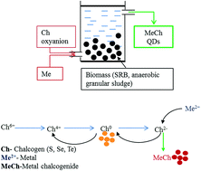
Metal chalcogenide (metal sulfide, selenide and telluride) quantum dots (QDs) have attracted considerable attention due to their quantum confinement and size-dependent photoemission characteristics. QDs are one of the earliest products of nanotechnology that were commercialized for tracking macromolecules and imaging cells in life sciences. An array of physical, chemical and biological methods have been developed to synthesize different QDs. Biological production of QDs follow green chemistry principles, thereby use of hazardous chemicals, high temperature, high pressure and production of by-products is either minimized or completely avoided. In the past decade, significant progress has been made wherein a diverse range of living organisms, i.e. viruses, bacteria, fungi, microalgae, plants and animals have been explored for synthesis of all three types of metal chalcogenide QDs. However, better understanding of the biological mechanisms that mediate the synthesis of metal chalcogenides and control the growth of QDs is needed for improving their yield and properties as well as addressing issues that arise during scale-up. In this review, we present the current status of the biological synthesis and applications of metal chalcogenide QDs. Where possible, the role of key biological macromolecules in controlled production of the nanomaterials is highlighted, and also technological bottlenecks limiting widespread implementation are discussed. The future directions for advancing biological metal chalcogenide synthesis are presented.


1. Introduction

Research and development on inorganic nanomaterials such as metal, metal oxide and semiconductor nanoparticles has emerged into a cutting edge multidisciplinary nanotechnology.1,2 Particularly, semiconductor metal chalcogenide quantum dots (QDs) have attracted wide interest for applications from material science to medicine.3,4 Physical and chemical methods exist for synthesis of highly stable metal chalcogenide nanoparticles (MeCh NPs) with defined properties.5,6 But most of these methods use energy intensive procedures with highly toxic precursors and reagents. On the other hand, microorganisms are known to create a variety of nanomaterials in a most energy efficient manner using non-toxic, inexpensive and renewable reagents. Therefore, biotechnological synthesis of nanomaterials is increasingly considered for the production of nanomaterials in an environmental friendly way following green chemistry principles.1,7,8 This review presents the state of art research on microbial synthesis of metal chalcogenide nanoparticles, the properties of biogenic metal chalcogenides and their applications. The challenges and perspectives of microbial synthesis of metal chalcogenides are also discussed.

2. Quantum dots and their properties

QDs are semiconductor nanoparticles made up of elements from groups II–VI or III–V of the periodic table and characterized as inorganic colloids with physical dimensions of 1 to 20 nm, smaller than the bulk-exciton Bohr radius, i.e. the distance in an electron–hole pair.9 Due to this very small size, the QDs exhibit distinct optical and electronic properties.10,11 Since the bandgap energy depends on the particle size, when the particle size is in the nano-scale, optical properties such as fluorescence excitation and emission can be “tuned” by altering the particle size, shape or surface structure.12 QDs have attracted applications as inorganic fluorophores because of bright, “size-tunable” fluorescence with narrow symmetric emission bands, and high photostability.13 The distinct separation between the excitation and emission spectra of the QDs make them better with the detection sensitivity, as the entire emission spectra of QDs can be detected.14

3. Metal chalcogenide QDs

Chalcogens are the chemical elements of the group 16 of the periodic table such as oxygen, sulfur (S), selenium (Se), tellurium (Te) and polonium (Po). The metalloid chalcogens such as S, Se and Te are used in semiconductors and in the preparation of MeCh. Among the various colloidal semiconductor nanoparticles, MeCh NPs such as CdS, ZnS, CdSe, and CdTe have attracted considerable attention due to their quantum confinement effects and size dependent photoemission characteristics.15 The fluorescent MeCh NPs are superior to organic fluorophores in terms of narrow absorption and emission spectra, quantum yield and photostability.6 The photooptical and photovoltaic properties of MeCh are particularly suited for their use in solar cells and optoelectronic sensors.16,17 QDs are widely used in the field of biology and medicine for imaging, sensing18–24 and tracking particles or cells25,26 including fluorescent biolabelling and cancer detection.27

MeCh NPs are synthesized using various methods such as microwave heating,28,29 microemulsion,30,31 ultrasonic irradiation32 and several other chemical methods.33–35 Newer methods are being developed to produce high quality MeCh NPs and to make the synthetic procedures simpler and scalable. In general, most of the current synthetic procedures use hazardous solvents, explosive precursors, and high temperatures (230–250 °C).10,36 In order to meet the requirements and exponentially growing demand, there is a need to develop green methods which are environmental friendly by using renewable materials instead of toxic and hazardous chemicals. In this context, synthesis of metal chalcogenide nanoparticles by microorganisms has attracted interest as a viable alternative for the chemical synthesis routes.

4. Biological metal chalcogenides

Almost all organisms (viruses, bacteria, fungi, algae and animals) and some biomolecules have been studied for their role as catalysts or nucleating agents for synthesis of MeCh. The vast majority of the studies focused on the use of microorganisms such as bacteria and fungi because of their rapid growth, ease of maintenance and handling.1 In anaerobic conditions, microorganisms oxidize organic compounds by utilizing chalcogen oxyanions (e.g. sulfate or selenate) as terminal electron acceptors in their metabolism. In this process, sulfur, selenium and tellurium oxyanions are reduced to the corresponding chalcogenides sulfide, selenide and telluride, respectively. The microbially produced chalcogenide reacts with the dissolved metal ions (e.g. Zn, Cd, and Pb) to form MeCh NPs (Fig. 1).
image file: c6ra08447h-f1.tif
Fig. 1 General schematic for biological synthesis of metal chalcogenide QDs.

Biological methods for manufacturing nanomaterials are in general considered safe, cost-effective and environment-friendly processes.37,38 The main aim is to develop a protocol to produce nanomaterials that can be incorporated into high-performance products that are less hazardous to human health or to the environment. The biological methods offer advantages such as synthesis at ambient temperatures and pressures, use of renewable materials as electron donors, use of inexpensive microorganisms, production of biocompatible nanomaterials and a possibility to use waste as precursor materials. The disadvantages include poor control on size, shape and crystallinity, scalability and separation of the nanoparticles from the microorganisms are sometimes difficult task. Another major drawback is the slow production rate compared to the chemical synthesis techniques, making the biological production more time consuming.39 The limitations of biogenic methods can be improved by proper selection of microorganisms, optimizing the reaction conditions such pH, incubation temperature and time, concentration of metal ions, and biomass content.1

A large numbers of reviews on the chemical and biological synthesis of metal nanoparticles have been published, but information on biogenic metal chalcogenides is scattered and the challenges in the production of biogenic MeCh are not addressed.39,40 The review on microbial synthesis of chalcogenide by Jacob et al.40 is a minireview that mainly focused on MeSe QDs. In the present review, details on the recent developments in biological synthesis of three kinds of MeCh, i.e. MeS, MeSe and MeTe QDs are presented for the first time. This manuscript is also the first comprehensive review on biological synthesis MeTe QDs.

Another important aspect is that more focus has been put to distinguish two different mechanisms of biological synthesis of MeCh QDs: (i) using hazardous or reactive chemicals and (ii) synthesis of MeCh QDs by converting environmentally toxic wastes into MeCh QDs. So, more emphasis was given on the biological synthesis of MeCh QDs by combining the bioremediation approach with synthesis of MeCh QDs, which makes this review distinctly different from other, previously published review papers. We critically reviewed almost all significant research in the field and provided a hypothesis of the biological mechanisms for the synthesis of MeCh QDs and to control the growth of QDs. This will certainly help in biological synthesis of “Cd-free” QDs (e.g. InSe) also in the near future.

5. Sulfur based chalcogenides

Sulfur is one of the most abundant elements on the Earth. The sulfur cycle is complex, as it exists in different oxidation states, varying from sulfide (−II) (completely reduced) to sulfate (+VI) (completely oxidized).41 Sulfate reducing bacteria (SRB) play an important part in the sulfur, carbon and nitrogen cycles. SRB are cosmopolitan in distribution, often found in a variety of environments such as soils, sediments and domestic, industrial and mining wastewaters. They are important members of microbial communities with economic, environmental and biotechnological interest.42 SRB reduce sulfate to sulfide, which reacts with divalent metals, such as cadmium and zinc, forming insoluble metal sulfide (MeS) precipitates.43

5.1. Sulfur oxyanion reduction mechanisms

SRB are anaerobic microorganisms that are widespread in anoxic habitats and use sulfate as a terminal electron acceptor for their growth.44 In the dissimilatory sulfate reduction pathway, prior to reduction, sulfate is activated by an ATP sulfurylase, resulting in the formation of adenosine-phosphosulfate (APS) and pyrophosphate. The formation of APS is an endergonic reaction and is driven by hydrolysis of the pyrophosphate by pyrophosphatase to form 2-phosphate.41,44 Two ATP molecules are consumed in the activation of sulfate to APS. Subsequently, APS is reduced to sulfite by the APS reductase (AprBA), a heterodimeric iron–sulfur flavoenzyme. The final step of the sulfate reduction pathway is the conversion of sulfite to sulfide, which is catalyzed by dissimilatory sulfite reductase (DsrAB) with the involvement of the small protein DsrC. Some important biological sulfur conversions in the presence of various electron donors are summarized in Table 1.41
Table 1 Energetics of biological sulfate reduction and sulfur disproportionation reactions41
Redox reaction ΔGr (kJ mol−1)
SO42− + 4H2 + H+ → HS + 4H2O −151.9
0.5SO42− + (C6H5O3) → 0.5HS + HCO3 + (C2H3O2) −80.2
SO42− + (C2H3O2) → HS + 2HCO3 −47.6

Conversion and disproportionation ΔGr (kJ mol−1)
4S(0) + 4H2O → 3HS + SO42− + 5H+ −4.6


5.2. Biological synthesis of metal sulfide QDs

Biosynthesis of metal sulfides (MeS), an important type of semiconductor nanomaterials, has been one of the most appealing research fields. The higher stability of sulfides in contrast to other chalcogenide compounds, and their wide band gap makes them more suitable for industrial applications including high-temperature operations, high voltage optoelectronic devices, and high efficiency electric energy transformers and generators.45 The first demonstration of biological synthesis of quantum semiconductor crystallites of CdS was reported using Bacillus cereus.46 Subsequent studies have used several bacteria, fungi, yeast, algae, plants and viruses for the biogenic production of MeS NPs (Table 2).47
Table 2 Summary of microorganisms, reaction conditions, and properties of biogenic metal sulfide nanoparticles
Microorganisms Precursor materials Temperature (°C) pH NPs Size (nm) Morphology Location of synthesis Ref.
Bacteria
K. pneumoniae Cd(NO3)2, SO42− 37 7.6 CdS 200 Sphere Extracellular 57
Thermoanaerobacter sp. CdCl2, S2O32− 65 6.9 CdS 10 Hexagonal Extracellular 175
Pseudomonas spp. CdCl2, S2O32− 15 7.0 CdS 10–40 Spherical Cell envelope 65
B. casei SRKP2 CdCl2, Na2S 25 7.0 CdS 10–20 Spherical Intracellular 48
B. amyloliquefaciens KSU 109 Cd(NO3)2, Na2S 30 7.2 CdS 3–4 Spherical 176
E. coli CdCl2, Na2S 25 7.0 CdS 6 Intracellular 127
Lactobacillus sp. CdCl2, H2S CdS 5 Spherical Extracellular 49
R. palustris CdSO4 30 7.0 CdS 8 Extracellular 59
E. coli CdCl2, Na2S 37 7.2 CdS 2–5 Spherical Intracellular 50
Bacillus cereus CdSO4 37 CdS 30–100 Intracellular 177
Bacillus subtilis CdCl2, Na2S 35   2.5–5.5 Spherical Extracellular 178
Phormidium tenue CdCl2, Na2S CdS 5 Spherical Extracellular 179
Desulforibrio caledoiensis Zn(NO3)2, Na2SO4 30 7.4 ZnS 30 Spherical Extracellular 180
Desulfovibrio desulfuricans ZnSO4 22 7.2 ZnS 47
Desulfobacteraceae sp. ZnSO4 ZnS 2–5 Spherical Intracellular 181
Desulfovibrio desulfuricans ZnSO4 25 7.0 ZnS 20–30 Spherical Extracellular 182
Thermoanaerobacter sp. ZnCl2, H2S 65 7.8 ZnS 2–10 Spherical Extracellular 63
R. sphaeroides ZnSO4 30 6.8 ZnS 8 Spherical Extracellular 60
S. nematodiphila ZnSO4 35 ZnS 80 Spherical Extracellular 183
R. sphaeroides PbCl2 30 7.0 PbS 10.5 Spherical Extracellular 61
[thin space (1/6-em)]
Fungi/yeast
S. pombe CdSO4 30 5.6 CdS 1–1.5 Spherical Intracellular 70
S. cerevisiae CdCl2, H2S CdS 3.5 Spherical Extracellular 49
C. glabrata CdCl2, SO42− 30 5.8 CdS Intracellular 67
S. pombe CdCl2, SO42− 30 5.8 CdS Intracellular 67
P. chrysosporium Cd(NO3)2, C2H5NS 37 7.0 CdS 2.5 Spherical Extracellular 72
S. pombe CdSO4 5.6 CdS 2–2.5 Intracellular 68
C. versicolor Cd(NO3)2, Na2S 25 5.6 CdS 5–9 Spherical Extracellular 75
S. cerevisiae ZnSO4 25 ZnS 30–40 Spherical Intracellular 184
R. diobovatum Pb(NO3)2 30 5.6 PbS 2–5 Intracellular 74
Torulopsis sp. Pb(NO3)2 30 5.6 PbS 4–8 Spherical Intracellular 185
[thin space (1/6-em)]
Virus
Bacteriophage P22 VLP CdCl2, C2H5NS 30 7.6 CdS 40 Spherical Intracellular 186
[thin space (1/6-em)]
Algae
C. reinhardtii CdCl2, K2SO4 28 3.5 CdS Intracellular 187
C. merolae CdCl2, K2SO4 45 3.5 CdS Intracellular 187
[thin space (1/6-em)]
Plant biomass
S. lycopersicum CdSO4 25 5.8 CdS 4–10 Spherical Intracellular 188


5.2.1. MeS QDs synthesis using bacteria. Microbial synthesis of MeS NPs usually requires raw materials of metal and sulfide ions as precursors which can be supplied as soluble salts. In several studies of MeS synthesis, sodium sulfide or hydrogen sulfide was used as the precursor material.48,49 For example, E. coli, B. casei SRKP2 and Lactobacillus sp. were incubated with Na2S and CdCl2 to form CdS nanoparticles.48–50 Since sulfide was used as the source of sulfur, there was no requirement for a reducing agent or SRB. The microorganisms acted as the support for complexation, nucleating centers and templates for metal sulfide seeds and growth of nanoparticles. Metal ions cause stress conditions due to which microorganisms secret stress proteins as a defense tool. These secreted proteins bind with the metal cations, and subsequently with HS in solution leading to growth of MeS nuclei and formation of MeS NPs.

The synthesis of CdS NPs strongly depends on the growth phase of the cells. A higher yield of CdS NPs was observed in the stationary phase of the bacterial cultures. The reason could be the enhanced production of proteins and polyphosphate during the stationary phase which plays a role in CdS nanocrystal formation and growth.51,52 An experiment with four different E. coli strains (ABLE C, TG1, RI89, and DH10B) for synthesis of CdS showed the effect of genetic differences among bacterial strains on the nucleation of nanocrystals.50 Only the E. coli ABLE C strain was able to produce CdS NPs suggesting that genetic differences among bacterial strains strongly affect the nucleation of nanocrystals. Although, not much information was available, it was reported that synthesis of polyphosphate increased at stationary phase in E. coli ABLE C strain.50 Polyphosphate is an in vitro nanocrystal capping agent and may possibly act as a nanocrystal templating agent and could be one reason why only the E. coli ABLE C strain was able to produce CdS NPs.

In contrast, when sulfate is used as the sulfur source, the mechanism of MeS NPs formation is different depending on assimilatory or dissimilatory reduction of the dissolved sulfate ions. Several microorganisms mediate sulfur transformations using dissimilatory reduction pathways and generate thus sulfide from mine waters and industrial effluents which contain high concentration of sulfate and heavy metals. These waters can be treated using combinations of bacterial sulfate reduction to generate sulfide, followed by removal of heavy metals as MeS precipitates.53–56 Sulfate-reducing biofilms and suspensions have been successfully applied for the production of the ZnS in a fluidized-bed reactor of acidic wastewater containing sulfate and zinc.55

Some bacteria follow a different mechanism wherein they use other sulfur source for forming MeS nanocrystals. Bacterial strains such as Klebsiella pneumonia57 and K. planticola Cd-1[thin space (1/6-em)]58 are able to reduce thiosulfate and form CdS nanoparticles when grown in a medium amended with sodium thiosulfate and Cd(II) ions. Rhodopseudomonas palustris, a purple non-sulfur bacterium was used to synthesize CdS,59 while Rhodobacter sphaeroides was used for production of ZnS60 and PbS61 NPs using cysteine as sulfur source. Cysteine desulfhydrase (C-S-lyase) of phototropic bacteria plays a role in CdS nanocrystal formation. Cysteine rich proteins can produce HS through the action of C-S-lyase.62 The production of CdS nanocrystals was higher during the stationary phase because total C-S-lyase activity was almost double in stationary phase cells as compared to other growth phases.59 But overproduction of C-S-lyase can be toxic to cell growth, which makes C-S-lyase production a key parameter in optimizing the sulfide production. In addition, Cd also strongly influences the production of cysteine and activity of the C-S-lyase. Sulfide production and cadmium removal both become adversely affected at higher concentrations of Cd (100 and 125 μM).62 Hence, along with production of the C-S-lyase, the Cd concentration also needs to be optimized for maximum sulfide production and CdS precipitation.

A metal reducing thermophilic bacterium Thermoanaerobacter was used for scalable production of ZnS at 65 °C63 using thiosulfate as the sulfur source. Thermophilic strains confer certain advantages including fast reaction and HS formation as compared to mesophilic or psychrotolerant microorganisms.64 However, the energy incurred to maintain high temperatures should be considered for cost effective production. On the contrary, Gallardo et al.65 demonstrated the production of CdS QDs from thiosulfate by using a psychrophilic Antarctic bacterium (Pseudomonas spp.) at 15 °C. A time-dependent variation of fluorescence color was also observed at 15 °C, switching from green to red emission. QDs synthesis at low-temperature provides certain advantages such as a better control on size and polydispersity of NPs based on a kinetic dependent-nucleation process.66 Moreover, if it is possible to biosynthesize QDs at low temperatures with the same yield as compared to high temperature, the energy costs would decrease enormously.

5.2.2. MeS QDs synthesis using yeast/fungi. Schizosaccharomyces pombe has been studied extensively for the formation of CdS NPs using sulfate as the sulfur source.67–70 Earlier studies suggested that timing of addition of heavy metals is very important for MeS production. Addition of heavy metals during the early-exponential phase can affect the growth and cellular metabolism and may cause an enhanced efflux of metals from the cell, possibly in the form of unstable MeS NPs. Heavy metal addition during the stationary phase does not result in significant MeS NPs production as the metal uptake and intracellular sulfide production is much less during the stationary phase. It was suggested that heavy metal addition during the mid-exponential growth phase is desired for the formation of stable MeS NPs with a good yield.69

The formation of biogenic MeS NPs by fungi also depends on factors such as high heavy metal uptake rate, appropriate intracellular heavy metal storage and a large biomass production.69,71 The initial glucose concentration and glucose consumption strongly affects the biomass yield. Although an increase in the initial glucose concentration leads to a higher final biomass concentration during the stationary phase, much of the glucose is converted to ethanol leading to negative effects on the cellular metabolism. Interestingly, the excess glucose concentration results in higher specific cadmium uptake rates which lead to higher CdS NPs production.67,71 The glucose concentration thus needs to be optimized for minimal glucose repression and ethanol production, but maximal biomass production and metal uptake to achieve a maximum MeS QDs yield. Thus, fed-batch fermentation of yeast is ideal for the synthesis of MeS QDs production as it can minimize glucose repression under a controlled specific growth rate for the cells, enabling high cell densities and product formation.71 Williams et al.71 reported that the final S. pombe biomass concentration (dry weight) of 18.2 g L−1 achieved under fed-batch conditions was four times higher than the final biomass concentration in batch experiments.

The formation of metal sulfides in fungal cells generally takes place through a few independent steps. First is the formation of low molecular weight phytochelatin–metal ion complexes as phytochelatin chelate with cytoplasmic cadmium ions. It prevents aggregation of toxic metal ions and their accumulation in specific cell organelles.67,72 Then, the chelating compounds are either complexed to form metal-binding particles on the cell wall or transported across the vacuolar membrane via an ATP binding cassette (ABC)-type vacuolar membrane protein, i.e. HMT1. Finally, HS is generated by enzymes in the purine biosynthesis pathway and reacts with the phytochelatin–metal complexes to form phytochelatin–metal sulfide complexes or metal sulfide NPs.68,73,74 Apart from metal–peptide interactions, at the same time, other carboxylic acid-containing biomolecules such as proteins or polypeptides might also be responsible for capping the nanocrystals via hydrogen bonding and electrostatic interactions.75 Interestingly, Candida glabrata has a slightly different particle formation mechanism. C. glabrata employs detoxification of heavy metals (e.g. Cu and Zn) by metallothioneins except for Cd. Moreover, the number of (γ-GluCys) residues in its phytochelatins is different. This could be the main reason behind the lower accumulation of CdS NPs in C. glabrata.76

One major drawback of fungal synthesis of sulfur based NPs is that the NPs are produced intracellularly and the timing of the recovery of intracellular NPs from batch cultures is a very critical step because of cell lysis. If delayed, QDs could be “lost” to the growth medium, making retrieval and purification more difficult.69 In contrast, Phanerochaete chrysosporium72 has been successfully exploited for extracellular CdS NPs production, which makes it much easier for harvesting the QDs (Fig. 2). HRTEM images (Fig. 3) showed that P. chrysosporium was able to produce uniform sphere-shaped CdS QDs with an average particle size of 1.96 ± 0.1 nm and the mycelial surface is a superior place for the self-assembly of the CdS nanoparticles. Although reports on the fungal synthesis of nanoparticles are available, focusing more on the biosynthesis mechanism could enable better control over the biosynthesis process allowing the preparation of high quality nanomaterials.


image file: c6ra08447h-f2.tif
Fig. 2 SEM-EDX micrograph of mycelium pellets: (a) native; (b) and (c) treated with Cd2+; (d) expanded image of the nanocrystals in (b); (e) showing plenty of nanoparticles adsorbed on the mycelia surface (adapted from ref. 72). Reprinted by permission from Elsevier.

image file: c6ra08447h-f3.tif
Fig. 3 (A) HRTEM images of extracellularly biosynthesized CdS QDs. (Inset) Size distribution of the QDs. (B) Expanded image of QDs. (2 2 0) lattice fringes of denoted area (d220 = 2.1 Å) (adapted from ref. 72). Reprinted by permission from Elsevier.

6. Selenium based chalcogenides

Selenium has been referred to as an “essential toxin” due to its requirement as a trace element in living systems and potential toxicity at only slightly higher concentrations.77 Selenium is a potential contaminant of concern in natural aquatic environments primarily because of large scale anthropogenic activities. In natural environments, Se exists in four different oxidation states: Se(VI), Se(IV), Se(0) and Se(−II). Microbial activities significantly contribute to the natural selenium cycle and are responsible for Se transformations in different oxic, anoxic and anaerobic environments.78 In fact, microbial reduction of soluble selenium oxyanions to insoluble elemental selenium has emerged as a leading technology for bioremediation and treatment of Se wastewaters.79 Further reduction of Se will lead to the formation of Se(−II) in natural and engineered settings. Under reducing conditions, selenide reacts with the co-existing heavy metal ions and forms insoluble metal selenide precipitates (e.g., PbSe, CdSe, ZnSe, and FeSe) or polysulfideselenide complexes (–SnSen–).80,81

6.1. Selenium oxyanion reduction mechanisms

Reduction of Se oxyanions (selenate and selenite) is widespread in natural environments. It is more likely that biotic mechanisms, such as assimilatory and dissimilatory selenium reduction, are responsible for the presence of selenide in the environment.78,81 Dissimilatory reduction and assembly of selenium oxyanions through anaerobic respiration is a two-step process involving the formation of elemental selenium nanoparticles: reduction of selenate to selenite is catalyzed by a trimeric molybdoenzyme, SerABC selenate reductase, located in the periplasmic space. Selenite is then reduced to elemental selenium, mediated by multiple mechanisms that include glutathione and glutaredoxin.78 It appears that elemental selenium nanoparticles are formed via divergent mechanisms and localized both in the cytoplasm and outside the cells. In several studies, elemental selenium nanospheres were observed as the stable end products of microbial reduction.82,83

Enzymes such as nitrite reductase, sulfite reductase, fumarate reductase, and hydrogenase I catalyze the reduction of selenite to elemental selenium.78,84,85 For example, B. selenitireducens mediated selenite reduction involves energy conservation by lactate oxidation coupled to growth via respiratory reduction of selenate.86 Fe(III) reducers such as Shewanella oneidensis87 and Geobacter sulfurreducens reduce Se(IV) to Se(0) via c-type cytochromes. Unlike S. oneidensis, Veillonella atypica produces Se nanospheres from selenite via a hydrogenase coupled reduction, mediated by ferredoxin.88

Microorganisms conserve energy when selenate is used as the terminal electron acceptor of anaerobic respiration. The Gibbs free energy change for dissimilatory reduction of selenate, selenite and elemental Se(0) is given in Table 3.89,90 Clearly, the reduction of Se(VI), Se(IV) and Se(0) are exergonic reactions favoring energy conservation and growth of microorganisms. Although the reduction of Se(0) yields less potential energy than similar respiratory reduction reactions of Se(VI) or Se(IV), the energy yields increase at alkaline pH and in the presence of Fe(II). However, the disproportionation reactions for Se are clearly unfavorable at either neutral or alkaline pH, as well as in the presence of Fe(II) suggesting that disproportionation does not contribute significantly to Se(−II) formation in natural environments and microbial cultures.90

Table 3 Energetics of biological selenium reduction and disproportionation reactions81
Redox reaction ΔGr (kJ mol−1 e)
2SeO32− + (C6H5O3) + H+ → SeAmorp + (C2H3O2) + HCO3 + H2O −64.7
2SeO42− + (C6H5O3) → SeAmorp + (C2H3O2) + HCO3 + H+ −107.4
2SeAmorp + (C6H5O3) + 2H2O → 2HSe + (C2H3O2) + HCO3 + 3H+ −11.9
2SeAmorp + (C6H5O3) + 2Fe2+ + 2H2O → 2FeSe + (C2H3O2) + HCO3 + 5H+ −30.1

Conversion and disproportionation ΔGr (kJ mol−1 e)
SeAmorp → Seblack hex −3.35 kJ mol−1
4SeAmorp + 4H2O → 3HSe + SeO42− + 5H+ +61.3
4SeAmorp + 3Fe2+ + 4H2O → 3FeSe + SeO42− + 8H+ +43.0


Reduction of selenate and selenite has been observed in several microorganisms, but Se(−II) formation was noticed only in a few selenite reducing bacterial cultures. For example, formation of trace amounts of selenide was observed in experiments on selenate reduction using Salmonella enterica serovar Heidelberg, Clostridium pasteurianum, Desulfovibrio desulfuricans and cell extracts of Micrococcus lactilyticus.91,92 Zehr and Oremland showed that the sulfate-reducing bacterium D. desulfuricans and anoxic estuarine sediments reduced trace amounts of Se(VI) to Se(−II) in the presence of sulfate.93 Herbel et al.81 reported that B. selenitireducens, a Se(VI)-respiring bacterium, produced significant amounts of Se(−II) from Se(0) or Se(VI). Pearce et al.94 reported that G. sulfurreducens and V. atypica are capable of reducing Se(IV) to Se(−II), but S. oneidensis can reduce selenite only up to Se(0). While G. sulfurreducens exhibited a continuous reduction of Se(IV) to Se(−II), V. atypica followed a biphasic reduction reaction. The production of Se(−II) occurred only after complete reduction of Se(IV) to Se(0). Surprisingly, the reduction of Se(0) to Se(−II) is not observed in several other Se(VI) reducing bacteria, i.e. Sulfurospirillum barnesii, B. arseniciselenatis, and Selenihalanaerobacter shriftii. It remains unclear why Se(0) reduction to selenide is not observed in these Se(VI)-respiring bacteria. Compared to the soluble selenate or selenite, reduction of insoluble Se(0) is challenging and bacteria may need to employ specific electron transport systems to perform reduction of nanosized Se(0) deposits. Selection of bacteria with particular attributes is thus a vital factor for microbial production of Se(−II) required for metal selenide synthesis.

6.2. Biological synthesis of metal selenide QDs

Unlike metal sulfide nanoparticles, there are rather few studies on the microbial production of metal selenide nanoparticles (MeSe NPs) (Table 4). This is mainly because the microbial production of Se(−II) is challenging and the aqueous selenide (HSe) is rapidly reoxidized to Se(0) under oxic conditions. Therefore, the use of Se oxyanions as precursor material typically requires the addition of a strong reducing agent such as sodium borohydride to produce the required HSe.95,96 There are few microorganisms known that can extend the reduction pathway beyond Se(0) to form HSe as the end product.77,78 An exogenous redox mediator, anthraquinone-2,6-disulfonate (AQDS) has also been used to facilitate the reduction of selenite up to selenide. In the presence of AQDS, a five-fold increase in selenide yield was reported, suggesting the possibility of linking the biosynthesis of QDs precursors to the bioremediation of selenium contaminated waste streams without addition of strong reducing agents.97
Table 4 Summary of microorganisms, reaction conditions, and characteristics of metal selenide nanoparticles
Microorganism Precursor materials Temperature (°C) pH NPs Size (nm) Morphology Location of synthesis Ref.
Bacteria
V. atypica Cd(ClO4)2, Na2SeO3 37 7.5 ZnSe 30 Spherical Extracellular 98
V. atypica Cd(ClO4)2, Na2SeO3 37 7.5 CdSe 2.3 ± 1.3 Spherical Extracellular 97
Pseudomonas sp. CdCl2, Na2SeO3 37 6.8 CdSe 10–20 Intracellular 100
E. coli CdCl2, Na2SeO3 37 CdSe 8–11 Spherical Intracellular 102
[thin space (1/6-em)]
Fungi
H. solani CdCl2, SeCl4 37 CdSe 5.5 ± 2 Spherical Extracellular 102
S. cerevisiae CdCl2, Na2SeO3 30 CdSe 2.69–6.34 Spherical Intracellular 104
F. oxysporum CdCl2, SeCl4 25 CdSe 11 ± 2 Spherical 38
Aspergillus niger Na2O3SSe PbSe 59 (A.R.: 5–10) Rod Extracellular 103


6.2.1. MeSe QDs synthesis using bacteria. Formation of MeSe was reported in bacterial cultures for the first time using the selenite reducing bacterium V. atypica.98 This microorganism is able to reduce selenite up to HSe, which was used as the precursor for synthesizing CdSe and ZnSe nanoparticles. Addition of heavy metals after the complete reduction of selenite up to selenide was recommended to avoid the formation of a mixture of metal selenide and Se(0) nanoparticles. The formed ZnSe nanoparticles were mainly distributed in the extracellular polymeric substances (EPS) associated with the cells and in the extracellular medium98 (Fig. 4). The drawback of this synthesis route was the larger particle size (30 nm) of the formed ZnSe nanoparticles. These ZnSe particles are not suitable for fluorescence applications as the quantum confinement effect requires ZnSe particles with a diameter below 20 nm. The ZnSe nanoparticles formed were also unstable and became a non-fluorescent precipitate upon exposure to oxygen.
image file: c6ra08447h-f4.tif
Fig. 4 TEM of V. atypica, showing (A) Se0 spheres associated with the cells, along with (B) EDX of the particles and also showing (C) ZnSe precipitated outside the cells, along with (D) EDX of the particles. TEM (thin sections) of washed cells of V. atypica, showing (E) intracellular Se0 spheres and also showing (F) extracellular ZnSe particles (adapted from ref. 98). Reprinted by permission from IOP Publishing.

Fellowes et al.97 used a similar two-step procedure for the synthesis of CdSe which involved production of biogenic selenide(−II) by V. atypica. The biogenic Se(−II) was filter sterilized and the pH was increased to 11.2 with NaOH. A solution containing 10 mM Cd(ClO4)2 and 30 mM reduced glutathione (GSH) was added to the biogenic selenide(−II) for maintaining a final molar ratio of 2[thin space (1/6-em)]:[thin space (1/6-em)]1[thin space (1/6-em)]:[thin space (1/6-em)]3 for Cd[thin space (1/6-em)]:[thin space (1/6-em)]Se[thin space (1/6-em)]:[thin space (1/6-em)]GSH. This approach was successful in forming spherical, smaller and crystalline CdSe particles with diameters below 8 nm.97

Biogenic selenide is advantageous as it allows slower growth of metal selenide particles leading to the formation of smaller nanoparticles with monodispersity. Biogenic selenide is more stable and less susceptible to oxidation compared to chemically synthesized selenide.97 The protein components associated with the biogenic selenide likely contributed to the slower growth rate of the particles and higher stability of the biogenic selenide. One of the proteins from the biogenic Se(−II) solution was identified as the α-subunit of methylmalonyl-CoA decarboxylase, originating from V. atypica cells.97 Another study showed the presence of amide I and amide II bands characteristic of protein molecules associated with the CdSe nanoparticles synthesized by E. coli.99 The association of proteins on the particle surface can contribute to the biocompatibility of nanoparticles in life science applications.

Recently, Ayano et al.37,100 used a single step procedure for CdSe synthesis. A cadmium resistant selenite reducing Pseudomonas sp. strain RB isolated from a soil sample was able to reduce selenite up to selenide in the presence of 1 mM of cadmium. However, separation of large CdSe particles and preventing the excess growth of the particles was necessary as both smaller (∼10 nm) and larger (70–100 nm) spherical particles were observed, respectively, inside and outside the cells. The effect of pH, temperature and salinity on CdSe synthesis was investigated. Mesophilic temperature (30 °C) and alkaline pH were found to be optimum for CdSe formation. Variation in salinity (from 0.05 to 10 g L−1 NaCl) did not significantly affect the CdSe formation. By controlling the selenite and heavy metal concentration, it may be possible to control the growth of the NPs. However, the role of various other factors such as the physiological status and growth phase of bacteria, speciation of heavy metals and incubation time on the MeSe synthesis needs to be investigated in detail.99,101

In our recent paper, a selenium removal mechanism by anaerobic granular sludge in the presence of heavy metals was proposed.8 It was revealed that bioreduction of selenite by anaerobic granular sludge was not significantly inhibited in the presence of high concentrations of Pb(II) (150 mg L−1 of Pb) and Zn(II) (400 mg L−1 of Zn). In contrast, selenite reduction was reduced to only 65–48% selenite in the presence of 150–400 mg L−1 Cd(II). It also shows that formation of Se(0) or HSe depends on the heavy metal type and its concentration. Hence choosing of heavy metal and optimization of its concentration is necessary for the formation of HSe which eventually precipitates with heavy metals to form metal selenides. However, adsorption of heavy metals onto Se(0) nanoparticles can also be another reason for the partial removal of the heavy metals from aqueous phase.8 So further speciation studies using X-ray absorption spectroscopic techniques (e.g. XANES and EXAFS) are required to differentiate whether the metals are sorbed onto the Se(0) nanoparticles or bound as metal selenides (e.g. CdSe).

6.2.2. MeSe QDs synthesis using yeast/fungi. Kumar et al.38 demonstrated for the first time production of CdSe nanoparticles using Fusarium oxysporum. Fungal mycelium was incubated with selenium tetrachloride (SeCl4) and CdCl2 as selenium and cadmium sources, respectively. Formation of highly stable and monodisperse CdSe quantum dots with a broad fluorescence emission spectrum were observed in the extracellular medium. In a recent study, a plant pathogenic fungus, Helminthosporium solani was used for the biosynthesis of CdSe QDs.102 When the fungal mycelium was incubated with 1 mM of Cd(II) and Se(IV) under ambient conditions, formation of spherical and highly stable CdSe QDs (size 5.5 ± 2.0 nm) in the extracellular medium was observed.

These studies have noted the presence of proteins on CdSe nanoparticles. It was hypothesized that the proteins such as phytochelatins released by the fungus act as capping agents during CdSe nanocrystal growth and help in forming colloids and prevent aggregation.38 The formation of PbSe rods was demonstrated using the marine fungus Aspergillus terreus.103 The extracellularly formed PbSe rods had an average diameter of 57 nm with an aspect ratio between 5 and 10 (Fig. 5). However, a more reactive selenium compound, sodium selenosulfate (Na2O3SSe), was used as the precursor material. Similarly, in some of the studies on CdSe synthesis selenium tetrachloride was used as the Se source. The use of selenite or selenate as precursor should be considered different from other studies, because the toxicity and reduction mechanisms of these oxyanions are different from other more reactive selenium compounds like SeCl4 or Na2O3SSe. Since selenium principally exists in the form of oxyanions (selenite or selenate) in wastewaters, the use of oxyanions is preferred if green synthesis of MeSe QDs using inexpensive raw materials and coupled to bioremediation is aimed for.


image file: c6ra08447h-f5.tif
Fig. 5 (A) TEM image of the biosynthesized nanorods PbSe. (B) SEM image of the nanorods PbSe; EDAX of the particles also showing biogenic PbSe (inset) (adapted from ref. 103). Reprinted by permission from Elsevier Ltd.

Baker's yeast, Saccharomyces cerevisiae, was used for the production of smaller sized highly fluorescent CdSe QDs.104 Monodisperse CdSe nanoparticles with an average diameter of 2 nm were formed inside the cells when CdCl2 was added to selenite reducing yeast cells. CdCl2 was added during the stationary phase to avoid inhibition on growth, glutathione production and selenite reduction. In S. cerevisiae, selenite can be reduced by GSH into the selenotrisulfide derivative of glutathione and further reduced into selenocystine. It is suggested that among the selenocompounds, only selenocystine can react with Cd2+ and form CdSe QDs. Importantly, the relative activity of GSH-related enzymes in the yeast cells is decreased by addition of 1 mM CdCl2 slowing down the selenium reduction.104 Therefore, the time sequence of adding CdCl2 is crucial for the successful synthesis of CdSe QDs. Addition of CdCl2 during the stationary phase of the yeast culture is most suitable to avoid the inhibitory effect of the heavy metal on the reduction of selenite to selenocysteine. Although Cd is generally toxic and high Cd concentrations inhibit the growth of microorganisms, the addition of 1 mM Cd has been shown to yield smaller sized CdSe nanoparticles.38,100,104 Hence, research should focus on finding novel fungal strains with high cadmium resistance for the biosynthesis of CdSe nanoparticles.100

7. Tellurium based chalcogenides

Te is a P (positive)-type semiconductor and has unique optical and electrical properties. It is an important component in industrial steels, glasses and solar panels.105 In the last decade, research on Te has gained considerable interest due to the development of fluorescent CdTe quantum dots with a high quantum yield for in vivo cell imaging applications.106 A significant amount of research has recently been focused on telluride clusters and nanoparticles as an important tool for new solar cell technology and in biomedicine.107,108 Te has no known function in living systems. But, microorganisms are involved in the biotransformation of Te oxyanions to insoluble elemental tellurium (Te(0)) or telluride (Te(−II)).105 This bioreduction can be useful in bioremediation efforts of Te polluted wastewaters or soils and couple it to heavy metal removal via metal telluride (MeTe) formation.

7.1. Tellurium oxyanion reduction mechanisms

Conversion of Te oxyanion, i.e. tellurate (Te(VI)) or tellurite (Te(IV)) to black elemental tellurium (Te(0)) has been observed in several microorganisms isolated from diverse environments.105 Te oxyanions, particularly Te(IV), are extremely toxic to microorganism and it was used as antimicrobial agent in the pre-antibiotic era.109 Tellurite exerts its toxicity by generating reactive oxygen species in the cytoplasm. So, tellurite uptake is a prerequisite for exerting toxicity.110–112 Both the phosphate transporter and acetate permease transporter system may facilitate tellurite uptake by microbial cells. Inside the cells, Te(IV) is rapidly detoxified by reduction to elemental Te(0) precipitates, either by membrane-bound nitrate reductase113 or by the thioredoxin (Trx)–glutathione (GSH) system.112 Tellurite reduction observed in various microorganisms is mediated through respiratory or detoxification mechanisms.114 Dissimilatory reduction of the Te(IV)/Te(0) redox couple is thermodynamically favourable for anaerobic respiration, but evidence on the coupling of Te(IV) reduction and microbial growth is not substantial.114

7.2. Biological synthesis of cadmium telluride QDs

Te(IV) reduction to Te(0) was reported in different bacterial cultures such as R. capsulatus,115 R. sphaeroides,116 Pseudomonas pseudoalcaligenes KF707[thin space (1/6-em)]117 and B. selenitireducens.118 Information on reduction of tellurite beyond elemental Te and the formation of telluride is limited. Nevertheless, formation of stable CdTe nanoparticles has been observed in bacterial and fungal cultures (Table 5).
Table 5 Summary of microorganisms, reaction conditions and characteristics of biogenic CdTe nanoparticles
Microorganism Precursor materials Temperature (°C) pH Size (nm) Morphology Location of synthesis Ref.
Bacteria
Bacillus pumilus CdI2, Na2TeO3 30 7.0 6–10 Spherical Extracellular 122
E. coli CdCl2, K2TeO3 6 Spherical Intracellular 121
E. coli CdCl2, Na2TeO3 37 7.0 2.0–3.2 Spherical Extracellular 119
[thin space (1/6-em)]
Fungi
F. oxysporum CdCl2, TeCl4 25 7.0 15–20 Spherical Extracellular 124
Serratia marcescens CdCl2, Na2TeO3 30 7.0 2–3.6 Spherical Extracellular 122
S. cerevisiae CdCl2, Na2TeO3 35 2–3.6 Spherical Extracellular 123


7.2.1. CdTe QDs synthesis using bacteria. Bao et al.119 were the first to report biogenic synthesis of CdTe quantum dots using E. coli K12 cells. In the synthetic procedure, microbial cells were incubated with tellurite and CdCl2 along with a strong reducing agent, sodium borohydride. Formation of CdTe nanoparticles was observed in the extracellular medium and the reduction employed in this procedure is analogous to the chemical synthesis, microbial cells acted only as sites for complexation of metal ions and growth of CdTe nuclei. Interestingly, CdTe nanocrystals with tunable size-dependent emission from blue to green were produced by controlling the incubation time.119 With prolongation of the incubation time, the absorption edge and the photoluminescence maxima of the biosynthesized CdTe QDs shifted towards longer wavelengths, due to the increase in particle size.

The Ostwald ripening process was used to explain the growth and formation of crystalline CdTe quantum dots in microbial systems.119 Upon exposure to metal ions, E. coli cells produce metal binding proteins as part of a stress response mechanism and secrete them into the extracellular medium. The proteins secreted by bacterial cells bind to the CdTe nuclei and crystals and stabilize them as colloidal nanocrystals (Fig. 6). Association of proteins with CdTe improves the biocompatibility of the CdTe nanoparticles.119 However, the mechanism of formation and growth of CdTe nanocrystals, particularly the nature and origin of proteins involved in the nanocomposite formation in microorganisms are poorly understood.


image file: c6ra08447h-f6.tif
Fig. 6 (A) Schematic diagram of the mechanism for CdTe QDs biosynthesis. (B) Confocal image of E. coli after 7 days incubation. (C) TEM image of the synthesized CdTe QDs on bacterial surface after 7 days incubation (adapted from ref. 119). Reprinted by permission from Elsevier Ltd.

GSH, an abundant thiol in microorganisms, has a dual role in nanoparticle synthesis. It can act as both reducing and stabilizing agent. GSH mediates the reduction of selenite and tellurite to Se(0) and Te(0), respectively. The role of GSH in the formation of Te(−II) from tellurite was studied in E. coli cells.120 E. coli strains overexpressing gshA and gshB genes involved in glutathione synthesis were used for the production CdTe QDs.121 Despite the fact that gshA and gshB genes are both involved in GSH synthesis, GSH production was much higher in case of the gshA overexpressing E. coli strain which eventually produced CdTe QDs, but not in case of the wild or gshB overexpressing E. coli strain. Similar to the chemical synthesis of CdTe QDs, it is clear that a bacterial strain containing increased levels of GSH should be a good candidate for biosynthesizing this kind of NPs.121

Increasing the incubation temperature (42 °C) leads to the enhancement of the fluorescence of the CdTe QDs, while incubation of bacteria with citrate buffer changes the fluorescence color, suggesting that NPs with different spectroscopic properties can be produced by varying the incubation temperature as well as the buffer.121 This could be the consequence of size, shape and/or composition changes mediated by a specific cellular status or factor. However, the authors did not report the exact reason behind the change in fluorescence properties of CdTe QDs. Interestingly, significant effects of increased temperature offers a new horizon of using thermophiles to improve the biological synthesis of MeTe QDs.

The optimal conditions and factors to enhance NP biosynthesis by microorganisms are far from being understood. For example, although no proper explanation was found, it was reported that CdI2 was required for CdTe synthesis by a B. pumilus, while Serratia marcescens needed CdCl2. This suggests proper cadmium salts are required for the synthesis of CdTe depending on the microorganism used.122 However, more research on the cellular events underlying the biomolecular mechanism(s) involved in the bacterial production of CdTe QDs will allow the bacterial production of CdTe QDs with defined properties.

7.2.2. CdTe QDs synthesis using fungi/yeast. Bao et al.123 demonstrated a simple and efficient biosynthesis of CdTe QDs by using Saccharomyces cerevisiae. Highly fluorescent CdTe QDs with a particle size ranging from 2.0 to 3.6 nm were well dispersed in the cytoplasm and nucleus of the yeast cells. Although NaBH4 was used as a strong reducing agent in this protocol, yeast cells played a key role in secreting proteins which control the size and fluorescence properties of CdTe QDs. Proteins secreted by the yeast cells as part of a defense strategy in response to Cd and Te play a role in the formation and stabilization of CdTe colloids. Two proteins with 7.7 and 692 kD were identified from the CdTe nanoparticles.123 Interestingly, the quantum yield of the biogenic CdTe QDs was ∼33%, which is higher than what is achieved for high quality CdTe nanocrystals using a hydrothermal procedure performed at a temperature of 180 °C.123 This shows that the formation of high quality CdTe QDs suitable of fluorescence applications using biological systems is well possible.

Syed et al.124 demonstrated the synthesis of highly fluorescent extracellular CdTe QDs using the fungal strain F. oxysporum without using any chemical reducing agent. CdCl2 and TeCl4 were used as Cd and Te precursors, respectively, incubated with the fungal mycelium at room temperature for 96 h on a rotary shaker (200 rpm). Highly stable and water dispersible CdTe nanoparticles with a size ranging from 15 to 20 nm were produced. Given the growing knowledge on the interaction mechanisms between fungi and metal ions (Te/Cd), along with the important and desired applications in medicine, the field of microbially driven synthesis of CdTe QDs is expected to become a very active research area in the near future.

8. Mechanisms of biological synthesis of metal chalcogenides

8.1. Localization of metal chalcogenides

High resolution microscopy of MeCh synthesizing microorganisms showed the presence of MeCh in their cytoplasm, periplasm and in their extracellular medium. Although, the exact mechanisms and sites of MeCh synthesis in microorganisms are still unknown, based on microscopic evidence, the formation of MeCh in the cytoplasm and outside the cells is hypothesized (Fig. 7). Since the particle size is small, their transport across the cell membrane from the cytoplasm to the extracellular medium and vice versa was considered. Sweeney et al.50 demonstrated that when E. coli cells were incubated with CdS particles, the majority of the added particles were seen outside the cells suggesting a minimal transport of nanoparticles across the cell membrane. This observation only shows that the transport of externally added particles to the cytoplasm is limited, but does not exclude transport mechanisms of intracellularly formed particles, if any.
image file: c6ra08447h-f7.tif
Fig. 7 Mechanisms of biological synthesis of selenium and metal selenide nanoparticles.

A study on the microbial synthesis of CdS by R. palustris reported a totally opposite phenomenon, wherein CdS nanoparticles were biosynthesized in the cytoplasm and transported to the extracellular medium.59 ZnSe nanoprecipitates formed by V. atypica were observed in the medium and in the EPS surrounding the cells suggesting the formation of nanomaterials directly in the extracellular medium.98

8.2. Size and shape of biogenic metal chalcogenides

Control of the particle size and distribution during the synthesis of nanoparticles is one of the most important criteria as applications of metal chalcogenide nanoparticles are mostly influenced by their size and shape. By controlling the environmental parameters at which nanoparticles are synthesized, control over the sizes and shapes during the synthesis of nanoparticles can be achieved.125 Pandian et al.48 demonstrated that at pH 9, Brevibacterium casei SRKP2 showed maximum synthesis of smaller sized CdS NPs, when compared to other pH values investigated. The pH of the solution highly influences the reduction reaction of metal(loid)s. Since, the precipitation rate is inversely proportional to the proton concentrations,126 a higher pH increases the precipitation rate, which eventually facilitates nuclei formation and growth of smaller sized nanocrystals. The effect of the medium pH on the size of the nanocrystals could also be due to a balance between nucleation and crystal growth.

It is suggested that MeCh QDs with different size can be achieved by controlling the incubation time.104 With the increase in incubation time of yeast cells with CdCl2 from 10 to 40 h, the average size of CdSe NPs increased from 2.6 to 6.4 nm,104 while Moon et al.63 reported an increase in mean particle diameter with a broader particle size distribution when the incubation time of the microbial synthesis of ZnS was increased from 1 to 5 d.

Moon et al.63 showed that addition of a pulse dose of 1 mM Zn(II) per day to a culture medium containing 10 mM thiosulfate produced 12 nm sized ZnS particles, while smaller ZnS particles (6.5 nm) were formed with a pulse dose of 1 mM Zn(II) per day to 5 mM thiosulfate. An increase in precursor concentration from 0.5 to 10 mM (Cd2+ or S2−) caused an increase in the average particle size and a consequent shift towards red color in the emission wavelength.127 So, like the chemical synthesis, the concentration and molar ratio of precursor materials can affect the particle size of the MeCh QDs formed by microbial cultures.

9. Applications of chalcogenide QDs

The application of MeCh QDs, as a new technology for biosystems, has been mostly studied in mammalian cells. With the introduction of water-soluble and bioconjugated CdSe–ZnS QDs to cell labeling and imaging by Chan and Nie128 and Alivisatos and coworkers,11 the applications of QDs to approach biological problems received new momentum. There are now various advanced detection methods available for tracking and detection of QDs in living systems including confocal microscopy, total internal reflection microscopy, wide-field epifluorescence microscopy and fluorometry, which show an increasing tendency to apply QDs as markers in medicine and biology.128–130 Some of the applications of MeCh QDs are summarized in Fig. 8. However, as impressive as this is, toxicity concerns are still valid for longer periods and for biological applications.131 In the demand of using biocompatible and less toxic QDs, new developments include polyethylene glycol (PEG) coating or peptide conjugation for cytosol localization and nucleus targeting,132,133 and increased focus on the biological synthesis of less toxic and biocompatible MeCh QDs.
image file: c6ra08447h-f8.tif
Fig. 8 (A) Applications of QDs in medicine and biology, (B) fluorescence image of human prostate cancer implanted in a mouse. The tumor is targeted with anti-PSMA antigen conjugated CdSe/ZnS QDs.19 Reprinted by permission from Macmillan publishers Ltd. (C) Cellular internalization efficiency of silica-coated CdSe QDs in cancer using CLSM study; QDs were incubated in presence of HeLa cells. The CLSM micrographs confirmed the peak internalization of QDs at 4 h (adapted from ref. 167). Reprinted by permission from Springer publishers Ltd.

9.1. Cell imaging and tracking

9.1.1. In vitro imaging. QDs are used as superior fluorescent labels for visualizing cells in in vitro assays. For instance, water soluble PbS and PbSe were used to label human colon cancer cells for imaging cancerous cells.134 Similarly, dihydrolipoic acid-capped CdSe and ZnS QDs were used for labelling HeLa cells.135 Li et al.136 demonstrated the labeling of Salmonella typhimurium cells by using 3-mercaptopropionic acid (3-MPA) capped CdS.136 This approach allows the development of an economic imaging method using CdS QDs for detection of S. typhimurium cells for practical applications.136 Moreover, with the help of surface modifications of MeCh QDs, the application of MeCh QDs can be increased vastly in in vitro and in vivo imaging. Chen et al.132 demonstrated the application of CdSe/ZnS nanocrystal–peptide conjugates as non-toxic, long-term imaging tool for observing nuclear trafficking mechanisms and cell nuclear processes.

The visualization of the movement of the CdSe/ZnS nanocrystal–peptide conjugates from the cytoplasm to the nucleus as well as the accumulation of the complex in the cell nucleus over a long observation time period is also possible.132 Water-soluble, biologically compatible CdSe QDs with L-cysteine as capping agent has been used to label serum albumin (BSA) and E. coli cells.137 Long-term live cell imaging to track whole cells or intracellular biomolecules by QDs is also possible and was demonstrated by Hasegawa et al.138 Jaiswal et al.135 also demonstrated that labeling living cells using CdSe/ZnS QDs does not affect the growth of normal cells and cell signaling. Importantly, the fluorescence performance of MeCh QDs in the cells can last over a week and does not affect cell morphology.

9.1.2. In vivo imaging. MeCh QDs have attracted interest for their applications for in vivo imaging.139–141 In vivo application requires MeCh QDs with low toxicity, high contrast, high sensitivity and photostability. In 2002, Akerman et al.142 reported the in vivo application of MeCh QD by injecting a peptide coated CdSe–ZnS QD into the tail vein of a mouse and demonstrated the specificity of the conjugate to endothelial cells in the lung blood vessels. During the same time period, Dubertret et al.143 demonstrated in vivo application of micelle-coated CdSe/ZnS QDs for fluorescent imaging of Xenopus embryos by microinjecting QD into a Xenopus embryo. These two in vivo experiments brought radical changes in the biological applications of MeCh QDs. Tumor targeting for early diagnosis of cancer by using CdSe/CdS/ZnS quantum rods (QRs) coated with PEGylated phospholipids and arginine–glycine–aspartic acid (RGD) peptide is possible and now offering new opportunities for imaging of early tumor growth.144 Till now, many reports are available on in vivo applications such as locating draining lymph nodes, visualizing blood cells10,145 targeting vasculatures, and imaging tumors.146–152
9.1.3. Cell tracking. QDs are insensitive to photobleaching even after prolonged exposure to light sources as opposed to organic fluorophores which makes it easier to track a particular stained cell. For specific cell labeling, imaging and tracking by using QDs, functionalization of QDs with biomolecules is a critical step. Functionalized QDs have been frequently used for multi-color imaging, through which multiple targets can be simultaneously detected and profiled.153,154 It has also been extensively applied to cell surface receptor labeling,155–157 intracellular biomolecule tracking and sensing158,159 and organelle targeting.160 Recent progress of QD-based cell imaging and tracking includes multiplex protein tracking, monitoring intracellular protein interaction dynamics, stem cell labeling and imaging as well as detection of gene expression.158,161–163 Moreover, it is also quite useful for single molecule imaging and tracking, which allows to follow a single molecule in real time and to visualize the actual molecular dynamics in their habitat environment.164,165

9.2. Cancer imaging

The use of MeCh QDs for cancer imaging is one of the most promising applications of QDs.166,167 One classical example is the localization of a QD-secondary antibody conjugated to Her2 (hairy-related 2 protein).168 Conjugates of antibodies and peptides with QDs have received much attention as potential markers of various cancers. Gao et al.20 successfully demonstrated imaging of prostate cancer in nude mice by using a MeCh QD conjugated with an antibody raised for prostate specific membrane antigen (PSMA). Akerman et al.142 showed application of ZnS-capped CdSe QDs conjugated with specific peptides for in vitro and in vivo targeted imaging of lung endothelial cells, brain endothelial cells, and breast carcinoma cells. QD-assisted mapping of lymph nodes has been considered a promising technique for staging certain types of cancers.142

Frangioni and coworkers reported fluorescence imaging of lymph nodes in animal models using CdTe/CdSe core/shell type QDs.169 The near-infrared (NIR) imaging employing MeCh QDs with accessibility to distant lymph nodes and specificity to lymph node metastasis is promising for image-guided pre-surgical and surgical oncology of gastrointestinal tumors, metastasis of spontaneous melanoma, breast cancer and non-small cell lung cancer.139,170–172

MeCh QDs with high quantum yields can be used as labels to improve imaging of fluorescence in situ hybridization (FISH) analysis of human chromosomal changes. Xiao and Barker have investigated coated (CdSe)ZnS QDs as fluorescence labels for FISH of biotinylated DNA to human lymphocyte metaphase chromosomes and demonstrated the detection of the clinically relevant HER2 locus in breast cancer cells by FISH.173 Recently, Ren et al.174 demonstrated that it is possible to link anti-epidermal growth factor receptor (EGFR) antibodies with CdSeTeS QDs modified with alpha-thioomega-carboxy poly(ethylene glycol). Moreover, these newly synthesized quaternary-alloyed QDs showed significantly long fluorescence lifetimes (>100 ns) as well as excellent photostability. The authors also showed that modified CdSeTeS QDs with the EGFR antibodies can be applied as labeling probes for targeted imaging of EGFR on the surface of SiHa cervical cancer cells through conjugation of QDs with the anti-EGFR antibodies.

9.3. Cytotoxicity of MeCh QDs

With the rapid development in the synthesis and commercialization of MeCh QDs, their release into the environment is also inevitable. This may pose hazards to ecosystem well-being and human health.189–191 One of the main reasons for QD cytotoxicity is the desorption of Cd (i.e. QD core degradation), free radical formation, interaction with intracellular components or bioavailability (uptake) of QDs.192 Exposure of the CdSe core to an oxidative environment can cause decomposition and desorption of Cd ions, which play an important role in subsequent toxicity.193 The generation of intracellular reactive oxygen species (ROS) has also been shown to be a controlling factor of the toxicity.3 Although Cd can generate free radicals, it is not clear whether or not the generation of free radicals depends on Cd desorption from QDs.194 In addition to the effects of the MeCh QD core components, ligands or the surface coating added to the core MeCH QDs to stabilize and make it biologically active may also exert toxic effects on cells. Mercaptopropionic acid and mercaptoacetic acid are mildly cytotoxic,189 while mercaptoundecanoic acid and TOPO have the ability to damage DNA in the absence of the QD core.195

In one of our studies, biogenic nano-Se (nano-Seb) synthesized by anaerobic granular sludge was 10-fold less toxic than chemically synthesized nano-Se (nano-Sec).196 The differences in toxicity can be due to the presence of different surface stabilizing agents of nano-Seb and nano-Sec. Nano-Sec is stabilized by a single protein, BSA, while nano-Seb is stabilized by extracellular polymeric substances (EPS) present on the surface of nano-Seb, originating from the microorganisms present in the anaerobic granular sludge. It is suggested that the presence of EPS increases the physiochemical stability of NPs and prevents their dissolution.197,198

The presence of humic acids can inhibit the generation of intracellular ROS, which could be responsible for the lower toxicity of NPs.199 Recently, Bondarenko et al. showed that levan (a fructose-composed biopolymer of bacterial origin) as a surface coating significantly reduced the toxic effects of Se-NPs in an in vitro assay on the human cell line Caco-2 (colorectal adenocarcinomatous tissue of the human colon).200 There are clear indications that the presence of EPS of bacterial origin can alleviate the toxicity of the NPs and more emphasis should be given to the use of biological synthesis for commercial production of MeCh QDs. Also, there is no need to use a hazardous surface stabilizer e.g. TOPO when QDs are formed by a biological route.

Groups III–V QDs have a lower cytotoxicity and may provide a more stable alternative to groups II–VI QDs.201 However, these QDs tend to have much lower quantum efficiencies and synthesis of these QDs is also difficult on a competitive time scale. Moreover, as described above, toxicity of QDs not only depends on its core (i.e. Cd), but on several other factors. In contrast, the use of microorganisms for production of MeCh QDs not only provides a low cost, environmentally friendly method, it also combines the bioremediation approaches, i.e. chalcogen oxyanion reduction, which tackle natural processes to convert environmentally toxic wastes into less toxic forms. With the help of the biological MeCh QDs synthesis routes, it is possible to convert industrial waste streams to ‘high-end’ industrial products like MeCh QDs.

10. Challenges and future directions in microbial synthesis of chalcogenides

Increased awareness towards development of reliable and ecofriendly processes for synthesis of metallic nanoparticles has led to a desire to use natural catalysts, i.e. microorganisms and renewable agents for the synthesis of nanocomposites. This review highlighted the recent developments in biological synthesis of MeCh NPs and identified bottlenecks impeding their advancement. Due to their rich diversity, microorganisms have the innate potential for the synthesis of nanoparticles. Although reduction of sulfate to sulfide by sulfate reducing bacteria is well studied, little information exists concerning the bioreduction of selenium and tellurium oxyanion. Despite the similarities in the chemical properties of selenium and tellurium, their cellular reduction mechanisms seem to differ significantly and divergent mechanisms operate in phylogenetically diverse microorganisms. Evidence showed that biosynthesis of MeCh NPs could occur extracellularly as well as intracellularly. Certainly, additional studies are required to improve our understanding of the reduction mechanisms, the location of the reduction sites and export mechanisms (in case of intracellular reduction) of microbial chalcogenide synthesis.

Biological synthesis of MeCh NPs has several challenges, mainly regarding control over size and shape, and scale-up of the process for bulk preparations, yet to be addressed prior to contemplating commercial scale applications. To improve the rate of synthesis and monodispersity of nanoparticles, factors such as microbial cultivation methods and downstream processing techniques need to be improved. The identification of specific genes and characterization of enzymes involved in the biosynthesis of nanoparticles is also required. Few studies reported to use AQDS as redox mediator to increase the reduction rate,94,97,98 but more research on electron-shunting pathways involved in the chalcogen conversion in microorganisms is required. Importantly, recent reports show that specific reducing agent with proper strain selection or with the inclusion of genetically engineered strains can be overexpressed. Thus, the directed microbial synthesis of MeCh NPs is a promising field of research.

Alongside these developments, it is important that new studies emphasizing the optimization and scale-up of these bioprocesses are carried out. Although efforts for large-scale NP synthesis have been recently initiated, the search for scalable and environmentally friendly NP synthesis addressing economic considerations (including cost-benefit analyses compare to more conventional synthesis routes) and the impact of scale-up parameters on the structure and properties of the biogenic NPs are much required.

Since the microbial synthesis of nanoparticles follows the Ostwald ripening process, the size of the NPs produced through microbial reduction increases with time. Biogenic MeCh NPs are associated with biogenic capping materials which enhance the compatibility and stability of the NPs under environmental conditions. Capping agents are specific to the microorganisms used for biosynthesis. Further research into the characterization and optimization of the role of bacterially derived capping agents in MeCh QD formation is warranted. The elucidation of the exact mechanism of secretion of proteins as capping agents due to stress condition at the molecular level may eventually foster better control over size, shape, and crystallinity as well as monodispersity in the future.

The potential applications of MeCh QDs in life science are numerous including the wide areas of imaging, therapy, drug delivery and nanodiagnostics. Despite all their promise, MeCh QDs are still away from large scale use in medicine, mainly due to toxicity concerns. Improvements in biological MeCh QD properties, such as reduced size, biocompatibility, photostability, and monovalent conjugation will further increase their utility. The challenge associated with QD-based medical applications, including the commercialization of the products and the development of appropriate regulations, also needs to be addressed. Considering the increased interest and growth in NPs biosynthesis research in recent years, the field appears to be on the threshold of much more widespread and intensive research on QDs applications.

Acknowledgements

This research was supported through the Erasmus Mundus Joint Doctorate Environmental Technologies for Contaminated Solids, Soils, and Sediments (ETeCoS3) (FPA no. 2010-0009) and BioMatch project No. 103922 (Role of biofilm-matrix components in the extracellular reduction and recovery of chalcogens), funded by the European Commission Marie Curie International Incoming Fellowship (MC-IIF).

References

  1. K. B. Narayanan and N. Sakthivel, Adv. Colloid Interface Sci., 2010, 156, 1 CrossRef CAS PubMed.
  2. A. Schröfel, G. Kratošová, I. Šafařík, M. Šafaříková, I. Raška and L. M. Shor, Acta Biomater., 2014, 10, 4023 CrossRef PubMed.
  3. M. E. H. Azzazy, M. H. M. Mansour and C. S. Kazmierczak, Clin. Biochem., 2007, 40, 917 CrossRef PubMed.
  4. A. Valizadeh, H. Mikaeili, M. Samiei, S. M. Farkhani, N. Zarghami, M. Kouhi, A. Akbarzadeh and S. Davaran, Nanoscale Res. Lett., 2012, 7, 480 CrossRef PubMed.
  5. V. Biju, T. Itoh, A. Anas, A. Sujith and M. Ishikawa, Anal. Bioanal. Chem., 2008, 391, 2469 CrossRef CAS PubMed.
  6. S. Mussa Farkhani and A. Valizadeh, IET Nanobiotechnol., 2012, 8, 59 CrossRef PubMed.
  7. J. R. Lloyd, J. M. Byrne and V. S. Coker, Curr. Opin. Biotechnol., 2011, 22, 509 CrossRef CAS PubMed.
  8. J. Mal, Y. V. Nancharaiah, E. D. van Hullebusch and P. N. L. Lens, Bioresour. Technol., 2016, 206, 1 CrossRef CAS PubMed.
  9. Semiconductor Nanocrystal Quantum Dots, ed. A. L. Rogach, Springer-Verlag, Vienna, 2008 Search PubMed.
  10. C. B. Murray, D. J. Noms and M. G. Bawendi, J. Am. Chem. Soc., 1993, 115, 8706 CrossRef CAS.
  11. A. P. Alivisatos, Science, 1996, 271, 933 CAS.
  12. H. Zhang, L. Wang, H. Xiong, L. Hu, B. Yang and W. Li, Adv. Mater., 2003, 15, 1712 CrossRef CAS.
  13. B. A. Rzigalinski and J. S. Strobl, Toxicol. Appl. Pharmacol., 2009, 238, 280 CrossRef CAS PubMed.
  14. J. K. Jaiswal and S. M. Simon, Trends Cell Biol., 2004, 14, 497 CrossRef CAS PubMed.
  15. J. Joo, H. B. Na, T. Yu, H. J. Yu and T. Hyeon, J. Am. Chem. Soc., 2003, 125, 11100 CrossRef CAS PubMed.
  16. T. Trindade, P. O'Brien and N. L. Pickett, Chem. Mater., 2001, 13, 3843 CrossRef CAS.
  17. N. Gaponik, D. V. Talapin, A. L. Rogach, K. Hoppe, E. V. Shevchenko, A. Kornowski, A. Eychmuller and H. Weller, J. Phys. Chem. B, 2002, 106, 7177 CrossRef CAS.
  18. X. Michalet, F. F. Pinaud and L. A. Bentolila, Science, 2005, 307, 538 CrossRef CAS PubMed.
  19. I. L. Medintz, H. T. Uyeda, E. R. Goldman and H. Mattoussi, Nat. Mater., 2005, 4, 435 CrossRef CAS PubMed.
  20. X. Gao, Y. Cui, R. M. Levenson, L. W. K. Chung and S. Nie, Nat. Biotechnol., 2004, 22, 969 CrossRef CAS PubMed.
  21. K. Sapsford, T. Pons, I. Medintz and H. Mattoussi, Sensors, 2006, 6, 925 CrossRef CAS.
  22. J. Gao, K. Chen and R. Xie, Bioconjugate Chem., 2010, 21, 604 CrossRef CAS PubMed.
  23. B. R. Smith, Z. Cheng, A. De, A. L. Koh, R. Sinclair and S. S. Gambhir, Nano Lett., 2008, 8, 2599 CrossRef CAS PubMed.
  24. J. Park, C. Dvoracek and K. H. Lee, Small, 2011, 7, 3148 CrossRef CAS PubMed.
  25. Y.-P. Chang, F. Pinaud, J. Antelman and S. Weiss, J. Biophotonics, 2008, 1, 287 CrossRef CAS PubMed.
  26. J. Maa, L. Wua, Z. Houa, Y. Songc, L. Wanga and W. Jiangb, Biomaterials, 2014, 35, 7042 CrossRef PubMed.
  27. G. F. Jie, L. Wang and S. S. Zhang, Chem.–Eur. J., 2011, 17, 641 CrossRef CAS PubMed.
  28. Y. Wada, H. Kuramoto, T. Anand and T. Tikamura, J. Mater. Chem., 2001, 11, 1936 RSC.
  29. L. Li, L. Wanga, T. Hub, W. Zhanga and X. Chen, J. Solid State Chem., 2014, 218, 81 CrossRef CAS.
  30. D. V. Talapin, S. K. Poznyak, N. P. Gaponik, A. L. Rogach and A. Eychmüller, Physica, 2002, 14, 237 CrossRef CAS.
  31. H. Ohde, M. Ohde, F. Bailey, H. Kim and C. M. Wai, Nano Lett., 2002, 2, 721 CrossRef CAS.
  32. Y. Zhua, C. Lia, Y. Xua and D. Wang, J. Alloys Compd., 2014, 608, 141 CrossRef.
  33. C. Yang, X. Zhou, L. Wang, X. Tian, Y. Wang and Z. Pi, J. Mater. Sci., 2009, 44, 3015 CrossRef CAS.
  34. M. R. Hodlur and K. M. Rabinal, Chem. Eng. J., 2014, 244, 82 CrossRef.
  35. X. Luoa, B. Guoa, L. Wanga, F. Denga and C. Au, Colloids Surf., A, 2014, 462, 186 CrossRef.
  36. J. Aguiera-Sigalat, S. Rocton, J. F. Sánchez-Royo, R. E. Galian and J. Pérez-Prieto, RSC Adv., 2012, 2, 1632 RSC.
  37. H. Ayano, M. Miyake, K. Terasawa, M. Kuroda, S. Soda, T. Sakaguchi and M. Ike, J. Biosci. Bioeng., 2013, 117, 576 CrossRef PubMed.
  38. S. A. Kumar, A. A. Ansary, A. Ahmad and M. I. Khan, J. Biomed. Nanotechnol., 2007, 3, 190 CrossRef CAS.
  39. R. M. Hosseini, M. Schaffie, M. Pazouki, A. Schippers and M. Ranjba, Mater. Sci. Semicond. Process., 2013, 16, 250 CrossRef.
  40. J. M. Jacob, P. N. L. Lens and R. M. Balakrishnan, Microb. Biotechnol., 2015, 9, 11 CrossRef PubMed.
  41. G. Muyzer and A. J. Stams, Nat. Rev. Microbiol., 2008, 6, 441 CAS.
  42. Y. J. Chang, A. D. Peacock, P. E. Long, J. R. Stephen, J. P. McKinley, S. J. Macnaughton, A. K. Hussain, A. M. Saxton and D. C. White, Appl. Environ. Microbiol., 2001, 67, 3149 CrossRef CAS PubMed.
  43. S. J. Benedetto, K. S. De Almeida, A. H. Gomes, F. R. Vazoller and A. C. D. Ladeira, Min. Eng., 2005, 18, 1341 CrossRef.
  44. F. Grein, A. R. Ramos, S. S. Venceslau and I. A. Pereira, Biochim. Biophys. Acta, 2013, 1872, 145 CrossRef PubMed.
  45. M. Hosseinia and N. M. Sarvi, Mater. Sci. Semicond. Process., 2015, 40, 293 CrossRef.
  46. C. T. Dameron, R. N. Reese, R. K. Mehra, A. R. Kortan, L. E. Bruse and D. R. Winge, Nature, 1989, 338, 596 CrossRef CAS.
  47. E. Peltier, P. Ilipilla and D. Fowle, Appl. Geochem., 2011, 26, 1673 CrossRef CAS.
  48. S. Pandian, V. Deepak, K. Kalishwaralal and S. Gurunathan, Enzyme Microb. Technol., 2011, 48, 319 CrossRef CAS PubMed.
  49. K. Prasad and K. A. Jha, J. Colloid Interface Sci., 2010, 342, 68 CrossRef CAS PubMed.
  50. Y. R. Sweeney, C. Mao, X. Gao, L. J. Burt, M. A. Belcher, G. Georgiou and L. B. Iverson, Chem. Biol., 2004, 11, 1553 CrossRef PubMed.
  51. C. E. Castuma, R. Huang, A. Kornberg and R. N. Reusch, J. Biol. Chem., 1995, 270, 12980 CrossRef CAS PubMed.
  52. N. N. Rao and A. Kornberg, J. Bacteriol., 1996, 178, 1394 CAS.
  53. T. Jong and D. L. Parry, Water Res., 2003, 37, 3379 CrossRef CAS PubMed.
  54. S. Azabou, T. Mechichi, B. K. Patel and S. Sayadi, J. Hazard. Mater., 2007, 140, 264 CrossRef CAS PubMed.
  55. A. H. Kaksonen, M. L. Riekkola-Vanhanen and J. A. Puhakka, Water Res., 2003, 37, 255 CrossRef CAS PubMed.
  56. M. Gallegos-Garcia, L. B. Celis, R. Rangel-Méndez and E. Razo-Flores, Biotechnol. Bioeng., 2009, 102, 91 CrossRef CAS PubMed.
  57. D. J. Holmes, J. D. Richardson, S. Saed, R. Evans-Gowing, A. D. Russell and R. J. Sodeau, Microbiology, 1997, 143, 2521 CrossRef PubMed.
  58. P. K. Sharma, D. L. Balkwill, A. Frenkel and M. A. Vairavamurthy, Appl. Environ. Microbiol., 2000, 66, 3083 CrossRef CAS PubMed.
  59. J. H. Bai, M. Z. Zhang, Y. Guo and E. G. Yang, Colloids Surf., B, 2009, 70, 142 CrossRef PubMed.
  60. H.-J. Bai, Z.-M. Zhang and J. Gong, Biotechnol. Lett., 2006, 28, 1135 CrossRef CAS PubMed.
  61. H.-J. Bai and Z.-M. Zhang, Mater. Lett., 2009, 63, 764 CrossRef CAS.
  62. C. L. Wang, P. D. Maratukulam, A. M. Lum, D. S. Clark and J. D. Keasling, Appl. Environ. Microbiol., 2000, 66, 4497 CrossRef CAS PubMed.
  63. J.-W. Moon, N. I. Ivanov, C. P. Joshi and L. B. Armstron, Acta Biomater., 2014, 10, 4474 CrossRef CAS PubMed.
  64. J.-W. Moon, Y. Roh, R. J. Lauf, H. Vali, L. W. Yeary and T. J. Phelps, J. Microbiol. Methods, 2007, 10, 8298 Search PubMed.
  65. C. Gallardo, P. J. Monrása, O. D. Plaza, B. Collao, L. A. Saona, V. Durán-Toroa, F. A. Venegas and J. M. Pérez-Donoso, J. Biotechnol., 2014, 187, 108 CrossRef CAS PubMed.
  66. M. Meixner, E. Schöll, V. A. Shchukin and D. Bimberg, Phys. Rev. Lett., 2001, 87, 236101 CrossRef CAS PubMed.
  67. N. Krumov, S. Oder, I. Perner-Nochta, A. Angelov and C. Posten, J. Biotechnol., 2007, 132, 481 CrossRef CAS PubMed.
  68. M. Kowshik, N. Deshmukh, W. Vogel, J. Urban, K. S. Kulkarni and K. M. Paknikar, Biotechnol. Bioeng., 2002, 78, 583 CrossRef CAS PubMed.
  69. P. Williams, E. Keshavarz-Moore and P. Dunnill, J. Biotechnol., 1996, 48, 259 CrossRef CAS.
  70. P. Williams, E. Keshavarz-Moore and P. Dunnill, Enzyme Microb. Technol., 1996, 19, 206 CrossRef.
  71. P. Williams, E. Keshavarz-Moore and P. Dunnill, Enzyme Microb. Technol., 2002, 30, 354 CrossRef CAS.
  72. G. Chen, B. Yi, G. Zeng, Q. Niua, M. Yan, A. Chen, J. Dua, J. Huanga and Q. Zhang, Colloids Surf., B, 2014, 117, 199 CrossRef CAS PubMed.
  73. D. M. Speiser, D. F. Ortiz, L. Kreppel, G. Scheel, G. McDonald and D. W. Ow, Mol. Cell. Biol., 1992, 12, 5301 CrossRef CAS PubMed.
  74. S. Seshadri, K. Saranya and M. Kowshik, Biotechnol. Prog., 2011, 27, 1464 CrossRef CAS PubMed.
  75. R. Sanghi and P. Verma, Chem. Eng. J., 2009, 155, 886 CrossRef CAS.
  76. R. Mehra and D. Winge, J. Cell. Biochem., 1991, 43, 30 CrossRef PubMed.
  77. M. Lenz and P. N. L. Lens, Sci. Total Environ., 2009, 407, 3620 CrossRef CAS PubMed.
  78. Y. V. Nancharaiah and P. N. L. Lens, Microbiol. Mol. Biol. Rev., 2015, 79, 61 CrossRef CAS PubMed.
  79. Y. V. Nancharaiah and P. N. L. Lens, Trends Biotechnol., 2015, 33, 323 CrossRef CAS PubMed.
  80. O. Weres, A. R. Jaouni and L. Tsao, Appl. Geochem., 1989, 4, 543 CrossRef.
  81. M. J. Herbel, S. J. Blum and R. S. Oremland, Geomicrobiol. J., 2003, 20, 587 CrossRef CAS.
  82. W. Zhang, Z. Chen, H. Liu, L. Zhang, P. Gao and D. Li, Colloids Surf., B, 2011, 88, 196 CrossRef CAS PubMed.
  83. N. Srivastava and M. Mukhopadhyay, Powder Technol., 2013, 244, 26 CrossRef CAS.
  84. J. F. Stolz, P. Basu, J. M. Santini and R. S. Oremland, Annu. Rev. Microbiol., 2006, 60, 107 CrossRef CAS PubMed.
  85. D. B. Li, Y. Y. Cheng, C. Wu, W. W. Li, N. Li, Z. C. Yang, Z. H. Tong and H. Q. Yu, Sci. Rep., 2014, 4, 3735 Search PubMed.
  86. B. J. Switzer, B. A. Burns, J. Buzzelli, J. F. Stolz and R. S. Oremland, Arch. Microbiol., 1998, 171, 19 CrossRef.
  87. A. Klonowska, T. Heulin and A. Vermeglio, Appl. Environ. Microbiol., 2005, 71, 5607 CrossRef CAS PubMed.
  88. T. A. Johns, Biochem. J., 1951, 49, 559 CrossRef PubMed.
  89. M. Baumle, D. Stamou, J. M. Segura, R. Hovius and H. Vogel, Langmuir, 2004, 20, 3828 CrossRef PubMed.
  90. M. J. Herbel, J. Switzer Blum, R. S. Oremland and S. E. Borglin, Geomicrobiol. J., 2003, 20, 587 CrossRef CAS.
  91. N. A. Steinberg and R. S. Oremland, Appl. Environ. Microbiol., 1990, 56, 3550 CAS.
  92. C. A. Woolfolk and H. R. Whiteley, J. Bacteriol., 1962, 84, 647 CAS.
  93. J. P. Zehr and R. S. Oremland, Appl. Environ. Microbiol., 1987, 53, 1365 CAS.
  94. C. I. Pearce, R. A. D. Pattrick, N. Law, J. M. Charnock, V. S. Coker, J. W. Fellowes, R. S. Oremland and J. R. Lloyd, Environ. Technol., 2009, 30, 1313 CrossRef CAS PubMed.
  95. Y. G. Zheng, Z. C. Yang and J. Y. Ying, Adv. Mater., 2007, 19, 1475 CrossRef CAS.
  96. Y. L. Wang, J. P. Lu and Z. F. Tong, Bull. Mater. Sci., 2010, 33, 543 CrossRef CAS.
  97. J. W. Fellowes, R. A. D. Pattrick, J. R. Lloyd, J. M. Charnock, V. S. Coker, W. Mosselmans, T. C. Weng and C. I. Pearce, Nanotechnology, 2013, 24, 145603 CrossRef CAS PubMed.
  98. C. I. Pearce, V. S. Coker, J. M. Charnock, R. A. D. Pattrick, J. F. W. Mosselmans, N. Law and J. R. Lloyd, Nanotechnology, 2008, 19, 156603 Search PubMed.
  99. Z. Yan, J. Qian, Y. Gu, Y. Su, X. Ai and S. Wu, Mater. Res. Express, 2014, 1, 15401 CrossRef.
  100. H. Ayano, M. Kuroda, S. Soda and M. Ike, J. Biosci. Bioeng., 2014, 119, 440 CrossRef PubMed.
  101. S. Silver, Gene, 1996, 179, 9 CrossRef CAS PubMed.
  102. K. A. Suresh, Spectrochim. Acta, Part A, 2014, 130, 344 CrossRef PubMed.
  103. M. J. Jacob, R. B. Mohan and K. U. Bhat, Mater. Lett., 2014, 124, 279–281 CrossRef.
  104. R. Cui, H. H. Liu, H. Y. Xie, Z. L. Zhang, Y. R. Yang, D. W. Pang, Z. X. Xie and P. Shen, Adv. Funct. Mater., 2009, 19, 2359 CrossRef CAS.
  105. R. J. Turner, R. Borghese and D. Zannoni, Biotechnol. Adv., 2012, 30, 954 CrossRef CAS PubMed.
  106. Z. T. Deng, Y. Zhang, J. C. Yue, F. Q. Tang and Q. Wei, J. Phys. Chem. B, 2007, 111, 12024 CrossRef CAS PubMed.
  107. J. Wachter, Eur. J. Inorg. Chem., 2004, 1367 CrossRef CAS.
  108. B. Zhang, W. Y. Hou, X. C. Ye, S. Q. Fu and Y. Xie, Adv. Funct. Mater., 2007, 17, 486 CrossRef CAS.
  109. A. Fleming and M. Y. Young, J. Pathol. Bacteriol., 1940, 51, 29 CrossRef CAS.
  110. F. Borsetti, V. Tremaroli, F. Michelacci, R. Borghese, C. Winterstein, F. Daldal and D. Zannoni, Res. Microbiol., 2005, 156, 807 CrossRef CAS PubMed.
  111. T. G. Chasteen, D. E. Fuentes, J. C. Tantaleán and C. C. Vásquez, FEMS Microbiol. Rev., 2009, 33, 820 CrossRef CAS PubMed.
  112. D. Zannoni, F. Borsetti, J. J. Harrison and J. R. Turner, Adv. Microb. Physiol., 2008, 53, 1 CrossRef CAS PubMed.
  113. M. Sabaty, C. Avazeri, D. Pignol and A. Vermeglio, Appl. Environ. Microbiol., 2001, 67, 5122 CrossRef CAS PubMed.
  114. S. M. Baesman, T. D. Bullen, J. Dewald, D. H. Zhang, S. Curran and F. S. Islam, Appl. Environ. Microbiol., 2007, 73, 2135 CrossRef CAS PubMed.
  115. F. Borsetti, R. Borghese, F. Francia, M. R. Randi, S. Fedi and D. Zannoni, Protoplasma, 2003, 221, 152 CrossRef PubMed.
  116. M. D. Moore and S. Kaplan, ASM News, 1994, 60, 17 Search PubMed.
  117. G. Di Tomaso, S. Fedi, M. Carnevali, M. Manegatti, C. Taddei and D. Zannoni, Microbiology, 2002, 148, 1699 CrossRef CAS PubMed.
  118. S. M. Baesman, T. D. Bullen, J. Dewald, D. H. Zhang, S. Curran and F. S. Islam, Appl. Environ. Microbiol., 2007, 73, 2135 CrossRef CAS PubMed.
  119. H. Bao, Z. Lu, X. Cui, Y. Qiao, J. Guo, M. J. Anderson and M. C. Li, Acta Biomater., 2010, 6, 3534 CrossRef CAS PubMed.
  120. J. M. Pérez-Donoso, J. P. Monrás, D. Bravo, A. Aguirre and A. F. Quest, PLoS One, 2012, 7, 30741 Search PubMed.
  121. P. J. Monrás, V. Díaz, D. Bravo, A. R. Montes and G. T. Chasteen, PLoS One, 2012, 7, 48657 Search PubMed.
  122. V. Pawar, A. R. Kumar, S. Zinjarde and S. Gosavi, J. Nanosci. Nanotechnol., 2013, 13, 3826 CrossRef CAS PubMed.
  123. H. Bao, N. Hao, Y. Yang and D. Zhao, Nano Res., 2010, 3, 481 CrossRef CAS.
  124. A. Syed and A. Ahmad, Spectrochim. Acta, Part A, 2013, 106, 41 CrossRef CAS PubMed.
  125. S. Gurunathan, K. Kalishwaralal, R. Vaidyanathan, V. Deepak, S. R. K. Pandian and J. Muniyandi, Colloids Surf., B, 2009, 74, 328 CrossRef CAS PubMed.
  126. H. Zhang and J. F. Banfield, J. Phys. Chem. C, 2009, 113, 9681 CAS.
  127. C. Mi, Y. Wang, J. Zhang, H. Huang, L. Xu, S. Wang, X. Fang and S. Xu, J. Biotechnol., 2011, 153, 125 CrossRef CAS PubMed.
  128. W. C. W. Chan and S. M. Nie, Science, 1998, 281, 2016 CrossRef CAS PubMed.
  129. X. Michalet, F. F. Pinaud, L. A. Bentolila, J. M. Tsay, S. Doose and J. J. Li, Science, 2005, 307, 538 CrossRef CAS PubMed.
  130. M. Bruchez Jr, M. Moronne, P. Gin, S. Weiss and A. P. Alivisatos, Science, 1998, 281, 2013 CrossRef CAS.
  131. N. Chen, Y. He, Y. Su, X. Li, Q. Huang, H. Wang, X. Zhang, R. Tai and C. Fan, Biomaterials, 2012, 33, 1238 CrossRef CAS PubMed.
  132. F. Q. Chen and D. Gerion, Nano Lett., 2004, 4, 1827 CrossRef CAS.
  133. I. Yildiz, B. McCaughan, F. S. Cruickshank, F. J. Callan and M. F. Raymo, Langmuir, 2009, 25, 7090 CrossRef CAS PubMed.
  134. R. B. Hyun, Y. H. Chen, A. D. Rey, F. W. Wise and C. A. Batt, J. Phys. Chem. B, 2007, 111, 5726 CrossRef PubMed.
  135. J. K. Jaiswal, H. Mattoussi, J. M. Mauro and S. M. Simon, Nat. Biotechnol., 2003, 21, 47 CrossRef CAS PubMed.
  136. H. Li, Y. W. Shih and H. W. Shih, Ind. Eng. Chem. Res., 2007, 46, 2013 CrossRef CAS.
  137. P. Liu, Q. S. Wang and X. Li, J. Phys. Chem. C, 2009, 11, 7670 Search PubMed.
  138. U. Hasegawa, S. M. Nomura and C. S. Kaul, Biochem. Biophys. Res. Commun., 2005, 331, 917 CrossRef CAS PubMed.
  139. H. S. Choi, W. H. Liu and F. B. Liuetal, Nat. Nanotechnol., 2010, 5, 42 CrossRef CAS PubMed.
  140. C. K. Weng, O. C. Noble and B. Papahadjopoulos-Sternberg, Nano Lett., 2008, 8, 2851 CrossRef PubMed.
  141. Y. Hamada, K. Gonda, M. Takeda, A. Sato, M. Watanabe, T. Yambe, S. Satomi and N. Ohuchi, Blood, 2011, 118, 93 CrossRef PubMed.
  142. M. E. Akerman, W. C. Chan, P. Laakkonen, S. N. Bhatia and E. Ruoslahti, Proc. Natl. Acad. Sci. U. S. A., 2002, 99, 12617 CrossRef CAS PubMed.
  143. B. Dubertret, P. Skourides, D. J. Norris, V. Noireaux, A. H. Brivanlou and A. Libchaber, Science, 2002, 298, 1759 CrossRef CAS PubMed.
  144. R. I. Yong and R. Hu, ACS Appl. Mater. Interfaces, 2009, 1, 710 Search PubMed.
  145. W. J. Crookes-Goodson, J. M. Slocik and R. R. Naik, Chem. Soc. Rev., 2008, 37, 2403 RSC.
  146. B. Ballou, L. A. Ernst, S. Andreko, T. Harper, J. A. J. Fitzpatrick, A. S. Waggoner and M. P. Bruchez, Bioconjugate Chem., 2007, 18, 389 CrossRef CAS PubMed.
  147. W. B. Cai and X. Y. Chen, Nat. Protoc., 2008, 3, 89 CrossRef CAS PubMed.
  148. S. Chapman, K. J. Oparka and A. G. Roberts, Curr. Opin. Plant Biol., 2005, 8, 565 CrossRef CAS PubMed.
  149. J. Gao, X. Chen and Z. Cheng, Curr. Top. Med. Chem., 2010, 10, 1147 CrossRef CAS PubMed.
  150. B. A. Kairdolf, A. M. Smith and T. H. Stokes, Annu. Rev. Anal. Chem., 2013, 6, 143 CrossRef CAS PubMed.
  151. X. Li, D. Deng, J. Xue, L. Qu, S. Achilefu and Y. Gu, Biosens. Bioelectron., 2014, 15, 512–518 CrossRef PubMed.
  152. A. M. Walling, A. J. Novak and R. E. J. Shepard, Int. J. Mol. Sci., 2009, 10, 441 CrossRef PubMed.
  153. H. Kobayashi, Y. Hama and Y. Koyama, Nano Lett., 2007, 7, 1711 CrossRef CAS PubMed.
  154. J. B. Delehanty, C. E. Bradburne, K. Susumu, K. Boeneman, B. C. Mei, D. Farrell, J. B. Blanco-Canosa and P. E. Dawson, J. Am. Chem. Soc., 2011, 133, 10482 CrossRef CAS PubMed.
  155. P. Zrazhevskiy and X. Gao, Nat. Commun., 2013, 4, 1619 CrossRef PubMed.
  156. D. Ag, R. Bongartz, L. E. Dogan, M. Seleci, J. G. Walter, D. O. Demirkol, F. Stahl, S. Ozcelik and T. Timur, Colloids Surf., B, 2014, 1, 96–103 CrossRef PubMed.
  157. R. Jeyadevi, T. Sivasudha, A. Rameshkumar, A. D. Ananth, G. S. Aseervatham, K. Kumaresan, D. L. Kumar, S. Jagadeeswari and R. Renganathan, Colloids Surf., B, 2013, 112, 255 CrossRef CAS PubMed.
  158. B. Biermann, S. Sokoll, J. Klueva, M. Missler, J. S. Wiegert, J. B. Sibarita and M. Heine, Nat. Commun., 2014, 5, 3024 CAS.
  159. M.-Z. Zhang, Y. Yu, R.-N. Yu, M. Wan, R.-Y. Zhang and Y.-D. Zhao, Small, 2013, 9, 4183 CrossRef CAS PubMed.
  160. J. Xu, T. Teslaa, T.-H. Wu, P.-Y. Chiou, M. A. Teitell and S. Weiss, Nano Lett., 2012, 12, 5669 CrossRef CAS PubMed.
  161. L. Q. Chen, S. J. Xiao, P. P. Hu, L. Peng, J. Ma, L. F. Luo, Y. F. Li and C. Z. Huang, Anal. Chem., 2012, 84, 3099 CrossRef CAS PubMed.
  162. E. T. Rasmussen, L. Jauffred, J. Brewer, S. Vogel, E. Torbensen, C. B. Lagerholm, L. Oddershede and C. E. Arnspang, J. Mod. Phys., 2013, 4, 27 CrossRef.
  163. A. L. McConnachie, D. Botta, C. C. White, S. Weldy, J. Yu, R. Dills, X. Yu and J. T. Kavanagh, PLoS One, 2013, 8, 64165 Search PubMed.
  164. J. C. Chang and S. J. Rosenthal, Methods Mol. Biol., 2011, 726, 51 CAS.
  165. M. P. Clausen, E. C. Arnspang, B. Ballou, J. E. Bear and B. C. Lagerholm, PLoS One, 2014, 9, 97671 Search PubMed.
  166. M. Fang, C.-W. Peng, D.-W. Pang and Y. Li, Cancer Biol. Med., 2012, 9, 151 CAS.
  167. M. Vibin, R. Vinayakan, A. John, B. F. Fernandez and A. Abraham, Cancer Nanotechnol., 2014, 5, 1 CrossRef PubMed.
  168. X. Wu, H. Liu, J. Liu, K. N. Haley, J. A. Treadway and J. P. Larson, Nat. Biotechnol., 2003, 21, 41 CrossRef CAS PubMed.
  169. S. Kim, Y. T. Lim, E. G. Soltesz, A. M. De Grand, J. Lee, A. Nakayama, T. Mihaljevic and J. V. Frangioni, Nat. Biotechnol., 2004, 22, 93 CrossRef CAS PubMed.
  170. B. Xue, D. W. Deng, J. Cao, F. Liu, X. Li, W. Akers and Y. Q. Gu, Dalton Trans., 2012, 41, 4935 RSC.
  171. Y. Yuan, J. Zhang, L. An, Q. Cao, Y. Deng and G. Liang, Biomaterials, 2014, 35, 7881 CrossRef CAS PubMed.
  172. L. W. Wang, C. W. Peng, C. Chen and Y. Li, Breast Cancer Res. Treat., 2015, 151, 7 CrossRef.
  173. Y. Xiao and P. E. Barker, Nucleic Acids Res., 2004, 32, 28 CrossRef PubMed.
  174. F. Yang, Z. Xu, J. Wang, F. Zan, C. Dong and J. Ren, Luminescence, 2013, 28, 392 CrossRef CAS PubMed.
  175. J.-W. Moon, I. N. Ivanov, C. E. Duty, L. J. Love, A. J. Rondinone and W. Wang, J. Ind. Microbiol. Biotechnol., 2013, 40, 1263 CrossRef CAS PubMed.
  176. B. J. Singh, S. Dwivedi, A. Al-Khedhairy and J. Musarrat, Colloids Surf., B, 2011, 85, 207 CrossRef CAS PubMed.
  177. H. Harikrishnan, K. Shine, K. Ponmurugan, I. G. Moorthy and R. S. Kumar, J. Optoelectron. Biomed. Mater., 2014, 6, 1 Search PubMed.
  178. A. El-Shanshoury, E. S. Elsilk and E. M. Ebeid, Afr. J. Biotechnol., 2012, 11, 7957 CAS.
  179. D. MubarakAli, V. Gopinath, N. Rameshbabu and N. Thajuddin, Mater. Lett., 2012, 74, 8 CrossRef CAS.
  180. P. Qia, D. Zhang and Y. Wan, Anal. Chim. Acta, 2013, 800, 65 CrossRef PubMed.
  181. M. Labrenz, G. K. Druschel, T. Thomsen-Ebert, B. Gilbert, S. A. Welch, S. D. Kelly and J. F. Banfield, Science, 2000, 290, 1744 CrossRef CAS PubMed.
  182. P. J. da Costa, V. A. Girão, P. J. Lourenço, O. C. Monteiro, T. Trindade and M. C. Costa, Hydrometallurgy, 2012, 117–118, 57 CrossRef.
  183. C. Malarkodi and G. Annadurai, Appl. Nanosci., 2013, 3, 389 CrossRef CAS.
  184. J. Geraldine Sandana Mala and C. Rose, J. Biotechnol., 2014, 170, 73 CrossRef PubMed.
  185. M. Kowshik, W. Vogel, J. Urban, K. S. Kulkarni and K. M. Paknikar, Adv. Mater., 2002, 14, 815 CrossRef CAS.
  186. Z. Zhou, J. G. Bedwell, R. Li, P. Prevelige Jr and A. Gupta, Sci. Rep., 2014, 4, 1 Search PubMed.
  187. C. Edwards, C. J. Beatty, J. Loiselle, K. Vlassov and D. D. Lefebvre, BMC Microbiol., 2013, 13, 1 CrossRef PubMed.
  188. Z. Al-Shalabi, A. M. Stevens-Kalceff and M. P. Doran, Biochem. Eng. J., 2014, 84(36), 189 Search PubMed.
  189. C. Kirchner, T. Liedl, S. Kudera, T. Pellegrino, A. M. Javier and H. E. Gaub, et al., Nano Lett., 2005, 5, 331 CrossRef CAS PubMed.
  190. J. Lovric, H. S. Bazzi, Y. Cuie, G. R. A. Fortin and F. M. Winnik, J. Mol. Med., 2005, 83, 377 CrossRef PubMed.
  191. P. N. Wiecinski, K. M. Metz, T. C. King Heiden, K. M. Louis, A. N. Mangham, R. J. Hamers, W. Heideman, R. E. Peterson and J. A. Pedersen, Environ. Sci. Technol., 2013, 47, 9132 CrossRef CAS PubMed.
  192. T. Jamieson, R. Bakhshi, D. Petrova, R. Pocock, M. Imani and A. M. Seifalian, Biomaterials, 2007, 28, 4717 CrossRef CAS PubMed.
  193. S. T. Selvan, T. T. Tan and J. Y. Ying, Adv. Mater., 2005, 17, 1620 CrossRef CAS.
  194. S. H. Oh and S. C. Lim, Toxicol. Appl. Pharmacol., 2006, 212, 212 CrossRef CAS PubMed.
  195. A. Hoshino, K. Fujioka, T. Oku, M. Suga, Y. F. Sasaki and T. Ohta, et al., Nano Lett., 2004, 4, 2163 CrossRef CAS.
  196. J. Mal, W. J. Veneman, Y. V. Nancharaiah, E. D. van Hullebusch, W. J. G. M. Peijnenburg, M. G. Vijver, P. N. L. Lens, unpublished results.
  197. J. Y. Liu and R. H. Hurt, Environ. Sci. Technol., 2010, 44, 2169 CrossRef CAS PubMed.
  198. J. W. Moreau, P. K. Weber, M. C. Martin, B. Gilbert, I. D. Hutcheon and J. F. Banfield, Science, 2007, 316, 1600 CrossRef CAS PubMed.
  199. D. Lin, J. Ji, Z. Long, K. Yang and F. Wu, Water Res., 2012, 46, 4477 CrossRef CAS PubMed.
  200. O. M. Bondarenko, A. Ivask, A. Kahru, H. Vija, T. Titma, M. Visnapuu, U. Joost, K. Pudova, T. Visnapuu and T. Alamäe, Carbohydr. Polym., 2016, 20, 710 CrossRef PubMed.
  201. D. J. Bharali, D. W. Lucey, H. Jayakumar, H. E. Pudavar and P. N. Prasad, J. Am. Chem. Soc., 2005, 127, 11364 CrossRef CAS PubMed.

This journal is © The Royal Society of Chemistry 2016