Supramolecular nanofibers of self-assembling peptides and DDP to inhibit cancer cell growth

Xiaoli Zhanga, Can Lia, Youzhi Wanga, Caiwen Oub, Shenglu Jia, Minsheng Chen*b and Zhimou Yang*a
aState Key Laboratory of Medicinal Chemical Biology, Key Laboratory of Bioactive Materials, Ministry of Education and College of Life Sciences, Nankai University and Collaborative Innovation Center of Chemical Science and Engineering, Tianjin 300071, P. R. China. E-mail: yangzm@nankai.edu.cn
bDepartment of Cardiology, Zhujiang Hospital of Southern Medical University, Guangzhou 510280, P. R. China. E-mail: gzminsheng@vip.163.com

Received 1st April 2016 , Accepted 7th June 2016

First published on 8th June 2016


Abstract

We report in this paper a molecular hydrogel formed by adding cis-dichlorodiamineplatinum(II) (DDP) to a self-assembling taxol-peptide amphiphile. Our study provides a novel self-assembling nanomedicine and hydrogel to deliver two anti-cancer drugs simultaneously.

Peptide-based hydrogels have shown great potential in sensing,1–5 drug delivery,6–10 and regenerative medicine.11–16 For their application in drug delivery, hydrogels can be used as carriers to physically encapsulate drug molecules,17–19 and they can also serve as self-delivery systems through the covalent conjugation between drug molecules and self-assembling peptides.9,20–22 The hydrogels as self-delivery systems hold advantages such as high and designable drug loadings, constant and controllable drug release, better selectivity, and more potent efficacies to inhibit cancer cells. There are therefore extensive recent research efforts to develop such kind of novel drug delivery system. For example, self-assembling peptides bearing anti-cancer drug taxol have been reported by Xu, Cui, and Yang groups, and the reported nanomedicines exhibit potent anti-cancer efficacies.23–25 Anti-inflammatory drug can also be conjugated with self-assembling D-peptides, and the resulting nanodrugs show better selectivity to their targets.26 Other anti-cancer drugs including gemcitabine,27 curcumin,7 and doxorubicin28 have also been developed into self-delivery systems by connecting with self-assembling peptides. However, there are only very few examples of such delivery systems bearing two drug molecules, and especially there are no such kind of delivery system for cis-dichlorodiamineplatinum(II) (DDP). The drug DDP has been widely used for the treatment of several kinds of cancers in clinic (lung, breast, ovarian, colorectal cancers).29 We therefore opt to develop such delivery system for simultaneous deliver two anti-cancer drugs, taxol and DDP.

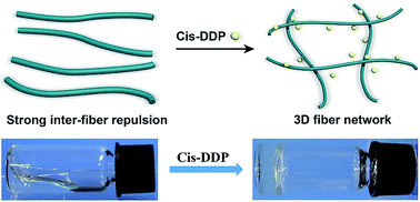
Our previous results have indicated that a taxol–peptide conjugate (Taxol-E-ss-EE) can form gels after the removal of hydrophilic dipeptide EE by glutathione from it.30 We have also showed that peptides with well-adjusted balance between hydrophobicity and hydrophilicity can only form nanofibers but not gels due to weak inter-fiber interactions, and the nanofiber dispersion can be converted to a gel by increasing inter-fiber interactions through the addition of a protein.31 These information indicate that the balance between hydrophobicity and hydrophilicity of a taxol–peptide conjugate is crucial for its self-assembling and hydrogelation ability. We therefore opted to make a taxol-peptide amphiphile only capable of self-assembling into nanofibers but not gels. The addition of DDP might decrease the solubility of the amphiphile and reduce the inter-fiber repulsion due to the chelation between carboxylic acid group and DDP, thus leading to the hydrogelations (Fig. 1). We then designed and prepared the compound Taxol-GRGD via a combination of solid phase and solution phase synthesis in a good yield (overall yield = 75%). We tested its self-assembling ability in a phosphate buffer saline (PBS, pH 7.4) solution at room temperature (20–25 °C). The compound could only form a clear solution at the concentration of 0.08 wt% (Fig. 2A). We then tested whether the addition of DDP to the PBS solution of Taxol-GRGD would result in hydrogelation or not (final concentration of Taxol-GRGD = 0.08 wt%). As shown in Fig. 2A, hydrogelations happened within 20 minutes after the addition of DDP, and the minimum equivalent of DDP to induce hydrogelation was about 0.05. We observed hydrogelations when 0.05–1.2 equiv. of DDP were added to the solution of Taxol-GRGD. Several previous reports had demonstrated that hydrogelations could happen by adding metal ions to peptide solutions due to charge screening or enhanced inter-fiber interactions,32–35 and our study provided a novel candidate of metal ion (an anti-cancer drug) to trigger hydrogelations in mild conditions. We then characterized the hydrogels by different techniques including rheology and TEM.


image file: c6ra08357a-f1.tif
Fig. 1 The schematic illustration for hydrogel formation.

image file: c6ra08357a-f2.tif
Fig. 2 (A) Optical images of gels formed by treating the solution containing 0.08 wt% of Taxol-GRGD with different equiv. of DDP: (a) 0, (b) 0.05, (c) 0.1, (d) 0.2, (e) 0.5, (f) 0.8, and (g) 1.2 equiv. to Taxol-GRGD; (B) G′ value of gels obtained by rheological measurements with the mode of dynamic frequency sweep at the strain of 0.5%.

The mechanical properties of hydrogels could be adjusted by adding different amounts of DDP to solutions of Taxol-GRGD. As shown in Fig. 2B, the results obtained by dynamic frequency sweeps indicated that the value of the storage modulus (G′) of the resulting gels was bigger when higher amounts of DDP were used. However, when the equiv. of DDP was bigger than 0.5, the G′ value of resulting gels became to decline. The G′ value at the strain of 0.5% was about 1642, 2626, 4142, 5579, 5130, and 4395 Pa for gels of Taxol-GRGD with 0.05, 0.1, 0.2, 0.5, 0.8, and 1.2 equiv. of DDP (relative to Taxol-GRGD), respectively. The DDP had been demonstrated to be capable of chelating with two carboxylic acid groups.36 Our observations suggested that DDP chelated with Taxol-GRGD through binding with two carboxylic acids of aspartic acid (D) on the molecule at low concentrations, thus reducing the inter-fiber repulsion and forming mechanically stronger gels. However, when the concentration of DDP was high, most of Taxol-GRGD chelated with DDP to form more water soluble peptide-DDP complex, leading to the decrease of the mechanical property of the gels. When the amount of DDP was more than 2.5 equiv., the samples remained as clear solutions and no gels would form.

We then used transmission electron microscopy (TEM) to investigate the nanofiber morphology in solution of Taxol-GRGD and the resulting gels. As shown in Fig. 3A, uniform nanofibers with the size of about 35 nm were observed in a PBS solution containing 0.08 wt% of Taxol-GRGD. These nanofibers could not form stable three dimensional (3D) networks that supported hydrogel formation probably due to the lack of strong interactions between nanofibers. After the addition of DDP to solution of Taxol-GRGD, the density of cross-linking of fibers was increased, leading to the hydrogel formation (Fig. 3B–D). We observed the dense and 3D networks of nanofibers in all hydrogels with different concentrations of DDP (Fig. 3B–D). However, the diameter and morphology of the nanofibers in these gels were different. As shown in Fig. 3 and S12, the diameter of the nanofibers in gels was about 32.4, 30.7, 27.6, 19.5, 18.9, and 18.4 nm when the concentration of DDP was 0.05, 0.1, 0.2, 0.5, 0.8, and 1.2 equiv. to Taxol-GRGD, respectively (50 nanofibers were measured in each gel by TEM observation). These observations also suggested that, with the increasing amounts of DDP, DDP chelated with Taxol-GRGD through binding with two carboxylic acids of aspartic acid (D) on the molecule, leading to an erosion effect of the nanofibers and creating smaller-sized nanofibers with stronger inter-fiber interactions.


image file: c6ra08357a-f3.tif
Fig. 3 TEM images of (A) the solution of Taxol-GRGD (0.08 wt%) and gels with different concentrations of cis-dichlorodiamineplatinum(II): (B) 0.1 equiv., (C) 0.2 equiv., (D) 0.5 equiv.

We determined the release profile of taxol from gels. The results in Fig. S16 indicated that taxol released slowly and about 10% of taxol got released from the gels during 12 h time period. The release speed of taxol was lower when the equiv. of DDP was bigger. We also obtained the IC50 value of the gels with different amounts of DDP against HepG2 cells (Fig. 4). After 48 hours of incubation with the cells, the solution of Taxol-GRGD without DDP had an IC50 value of about 86.2 nM, and the gels with different amounts of DDP (0.05, 0.1, 0.2, 0.5, 0.8, and 1.2 equiv. of DDP to Taxol-GRGD) exhibited an IC50 value of about 59.0, 47.4, 35.9, 29.5, 22.8 and 19.7 nM, respectively. The results indicated that the better inhibition capacity could be achieved with the more concentration of DDP. The IC50 value of DDP was about 5.3 μM. The enhanced inhibition capacity of nanofibers with DDP than those without DDP was probably due to the synergistic effect of both anti-cancer drugs. Taxol interrupted the dynamics of tubulin assembly and DDP chelated with DNA molecules, the combination of two drugs therefore inhibited cancer cells growth more efficiently. We also tested the IC50 value of the gels with different amounts of DDP against NIH 3T3 cells (Fig. S14). The IC50 values of most of gels were within the range of 30 to 110 μM, indicating that the toxicities of our nanomedicines were smaller to the normal cells, probably because of the targeting effect of Taxol-GRGD to cancer cells.


image file: c6ra08357a-f4.tif
Fig. 4 Cytotoxicity of Taxol-GRGD with different amounts of DDP against HepG2 cells.

In summary, we have developed a novel hydrogel system based on two anti-cancer drugs, taxol and DDP. The hydrogels can be prepared easily, only by mixing two solutions. Our hydrogels showed enhanced efficacies to inhibit cancer cells growth, suggest its big potential as a drug delivery system for chemotherapy. Our study provides a versatile method to prepare supramolecular hydrogels through the chelation between carboxylic acid of peptides and DDP. The resulting hydrogels might assist the delivery of DDP to cancer cells, and we envision that such nanofibers or hydrogels will be beneficial for cancer therapy.

Acknowledgements

This work is supported by International S&T Cooperation Program of China (ISTCP, 2015DFA50310), Science and Technology Guiding Project of Guangdong Province (2013B091500071), Tianjin MSTC (15JCZDJC38100), and Program for Changjiang Scholars and Innovative Research Team in University (IRT13023).

Notes and references

  1. T. Yoshii, S. Onogi, H. Shigemitsu and I. Hamachi, J. Am. Chem. Soc., 2015, 137, 3360 CrossRef CAS PubMed.
  2. M. Ikeda, T. Tanida, T. Yoshii, K. Kurotani, S. Onogi, K. Urayama and I. Hamachi, Nat. Chem., 2014, 6, 511 CrossRef CAS PubMed.
  3. C. Ren, H. Wang, D. Mao, X. Zhang, Q. Fengzhao, Y. Shi, D. Ding, D. Kong, L. Wang and Z. Yang, Angew. Chem., Int. Ed., 2015, 54, 4823 CrossRef CAS PubMed.
  4. C. Ren, J. Zhang, M. Chen and Z. Yang, Chem. Soc. Rev., 2014, 43, 7257 RSC.
  5. Q. Miao, Z. Wu, Z. Hai, C. Tao, Q. Yuan, Y. Gong, Y. Guan, J. Jiang and G. Liang, Nanoscale, 2015, 7, 2797 RSC.
  6. Y. Shi, Z. Wang, X. Zhang, T. Xu, S. Ji, D. Ding, Z. Yang and L. Wang, Chem. Commun., 2015, 51, 15265 RSC.
  7. C. Yang, Z. Wang, C. Ou, M. Chen, L. Wang and Z. Yang, Chem. Commun., 2014, 50, 9413 RSC.
  8. J. Li, Y. Gao, Y. Kuang, J. Shi, X. Du, J. Zhou, H. Wang, Z. Yang and B. Xu, J. Am. Chem. Soc., 2013, 135, 9907 CrossRef CAS PubMed.
  9. J. Li, Y. Kuang, J. Shi, J. Zhou, J. E. Medina, R. Zhou, D. Yuan, C. Yang, H. Wang, Z. Yang, J. Liu, D. M. Dinulescu and B. Xu, Angew. Chem., 2015, 127, 13505 CrossRef.
  10. J. Wang, W. Mao, L. L. Lock, J. Tang, M. Sui, W. Sun, H. Cui, D. Xu and Y. Shen, ACS Nano, 2015, 9, 7195 CrossRef CAS PubMed.
  11. Y. Tian, H. Wang, Y. Liu, L. Mao, W. Chen, Z. Zhu, W. Liu, W. Zheng, Y. Zhao, D. Kong, Z. Yang, W. Zhang, Y. Shao and X. Jiang, Nano Lett., 2014, 14, 1439 CrossRef CAS PubMed.
  12. T. Sun, H. Han, G. A. Hudalla, Y. Wen, R. R. Pompano and J. H. Collier, Acta Biomater., 2016, 30, 62 CrossRef CAS PubMed.
  13. R. R. Pompano, J. Chen, E. A. Verbus, H. Han, A. Fridman, T. McNeely, J. H. Collier and A. S. Chong, Adv. Healthcare Mater., 2014, 3, 1898 CrossRef CAS PubMed.
  14. C. M. R. Pérez, N. Stephanopoulos, S. Sur, S. S. Lee, C. Newcomb and S. I. Stupp, Ann. Biomed. Eng., 2015, 43, 501 CrossRef PubMed.
  15. D. Kalafatovic, M. Nobis, N. Javid, P. W. Frederix, K. I. Anderson, B. R. Saunders and R. V. Ulijn, Biomater. Sci., 2015, 3, 246 RSC.
  16. R. J. Mart, R. D. Osborne, M. M. Stevens and R. V. Ulijn, Soft Matter, 2006, 2, 822 RSC.
  17. A. Altunbas, S. J. Lee, S. A. Rajasekaran, J. P. Schneider and D. J. Pochan, Biomaterials, 2011, 32, 5906 CrossRef CAS PubMed.
  18. L. Chen, J. Wu, L. Yuwen, T. Shu, M. Xu, M. Zhang and T. Yi, Langmuir, 2009, 25, 8434 CrossRef CAS PubMed.
  19. J. Wu, A. Chen, M. Qin, R. Huang, G. Zhang, B. Xue, J. Wei, Y. Li, Y. Cao and W. Wang, Nanoscale, 2015, 7, 1655 RSC.
  20. H. Wang, J. Wei, C. Yang, H. Zhao, D. Li, Z. Yin and Z. Yang, Biomaterials, 2012, 33, 5848 CrossRef CAS PubMed.
  21. J. Li, Y. Kuang, Y. Gao, X. Du, J. Shi and B. Xu, J. Am. Chem. Soc., 2013, 135, 542 CrossRef CAS PubMed.
  22. L. L. Lock, Z. Tang, D. Keith, C. Reyes and H. Cui, ACS Macro Lett., 2015, 4, 552 CrossRef CAS.
  23. Y. Gao, Y. Kuang, Z.-F. Guo, Z. Guo, I. J. Krauss and B. Xu, J. Am. Chem. Soc., 2009, 131, 13576 CrossRef CAS PubMed.
  24. L. Mao, H. Wang, M. Tan, L. Ou, D. Kong and Z. Yang, Chem. Commun., 2012, 48, 395 RSC.
  25. R. Lin, A. G. Cheetham, P. Zhang, Y.-a. Lin and H. Cui, Chem. Commun., 2013, 49, 4968 RSC.
  26. J. Zhou, X. Du, J. Li, N. Yamagata and B. Xu, J. Am. Chem. Soc., 2015, 137, 10040 CrossRef CAS PubMed.
  27. C. Ren, C. Xu, D. Li, H. Ren, J. Hao and Z. Yang, RSC Adv., 2014, 4, 34729 RSC.
  28. Y. Yang, J. Aw, K. Chen, F. Liu, P. Padmanabhan, Y. Hou, Z. Cheng and B. Xing, Chem.–Asian J., 2011, 6, 1381 CrossRef CAS PubMed.
  29. L. Kelland, Nat. Rev. Cancer, 2007, 7, 573 CrossRef CAS PubMed.
  30. H. Wang, L. Lv, G. Xu, C. Yang, J. Sun and Z. Yang, J. Mater. Chem., 2012, 22, 16933 RSC.
  31. X. Zhang, X. Chu, L. Wang, H. Wang, G. Liang, J. Zhang, J. Long and Z. Yang, Angew. Chem., Int. Ed., 2012, 124, 4464 CrossRef.
  32. E. D. Spoerke, S. G. Anthony and S. I. Stupp, Adv. Mater., 2009, 21, 425 CrossRef CAS PubMed.
  33. C. M. Micklitsch, P. J. Knerr, M. C. Branco, R. Nagarkar, D. J. Pochan and J. P. Schneider, Angew. Chem., 2011, 123, 1615 CrossRef.
  34. P. J. Knerr, M. C. Branco, R. Nagarkar, D. J. Pochan and J. P. Schneider, J. Mater. Chem., 2012, 22, 1352 RSC.
  35. J. Shi, Y. Gao, Y. Zhang, Y. Pan and B. Xu, Langmuir, 2011, 27, 14425 CrossRef CAS PubMed.
  36. H. Xiao, D. Zhou, S. Liu, R. Qi, Y. Zheng, Y. Huang and X. Jing, Macromol. Biosci., 2012, 12, 367 CrossRef CAS PubMed.

Footnote

Electronic supplementary information (ESI) available: Details of synthesis and characterization of the compounds, details of experimental procedure, rheology, and congress curve of cell inhibition assay. See DOI: 10.1039/c6ra08357a

This journal is © The Royal Society of Chemistry 2016