Assessment of polydopamine coated magnetic nanoparticles in doxorubicin delivery

Radosław Mrówczyński*a, Justyna Jurga-Stopab, Roksana Markiewicza, Emerson L. Coya, Stefan Jurgaac and Anna Woźniaka
aNanoBioMedical Centre at Adam Mickiewicz University, ul. Umultowska 85, 61-614 Poznań, Poland. E-mail: radek.mrowczynski@gmail.com
bDepartment of Biomaterials and Experimental Dentistry, Poznan University of Medical Sciences, ul. Fredry 7, Poznań, Poland
cDepartment of Macromolecular Physics, Faculty of Physics, Adam Mickiewicz University, ul. Umultowska 85, 61-614 Poznań, Poland

Received 16th November 2015 , Accepted 5th January 2016

First published on 8th January 2016


Abstract

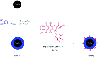
Magnetic nanoparticles (MNP) coated with bioinspired polydopamine (PDA) were obtained via a co-precipitation method and oxidative polymerization of dopamine. Nanoparticles were investigated by FTIR, TEM and SQUID. Loading capacity of anticancer drug doxorubicin was determined by UV-Vis spectroscopy. The nanocomposites exhibit a high drug loading capacity of 0.46 mg mg−1. Anticancer activity of the nanocomposites was proved in profound in vitro tests on HeLa cells. Cytotoxicity and internalization of nanoparticles were checked using various method, i.e. proliferation assay (WST-1), a two-colour fluorescence cell viability assay, and fluorescent and confocal microscopy.


Introduction

In recent years applications of nanomaterials have grown enormously. They have found application in material science,1–3 catalysis4,5 and environmental protection i.e. removal of heavy metal and organic waste from water.6–8 However, one of the most significant fields where nanomaterials have found application and attracted a lot of attention both in basic and applied research is nanomedicine.9–11 In the field of nanomedicine, the leading branch is drug delivery. The commonly utilized nanomaterials for drug delivery systems are inorganic nanoparticles – gold and iron oxides – but polymeric nanoparticles and micelles also play an important role in this field.12–14 Application of nanomaterials to drug delivery allows overcoming weak solubility of linked drugs, increase in its stability and altered drug biodistribution. Properly engineered surfaces of nanomaterials render them as “smart” and multifunctional materials, which can be used in cancer therapy, reducing side effects of traditional chemotherapy, and simultaneously they can act as probes for imaging in magnetic resonance.15

A very promising group of nanomaterials are nowadays magnetic nanoparticles, both Fe3O4 (magnetite) and Fe2O3 (maghemite). They have been applied as supports not only for drug delivery systems16 or multifunctional nanomaterials17–19 but also for preparation of gene20 and nucleic acid21 nanocarriers, supports for metalo22 and organocatalysts23 as well as analytical methods.24 Also extensive studies are conducted on application of magnetic nanoparticles in hyperthermia treatment.25,26

Nature still remains the inventible inspiration for scientist and stimulates development of new versatile materials. Hence, bio-inspired materials are the group of materials that has drawn a lot of attention in the last years. One of such materials is polydopamine (PDA),25 a polymer obtained by polymerization of neurotransmitter-dopamine in weakly basic environment. Indeed, the research performed on this polymer showed that it possesses strong adhesives properties toward myriad materials i.e. polymers, wood, noble metals and magnetic or gold nanoparticles.26 Since 2007 polydopamine has been used in many fields like polymer synthesis,27 catalysis,28 preparation of supercapacitors29 or extraction of heavy metals.30 Polydopamine have been used also as coating of dental tissue inducing enamel and dentin remineralization.31 The simplicity of preparation polydopamine on virtually any material, renders it as a versatile coating ready for post-modification i.e. deposition of metals (Au, Ag, Fe, Cu) and grafting various (bio)macromolecules by Michael reaction, Schiff base formation or thio-Michael reaction.26,28,32–35 Recently click reaction as a new way for polydopamine modification method was introduced improving its versatility.35–37

Polydopamine has been successfully utilized in drug delivery as well. It was shown that capsules made from polydopamine can serve as effective carriers for doxorubicin.38 Polydopamine spheres were used to carry camptothecin – cytotoxic quinoline alkaloid.39 Also electrospun poly(ε-caprolactone) (PCL) nanofibers40 and mesoporous silica nanoparticles coated with polydopamine could be applied in controlled released of doxorubicin.41 Biomimetic spheres made from polydopamine were reported to be excellent agent for photothermal therapy42 and for combined chemo- and photothermal therapy.43 Combination of polydopamine with magnetic nanoparticles delivered new materials which could serve as nanoplatform for intracellular mRNA detection and imaging – guided photothermal therapy and as MRI and photothermal agent rendering smart nanomaterials.44

Even though polydopamine have found wide application in preparation of various nanomaterials for bioapplications any studies have been reported on basic model consisting of polydopamine magnetite coated nanoparticles and its potential application in doxorubicin delivery. Thus, we disclose the results which covered the gap in this area and are of great importance in the field of nanomedicine and polydopamine chemistry.

Experimental

Reagents and instruments

All reagents were commercially available and used without further purification, unless otherwise stated.

Dopamine hydrochloride and tris(hydroxymethyl)aminomethane (TRIS) were purchased from Alfa Aesar. Doxorubicin hydrochloride was delivered by LC Laboratories. For polymerization reaction Milli-Q deionized water (resistivity 18 MΩ cm) was used.

Transmission electron microscopy (TEM) images were recorded on a JEM-1400 microscope made by JEOL (Japan). The accelerating voltage was 120 kV. A small amount of the sample, placed on a copper measuring grid (Formvar/Carbon 200 Mesh made by TedPella (USA)) after 5 minutes of sonication in deionized water. Then the sample was dried in a vacuum desiccator for 24 hours.

FTIR spectra were recorded on Bruker Tensor 27 spectrometer in KBr pallets. Magnetic measurements were performed on Quantum Design SQUID magnetometer at 5 K and 300 K.

UV-Vis measurements were performed on Perkin-Elmer lambda 950 UV/Vis/NIR.

Synthesis

Synthesis of MNP 1. 1 g of freshly prepared magnetite nanoparticles (obtained via co-precipitation method) were redispersed in TRIS buffer (pH = 8.5, 10 mmol, 500 ml) using ultrasound followed by addition of dopamine hydrochloride (1 g). Stirring was continued for 6 h. Then nanoparticles were collected by an external magnet and washed with deionized water.
Loading tests – MNP 2. 2 mg of MNP 1 were mixed with 2 ml of doxorubicin solution at concentration 1 mg ml−1. The mixture was shaken at 37 °C for 24 h. Resulting MNP were collected by an external magnet and washed two times with PBS buffer. Amount of linked doxorubicin was analysed by UV-Vis spectroscopy.
Drug release test. 2 mg of MNP 2 loaded with doxorubicin were mixed with citric buffer (2 ml) at pH 4.5 and 5.5. The mixture was shaken at 37 °C and sample was taken in appropriate time intervals refilling mixture with the fresh buffer.

Cell culture

Examinations were conducted on HeLa (cervical cancer) cell line. The cells were cultured in complete medium: DMEM (Dulbecco's Modified Eagle's medium, Sigma) with FBS (Fetal Bovine Serum, Sigma) to a final concentration of 10% supplemented with antibiotics (penicillin 100 μl ml−1, streptomycin 100 μg ml−1, Gibco) in humid atmosphere, 5% CO2 at 37 °C. When the culture reach needed confluence, cells were trypsinized (0.25% trypsin, 0.02% EDTA, Sigma) and seeded on culture plates in appropriate concentration. All activities were performed under sterile conditions (cabinet with laminar air flow). The growth rate and cell morphology were evaluated using an inverted microscope Leica DCM.

Cytotoxicity tests

To assess cytotoxicity profile of synthetized nanocomposites two tests were performed. WST-1 assay (water-soluble tetrazolium salt, Clonetech) – cells were seeded in standard 96-wells plate at densities 105 ml−1 in complete medium and after 24 h the medium was removed and then MNP 1 or MNP 2 were added. To examine different concentrations, the 1 mg ml−1 stock solution was prepared and then diluted with complete medium. Every sample (concentrations: 1, 8, 16, 32, 64, 100, 300 μg ml−1) was repeated twice, and positive (cells without nanocomposites) and negative (10% dimethyl sulfoxide) controls were carried out. Then, the cells were cultured in described conditions. The investigations were preceded in three time intervals – 24 h, 48 h or 72 h. After this time, 10 μl of WST-1 reagent were added into the individual well with cells and incubated for 2–4 h at 37 °C. The OD values (optical density) were read with plate reader (Zenyth) at wave length λ ∼ 450 nm and λ ∼ 620 nm as reference.

LIVE/DEAD® Viability/Cytotoxicity Kit for mammalian cells

(Molecular Probes) based on fluorescent staining with calcein AM and ethidium homodimer-1 (EthD-1). The HeLa cells were seeded in black polystyrene 96-wells flat bottom plate with micro-clear bottom (Greiner Bio-One GmbH) at densities 104 per well in complete medium. After 24 h medium was removed and MNP 2 were added. Every sample (seven mentioned above concentrations) was repeated 2–4 times, as well as positive and negative controls were carried out. Then the cells were cultured in described conditions in three time intervals: 24 h, 48 h, 72 h. After incubation, cells were stained with fluorescent dyes: calcein AM, ethidium homodimer-1 at working concentrations 100 μg ml−1. The cells were incubated 30 min at 5% CO2, 37 °C. Then fluorescent dyes were removed, cells were washed with HBSS (Hanks' Balanced Salt Solution, Sigma) and preceded with qualitative analyses using IN Cell Analyzer 2000 (GE Healthcare Life Sciences) and quantitative analyses with IN Cell Developer Toolbox Image Analysis Software.

Confocal bioimaging

Fluorescent staining with concanavalin A, Alexa Fluor 488 Conjugate (Con A, Molecular Probes) and detection of doxorubicin autofluorescence to determine internalization of MNP 2 and drug release study as well as internalization of free doxorubicin solution. The HeLa cells were seeded in Lab-Tek™ 8 Chamber Slide with removable, polystyrene media chamber attached to a standard glass microscope slide (25 × 75 mm) at densities 105 ml−1 in complete medium. After 24 h medium was removed and nanocomposites in seven described above concentrations were added. After 24 h, 48 h and 72 h of incubation in standard conditions, fluorescent dyes were washed, cells were fixed with 4% paraformaldehyde in medium at 37 °C for 15 min, then washed twice with PBS and cell membrane were permeabilized with 0.2% Triton X-100 (5 min). Fixed cells were stained with cell membrane dye, concanavalin A, at working concentration 100 μg ml−1. Confocal imaging was performed with Leica TCS SP5 system.

Results and discussion

MNP 1 were easily synthesized according to previously reported literature protocol.45 In the first step magnetite nanoparticles were obtained by well-established co-precipitation method followed by covering with polydopamine by oxidative polymerization of dopamine under basic conditions (Scheme 1). The FTIR spectra of the resulting MNP 1 show typical for polydopamine signals at 1440–1620 cm−1, proving successful functionalization of magnetic nanoparticles with polydopamine polymer (Fig. 1). In addition, the transformation of the initial dopamine anchored to the surface of the Fe3O4 into biomimetic polydopamine can be expected to render the otherwise eventually reversible surface coating46,47 irreversible.
image file: c5ra24222c-s1.tif
Scheme 1 Synthesis of magnetic nanoparticles covered with PDA and loaded with doxorubicin (red molecule).

image file: c5ra24222c-f1.tif
Fig. 1 FTIR spectra of MNP 1 and nanoparticles loaded with doxorubicin MNP 2.

Synthesized MNP 1 exhibit (Fig. 2a) high value of saturation magnetization Ms = 62 emu g−1 and 52 emu g−1 at 5 K and 300 K respectively. Detailed magnetic investigations revealed that coercion filed had value of 275 Oe in 5 K and 25 Oe in 300 K which suggests that almost all nanoparticles changed their state to superparamagnetic in the room temperature but still small fraction remained in ferromagnetic state (Fig. 2b).


image file: c5ra24222c-f2.tif
Fig. 2 (a) Hysteresis loop of MNP 1 at different temperatures. (b) Coercion field of MNP 1 recorded at different temperatures.

The morphology of MNP 1 was determined by TEM revealing spherical particles and a medium size of about 8 nm with medium polymer thickness around 1.5 nm (see Fig. 3 and 4). Small aggregation of particles was caused by sample preparation method.


image file: c5ra24222c-f3.tif
Fig. 3 TEM picture of MNP 1.

image file: c5ra24222c-f4.tif
Fig. 4 Histogram of particles size distribution and polymer thickness.

In such a material in hand its loading capacity of anticancer drug was investigated. Doxorubicin was used as a model, aromatic drug commonly used in chemotherapy. It was reported that doxorubicin hydrochloride has maximum stability at pH 4, while kept in pH 9 may degrade and loose therapeutic features.48 Thus, the loading tests were performed in phosphate buffer with pH 7.4 in order to minimize degradation of doxorubicin and ensure the highest loading possible. After incubation of nanocomposites with doxorubicin, they were collected by an external magnet and washed with PBS buffer resulting MNP 2. In order to determine how much doxorubicin was bound to the MNP 1, the eluate containing unbound doxorubicin was collected and analysed by UV-Vis spectroscopy by measuring the absorbance of doxorubicin at 490 nm. The amount of attached doxorubicin was calculated from the difference between initial concentration of doxorubicin and its concentration in eluate considering necessary dilution using calibration curve method. The loading experiments were repeated 3 times (see Fig. 1 in ESI for UV-Vis spectra from doxorubicin in eluate). The estimated average loading of doxorubicin loading on MNP 2 was 0.46 mg mg−1 which is higher than in case of other structure based on PDA.49 The high loading capacity of MNP 1 is due to the strong π–π stacking and hydrogen bond interactions of PDA shell of MNP 1 with doxorubicin because PDA possesses in the structure phenyls, amino, and hydroxyl groups.

Successful loading of doxorubicin onto surface of MNP 1 was also proved by FTIR50 (see Fig. 1). The new intensive peaks at 987 cm−1 was assigned to the bending vibration of C–C[double bond, length as m-dash]O, C–OH, C-Hx (ali) groups. Band present at 1210 cm−1 corresponded to bending vibration of CO–H and CHx groups from doxorubicin. Strong signal at 1280 cm−1 was due to the bending vibration of aromatic ring. Peak at 1407 cm−1 was assigned to vibration of aromatic ring too. Breathing vibrations of aromatic moieties were observed at 1578 cm−1. Signal at 1615 cm−1 is due to the stretching vibration of aromatic ring and CO–H group. Band at 1724 cm−1 belonged to N–H bending vibration from amino moiety. Signal observed at 2834 cm−1 and 2917 cm−1 corresponded to stretching vibration of CH2 groups. Signal from C[double bond, length as m-dash]O group which supposed to be at 1710 cm−1 was superimposed with other bands.

In the next step the drug release profile was determined by incubation of MNP 2 with the solution of citric buffer at pH 4.5 and 5.5 respectively. As can be seen in the Fig. 5 the cumulative release of doxorubicin within 10 h is around 3% and 7% for pH 5.5 and 4.5 respectively. The maximum doxorubicin release is achieved after 24 h in both cases and it varies from 4% to almost 9% dependent of the pH. Degradation of nanocarrier at pH 4.5 after 24 h was observed, thus the measurements were stopped in order to have reliable data (see Fig. 5).


image file: c5ra24222c-f5.tif
Fig. 5 Release profile of drug from MNP 2 dependent on time and pH.

Similar phenomenon of carrier degradation was described recently for silica particles covered with PDA.41 The carrier destroying was not observed when drug release was performed at pH 5.5 so longer investigations were performed. The pH dependent drug release from PDA coated materials is in agreement with literature data.49 Similarly to previously reported data also in our case the cumulative release of doxorubicin is almost two times higher at pH 4.5 than at pH 5.5. This may be due to the more efficient breaking π–π stacking and hydrogen bonds at lower pH. Nevertheless the percentage of cumulative release is lower than in other PDA derived materials where doxorubicin was attached.51 This phenomenon can be caused by covalent linking between amine group present in doxorubicin sugar moiety and quinone groups in PDA.

A crucial point is the toxicity of the obtained nanomaterials. The first test used to determine cytotoxicity profile of MNP 1 and MNP 2 on HeLa cell line in the culture medium containing nanocomposites was WST-1 proliferation assay. The tested concentrations ranged from 1 to 300 μg ml−1. MNP 1 after 24 h and 48 h of incubation did not decreased viability profile of HeLa cells comparing to the control (average 97% and 91%, respectively) (Fig. 6, for further information including comparison to positive and negative control see Fig. 2 at ESI). In the third day of incubation, ability to reduction of WST-1 salt decreased only at the highest concentrations: 100 μg ml−1 – 72% and 300 μg ml−1 – 50%. This indicated for very good biocompatibility of MNP 1.


image file: c5ra24222c-f6.tif
Fig. 6 HeLa cell line viability after 24 h, 28 h, 72 h of incubation with MNP 1.

After 24 h of incubations with MNP 2, HeLa cells showed survival rate above 90% (97 to 90% at all tested concentrations) accept from the highest concentration (300 μg ml−1) which represent 84% cells' viability comparing to the control (Fig. 7, for further information including comparison to positive and negative control see Fig. 3 at ESI). After 48 h of incubation, average cells' viability at concentration 1–64 μg ml−1 was 98%. This value was lower at concentration 100 μg ml−1 – 79% and 300 μg ml−1 – 44%.


image file: c5ra24222c-f7.tif
Fig. 7 HeLa cell line viability after 24 h, 48 h, 72 h of incubation with MNP 2 (WST-1 assay).

At the third day of incubation average cells' viability at concentration 1–64 μg ml−1 was 98%. At the concentration 100 μg ml−1 viability was at the level of 68% and dramatically decreased in concentration of 300 μg ml−1 to 6% (Fig. 7). Obtained results showed that doxorubicin was linked to MNP 2 and was effectively released from the carrier casing death of HeLa cells while the cytotoxic effect was not observed for MNP 1.

The second conducted cytotoxic test was based on LIVE/DEAD® Viability/Cytotoxicity, which uses fluorescent staining. Here the calcein AM can be transported through the cellular membrane into live cells, then intracellular esterases remove the AM group trapping inside the cells and causes green fluorescence. Ethidium homodimer enters into dead cells through permeable cell membrane and binds to DNA.52 This assay allows to determinate viability of the cells using IN Cell Analyzer 2000. The concentration of 100 and 300 μg ml−1 of MNP 2 resulted in the greatest decrease in the number of live cells in time dependent manner (Fig. 8–10).


image file: c5ra24222c-f8.tif
Fig. 8 Live/dead test results. % of live HeLa cells after 24 h, 48 h and 72 h of incubation with MNP 2.

image file: c5ra24222c-f9.tif
Fig. 9 HeLa cells after 24 h incubation with MNP 2 (concentration 1 μg ml−1). Live cells' cytoplasm – green (calcein AM), dead cells' nucleus – red (ethidium homodimer). IN Cell Analyzer 2000, magnification ×40.

image file: c5ra24222c-f10.tif
Fig. 10 HeLa cells after 72 h incubation with MNP 2 (concentration 300 μg ml−1). Live cells cytoplasm – green (calcein AM), dead cells' nucleus – red (ethidium homodimer). IN Cell Analyzer 2000, magnification ×40.

Cellular tracking was performed using fluorescent staining of the examined cells after incubation with MNP 2 and free doxorubicin solution (both 300 μg ml−1). Concanavalin A was electively bounded to α-mannopyranosyl and α-glucopyranosyl residues and labelled cell membrane (absorption/emission maxima at wave length λ ∼ 495/519 nm). Doxorubicin autofluorescence was examined at wavelength λ ∼ 488 nm. In all analysed concentrations and time intervals internalization process was observed as well as red-labelled cytoplasm/nucleus from released doxorubicin. After 24 h of incubation agglomerates of nanocomposites were visible (Fig. 11) (see ESI for video of nanocomposites internalization recorded on Leica TCS Sp 5 in Z-stack mode). In the second and third day of incubation, the release process of doxorubicin from the nanocomposites into the cytoplasm/nucleus was observed (Fig. 12 and 13). Internalization of free doxorubicin solution was also detected, starting form the first day of incubation (Fig. 14).


image file: c5ra24222c-f11.tif
Fig. 11 HeLa cells after 24 h incubation with MNP 2 (concentration 300 μg ml−1). Cell membrane – green (concanavalin A), doxorubicin – red. Leica TCS SP5, magnification 60×.

image file: c5ra24222c-f12.tif
Fig. 12 HeLa cells after 48 h incubation with MNP 2 (concentration 300 μg ml−1). Cell membrane – green (concanavalin A), doxorubicin – red. Leica TCS SP5, magnification 60×.

image file: c5ra24222c-f13.tif
Fig. 13 HeLa cells after 72 h incubation with MNP 2 (concentration 300 μg ml−1). Cell membrane – green (concanavalin A), doxorubicin – red. Leica TCS SP5, magnification 60×.

image file: c5ra24222c-f14.tif
Fig. 14 HeLa cells after 72 h incubation with free doxorubicin solution (concentration 121, 2 μg). Cell membrane – green (concanavalin A), doxorubicin – red. Leica TCS SP5, magnification 60×.

Conclusions

In conclusion, we have prepared and investigated magnetic nanoparticles covered with polydopamine and loaded with anticancer drug – doxorubicin. MNP 1 have been characterized by TEM, FTIR, SQUID confirming successful covering with polymer and revealing spherical particles with good magnetic properties and biocompatibility. We also presented that doxorubicin can be directly loaded on MNP 1 with high loading capacity 0.46 mg mg−1. In addition, two independent in vitro tests have been performed on MNP 2 pointed that concentration of 100 and 300 μg ml−1 resulted in the greatest decrease in the number of HeLa cells proving its therapeutic efficiency. Obtained material may be suitable candidate for the preparation of multifunctional materials where all attributes of polydopamine and magnetite are used for theragnostics but it requires further improvement in terms of drug release. Thus, work on development new polydopamine coated magnetic nanoparticles for drug delivery and their simultaneous application in MRI, photothermal therapy combined with chemotherapy is ongoing in our laboratory.

Acknowledgements

We thank for SQUID magnetic measurements to Dr Karol Zaleski from the NanoBioMedical Centre. Financial support from the National Science Centre under project number UMO-2014/13/D/ST5/02793 and from National Centre for Research and Development contract number PBS1/A9/13/2012 is gratefully acknowledged.

References

  1. M.-R. Gao, Y.-F. Xu, J. Jiang and S.-H. Yu, Chem. Soc. Rev., 2013, 42, 2986 RSC.
  2. X. Peng, L. Peng, C. Wu and Y. Xie, Chem. Soc. Rev., 2014, 43, 3303 RSC.
  3. H. Kim, H.-B.-R. Lee and W. J. Maeng, Thin Solid Films, 2009, 517, 2563 CrossRef CAS.
  4. D. Astruc, F. Lu and J. R. Aranzaes, Angew. Chem., Int. Ed., 2005, 44, 7852 CrossRef CAS PubMed.
  5. M. Stratakis and H. Garcia, Chem. Rev., 2012, 112, 4469 CrossRef CAS PubMed.
  6. L. Giraldo, A. Erto and J. Moreno-Piraján, Adsorption, 2013, 19, 465 CrossRef CAS.
  7. W. Yantasee, C. L. Warner, T. Sangvanich, R. S. Addleman, T. G. Carter, R. J. Wiacek, G. E. Fryxell, C. Timchalk and M. G. Warner, Environ. Sci. Technol., 2007, 41, 5114 CrossRef CAS PubMed.
  8. M. T. Amin, A. A. Alazba and U. Manzoor, Adv. Mater. Sci. Eng., 2014, 2014, 24 Search PubMed.
  9. B. M. Maciejewska, A. Warowicka, A. Baranowska-Korczyc, K. Załęski, T. Zalewski, K. K. Kozioł and S. Jurga, Carbon, 2015, 94, 1012 CrossRef CAS.
  10. M. Hałupka-Bryl, K. Asai, S. Thangavel, M. Bednarowicz, R. Krzyminiewski and Y. Nagasaki, Colloids Surf., B, 2014, 118, 140 CrossRef PubMed.
  11. Y. Min, J. M. Caster, M. J. Eblan and A. Z. Wang, Chem. Rev., 2015, 115, 11147 CrossRef CAS PubMed.
  12. K. S. Soppimath, T. M. Aminabhavi, A. R. Kulkarni and W. E. Rudzinski, J. Controlled Release, 2001, 70, 1–20 CrossRef PubMed.
  13. J. Kao, K. Thorkelsson, P. Bai, B. J. Rancatore and T. Xu, Chem. Soc. Rev., 2013, 42, 2654–2678 RSC.
  14. E. C. Dreaden, A. M. Alkilany, X. Huang, C. J. Murphy and M. A. El-Sayed, Chem. Soc. Rev., 2012, 41, 2740 RSC.
  15. C. Sun, C. Fang, Z. Stephen, O. Veiseh, S. Hansen, D. Lee, R. G. Ellenbogen, J. Olson and M. Zhang, Nanomedicine, 2008, 3, 495 CrossRef PubMed.
  16. O. Veiseh, J. W. Gunn and M. Zhang, Adv. Drug Delivery Rev., 2010, 62, 284 CrossRef PubMed.
  17. J. Gao, H. Gu and B. Xu, Acc. Chem. Res., 2009, 42, 1097 CrossRef CAS PubMed.
  18. J. R. McCarthy, K. A. Kelly, E. Y. Sun and R. Weissleder, Nanomedicine, 2007, 2, 153 CrossRef CAS PubMed.
  19. R. Hao, R. Xing, Z. Xu, Y. Hou, S. Gao and S. Sun, Adv. Mater., 2010, 22, 2729 CrossRef PubMed.
  20. J. Dobson, Gene Ther., 2006, 13, 283 CrossRef CAS PubMed.
  21. B. Grześkowiak, Y. Sánchez-Antequera, E. Hammerschmid, M. Döblinger, D. Eberbeck, A. Woźniak, R. Słomski, C. Plank and O. Mykhaylyk, Pharm. Res., 2015, 32, 103 CrossRef PubMed.
  22. V. Polshettiwar, R. Luque, A. Fihri, H. Zhu, M. Bouhrara and J.-M. Basset, Chem. Rev., 2011, 111, 3036 CrossRef CAS PubMed.
  23. R. Mrowczynski, A. Nan and J. Liebscher, RSC Adv., 2014, 4, 5927 RSC.
  24. J. S. Beveridge, J. R. Stephens and M. E. Williams, Annu. Rev. Anal. Chem., 2011, 4, 251 CrossRef CAS PubMed.
  25. Y. Liu, K. Ai and L. Lu, Chem. Rev., 2014, 114, 5057–5115 CrossRef CAS PubMed.
  26. H. Lee, S. M. Dellatore, W. M. Miller and P. B. Messersmith, Science, 2007, 318, 426 CrossRef CAS PubMed.
  27. B. Zhu and S. Edmondson, Polymer, 2011, 52, 2141 CrossRef CAS.
  28. R. Mrówczyński, A. Bunge and J. Liebscher, Chem.–Eur. J., 2014, 20, 8647 CrossRef PubMed.
  29. Y. Liang, H. Liu, Z. Li, R. Fu and D. Wu, J. Mater. Chem. A, 2013, 1, 15207 CAS.
  30. H. Gao, Y. Sun, J. Zhou, R. Xu and H. Duan, ACS Appl. Mater. Interfaces, 2013, 5, 425 CAS.
  31. Y.-Z. Zhou, Y. Cao, W. Liu, C. H. Chu and Q.-L. Li, ACS Appl. Mater. Interfaces, 2012, 4, 6901 CAS.
  32. H. Lee, J. Rho and P. B. Messersmith, Adv. Mater., 2009, 21, 431 CrossRef CAS PubMed.
  33. S. M. Kang, N. S. Hwang, J. Yeom, S. Y. Park, P. B. Messersmith, I. S. Choi, R. Langer, D. G. Anderson and H. Lee, Adv. Funct. Mater., 2012, 22, 2949 CrossRef CAS PubMed.
  34. O. Pop-Georgievski, D. Verreault, M.-O. Diesner, V. Proks, S. Heissler, F. Rypáček and P. Koelsch, Langmuir, 2012, 28, 14273 CrossRef CAS PubMed.
  35. R. Mrowczynski, L. Magerusan, R. Turcu and J. Liebscher, Polym. Chem., 2014, 5, 6593 RSC.
  36. C. Rodriguez-Emmenegger, C. M. Preuss, B. Yameen, O. Pop-Georgievski, M. Bachmann, J. O. Mueller, M. Bruns, A. S. Goldmann, M. Bastmeyer and C. Barner-Kowollik, Adv. Mater., 2013, 25, 6123 CrossRef CAS PubMed.
  37. B. Yameen, C. Rodriguez-Emmenegger, C. M. Preuss, O. Pop-Georgievski, E. Verveniotis, V. Trouillet, B. Rezek and C. Barner-Kowollik, Chem. Commun., 2013, 49, 8623 RSC.
  38. J. Cui, Y. Yan, G. K. Such, K. Liang, C. J. Ochs, A. Postma and F. Caruso, Biomacromolecules, 2012, 13, 2225 CrossRef CAS PubMed.
  39. C.-C. Ho and S.-J. Ding, J. Mater. Sci.: Mater. Med., 2013, 24, 2381 CrossRef CAS PubMed.
  40. J. Xie, P. L. Michael, S. Zhong, B. Ma, M. R. MacEwan and C. T. Lim, J. Biomed. Mater. Res., Part A, 2012, 100, 929 CrossRef PubMed.
  41. Q. Zheng, T. Lin, H. Wu, L. Guo, P. Ye, Y. Hao, Q. Guo, J. Jiang, F. Fu and G. Chen, Int. J. Pharm., 2014, 463, 22 CrossRef CAS PubMed.
  42. Y. Liu, K. Ai, J. Liu, M. Deng, Y. He and L. Lu, Adv. Mater., 2013, 25, 1353 CrossRef CAS PubMed.
  43. Y. Gao, X. Wu, L. Zhou, Y. Su and C.-M. Dong, Macromol. Rapid Commun., 2015, 36, 916 CrossRef CAS PubMed.
  44. L.-S. Lin, Z.-X. Cong, J.-B. Cao, K.-M. Ke, Q.-L. Peng, J. Gao, H.-H. Yang, G. Liu and X. Chen, ACS Nano, 2014, 8, 3876 CrossRef CAS PubMed.
  45. R. Mrówczyński, R. Turcu, C. Leostean, H. A. Scheidt and J. Liebscher, Mater. Chem. Phys., 2013, 138, 295 CrossRef.
  46. E. Amstad, A. U. Gehring, H. Fischer, V. V. Nagaiyanallur, G. Hahner, M. Textor and E. Reimhult, J. Phys. Chem. C, 2011, 115, 683 CAS.
  47. E. Amstad, T. Gillich, I. Bilecka, M. Textor and E. Reimhult, Nano Lett., 2009, 9, 4042 CrossRef CAS PubMed.
  48. M. J. H. Janssen, D. J. A. Crommelin, G. Storm and A. Hulshoff, Int. J. Pharm., 1985, 23, 1 CrossRef CAS.
  49. F.-F. Cheng, J.-J. Zhang, F. Xu, L.-H. Hu, E. S. Abdel-Halim and J.-J. Zhu, J. Biomed. Nanotechnol., 2013, 9, 1155 CrossRef CAS PubMed.
  50. G. Das, A. Nicastri, M. L. Coluccio, F. Gentile, P. Candeloro, G. Cojoc, C. Liberale, F. De Angelis and E. Di Fabrizio, Microsc. Res. Tech., 2010, 73, 991 CAS.
  51. F. Liu, X. He, Z. Lei, L. Liu, J. Zhang, H. You, H. Zhang and Z. Wang, Adv. Healthcare Mater., 2015, 4, 479 CrossRef CAS.
  52. É. Karászi, K. Jakab, L. Homolya, G. Szakács, Z. Holló, B. Telek, A. Kiss, L. Rejtô, S. Nahajevszky, B. Sarkadi and J. Kappelmayer, Br. J. Haematol., 2001, 112, 308 CrossRef.

Footnote

Electronic supplementary information (ESI) available. See DOI: 10.1039/c5ra24222c

This journal is © The Royal Society of Chemistry 2016