
Red blood cell membrane-mediated fusion of hydrophobic quantum dots with living cell membranes for cell imaging†
Abstract
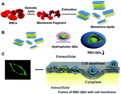
Nanoparticle fusion with cell membranes is an interesting phenomenon that may have crucial implications for their biomedical applications. Here, we proposed a biomimetic and controlled route to fusion of hydrophobic quantum dots (QDs) with the cell membranes of living cells, while preserving their sensing and optical properties and thus their capability of membrane imaging and single-nanoparticle tracking. Red blood cell (RBC) membrane lipids were extracted to phase transfer hydrophobic QDs and the resulting RBC-encapsulated QDs (RBC-QDs) can be well fused within cell membranes as membrane markers. The fusion was validated through single-nanoparticle imaging and different movement behaviours were reliably discriminated. RBC-QDs possessed some novel features, such as controllable selective membrane staining, no invasion, and high photobleaching resistance, which allowed for long-term imaging, and single-nanoparticle tracking. This approach provides a versatile platform for controlled hydrophobic QD-based fluorescence investigation of living cell membranes.
 Please wait while we load your content...

