White light emission by controlled mixing of carbon dots and rhodamine B for applications in optical thermometry and selective Fe3+ detection

Sonam Mandani, Bhagwati Sharma, Deepa Dey and Tridib K. Sarma*
Discipline of Chemistry, Indian Institute of Technology Indore, Simrol, Khandwa Road, Indore – 453552, India. E-mail: tridib@iiti.ac.in

Received 6th July 2016 , Accepted 31st August 2016

First published on 1st September 2016


Abstract

A simple mixing of rhodamine B with fluorescent carbon dots in water led to aggregation of the dye molecules on the carbon dot surface. Controlling the emission of free rhodamine B dye with that of the resultant carbon dot-aggregated rhodamine B composite resulted in efficient white light emission with the CIE coordinate (0.33, 0.32). The white light emitting system can be incorporated into a gel or polymer matrix for solid-state processibility. Further, selective sensing of Fe3+ ions as well as reversible and thermo-responsive emission in the temperature range of 25–80 °C in water shows the versatility in application potential of the nanocomposite.

Exploration of efficient and low-cost white light emitting materials has been a focal area of research due to the ever increasing demand for white light sources in areas such as lighting and displays. Due to advantages such as long working life time, low power consumption and compact size, white light emitting diodes (WLEDs) are promising illuminating sources to replace traditional incandescent lamps which suffer from serious environmental concerns due to their mercury content as well as the use of costly rare-earth based phosphors.1 Concerted efforts have been dedicated for the development of materials for use in WLEDs using a wide range of materials based on organic molecules,2a–c polymers,2d,e metal–organic frameworks,2f–h semiconducting quantum dots2i–k and natural pigments.2l Although single fluorophores with white light emission have been reported,3 the common approach has been to concisely blend two or three color luminophore components taking advantage of their tunable optical properties. The focus has been towards the development of hierarchically assembled white-light emitting materials stable in aqueous medium for their possible applications in optical sensors and biological imaging. Several composites such as lanthanide or silica hybrids, boronate microparticles or water-stable nanoparticles composed of π-conjugated components have been reported.4 However, there is a broad scope for development of economically viable, environmentally friendly and sustainable white light emissive composites in water for analyte monitoring and advanced technological applications.

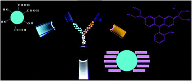
Fluorescent carbon dots (C-dots) have emerged as an alternative to semiconducting quantum dots owing to their splendid emission property, good biocompatibility, ease of bioconjugation, photo-stability and energy conversion abilities without incurring the burden of intrinsic toxicity or elemental scarcity.5 Easy methods of synthesis through microwave/hydrothermal procedures from commonly available precursors coupled with tunable excitation dependent and surface functionality dependent emission properties actuated C-dots for applications in bioimaging, sensing, drug delivery and opto-electronic devices.6 C-Dots and their composites have been exploited for carbon only white light emitting materials.7 For multidimensional applications, generation of white light through blending C-dot emission with another commonly available luminophore by simple mixing would be advantageous. In this direction, here we demonstrate how a full range of emission can be obtained by controlled mixing of C-dots and rhodamine B (RhB) in water. The interaction of C-dots with RhB and their photophysical properties have been studied,8a–e however the generation of white light emission by simple mixing of these two luminophores has not been reported. Considering the bluish green emission of C-dots when excited at 365 nm, RhB with their reddish orange emissions was an obvious choice as a dual cooperating emitter as they are complementary colors of white light. The addition of RhB to C-dots resulted in the aggregation of the dye molecules on the C-dot surface and controlling the emission of free RhB with the emission of C-dot-aggregated RhB composite led to fine tuning of the overall white light emission properties (Scheme 1).


image file: c6ra17306c-s1.tif
Scheme 1 A simple mixing of C-dots and rhodamine B leads to a mixture with white light emitting properties.

C-Dots were synthesized in aqueous medium from a natural precursor β-carotene through microwave irradiation as reported earlier.9 Briefly, a β-carotene dispersion in water when irradiated in a microwave reactor resulted in a pale yellow solution containing C-dots. The as synthesized C-dots showed maximum emission intensity at 470 nm while excited at 370 nm. The emission peak showed a bathochromic shift with increase in excitation wavelength (Fig. 1a). The morphology of C-dots was established using transmission electron microscopy (TEM) where spherical nanoparticles having diameter in the range 3–5 nm were observed (Fig. 1b).


image file: c6ra17306c-f1.tif
Fig. 1 (a) Emission spectrum of C-dots synthesized from β-carotene at different excitation wavelengths showing excitation dependent emission and (b) TEM image of C-dots (scale bar 20 nm).

Under UV light excitation (λex = 365 nm), the C-dots exhibited strong blue fluorescence which prompted us to exploit them as a component in pursuit of a white light emitting system. The CIE (Commission International de d'Eclairage) chromaticity coordinate for C-dots (0.21, 0.26) appear in the blue region. Therefore, we anticipated that a simple mixing of a fluorophore with orange emission might result in white light emitting composite. A commonly available dye, rhodamine B with chromaticity coordinate (0.54, 0.46) was selected for its low-cost, high quantum yield, good photostability and less toxicity compared to heavy metal based phosphors.10 RhB shows emission at 578 nm in water when excited at 365 nm (Fig. 2a). When aliquots of RhB were added to a 0.45 mg mL−1 dispersion of C-dots, the emission due to RhB increased without affecting the emission of C-dots (Fig. 2b). The calculation for CIE coordinates was performed from the fluorescence emission data. At certain composition of RhB (final concentration 20 μM) the resulting mixture exhibited white light emission with CIE coordinates (0.33, 0.32) (Fig. 2c). This is very close to that of pure white light emission (0.33, 0.33). The white light emission obtained was a mixture of blue-green and orange colors of its individual components as clearly observed from the digital images taken under UV illumination (Fig. 2d) and is tunable from blue to orange. Under daylight, C-dots in water showed pale yellow color while C-dot–RhB mixture and only RhB displayed two different shades of pink color (Fig. S1).


image file: c6ra17306c-f2.tif
Fig. 2 (a) Emission spectrum of rhodamine B in water, (b) emission spectrum of C-dots after addition of different concentrations of RhB, (c) chromaticity plot for colour coordinates after mixing C-dots (C) with different concentrations of rhodamine B (R) solution in water and (d) digital images of C-dots (blue), C-dot–RhB mixture (white) and RhB (orange) in water respectively under UV light (λex = 365 nm).

An interesting observation from the fluorescence spectra of a mixture C-dots with RhB (Fig. 2b) was that the emission owing to C-dots did not change significantly even with increasing concentration of the dye. The emission of C-dots showed an overlap with the absorption of RhB which suggests that they can form a donor–acceptor pair of a Förster resonance energy transfer (FRET) system (Fig. S2).11 In such a scenario, quenching of C-dot emission was a possibility upon addition of RhB. However, we did not observe any apparent quenching in the emission intensity of C-dots implying that no energy transfer took place between C-dots and RhB. To validate this, a mixture of C-dots with varying concentration of RhB was excited at 405 nm. At this excitation wavelength, there was significantly higher overlap between the emission of C-dot and absorption of RhB (Fig. S3). However, no quenching of C-dot emission was observed (Fig. S4). Absence of FRET was further confirmed by time correlated single photon counting (TCSPC) studies, where the lifetime of C-dots in presence and absence of RhB remained unaltered while excited at 375 nm and 405 nm indicating that the dye did not perturb the excited state of C-dots (Fig. S5).

In order to have an insight into the physico-chemical interaction between the C-dots and RhB molecules, a RhB solution (20 μM) was titrated against C-dots with various concentrations (Fig. 3a). The emission intensity of RhB at 578 nm decreased considerably with increasing C-dot concentration. Further the zeta (ζ) potential of pristine C-dots (−33.44 mV) decreased considerably to −27.36 mV upon addition of RhB (Fig. 3b). This was probably due to electrostatic interaction of RhB molecules on the negatively charged surface of C-dots having –COOH functionalities. The presence of –COOH functionality on C-dot surface was confirmed by FTIR (Fig. S6). The dynamic light scattering (DLS) measurements showed that the average particle size increased from ∼10 nm (Fig. S7) in case of C-dots to ∼180 nm for C-dot–RhB composite (Fig. 3c), clearly suggesting the formation of an agglomerated composite. In order to verify the fate of RhB upon interaction with C-dots, we compared the emission of same concentration of RhB in absence and presence of C-dots (Fig. S8).The difference in the emission at 578 nm suggested that RhB was adsorbed on the C-dot surface resulting in decrease of emission owing to free RhB molecules. UV-visible spectra of RhB upon interaction with various concentrations of C-dots showed a decrease in the intensity of the characteristic peak at 554 nm and an enhanced absorption at the short-wavelength shoulder at 512 nm (Fig. S9). By comparing the extinction spectrum of the mixed solutions with that of free RhB, the formation of H-aggregates could be ascertained.12 The aggregated RhB emits at 465 nm,13 which coincides with the emission of C-dots. TEM studies further confirmed the aggregation of RhB molecules on C-dot surface, as the resulting composite showed agglomerated particles (Fig. S10). High resolution TEM studies showed a cluster of C-dots wrapped with aggregated RhB resulting in larger aggregates (Fig. 3d). It is reported that RhB molecules aggregate at concentration higher than 10−4 M (ref. 14) that leads to a decrease in the emission peak at 578 nm. However, in the present case, the aggregation occurred at a much lower concentration of the dye molecules (20 μM), clearly suggesting the influence of negatively charged C-dot surfaces on RhB aggregation. Similar behavior of RhB aggregation on anionic surfaces is well reported.14


image file: c6ra17306c-f3.tif
Fig. 3 (a) Emission spectra of RhB with increasing concentration of C-dots, (b) zeta potential of C-dots before and after addition of RhB, (c) dynamic light scattering measurement of C-dot–RhB composite and (d) TEM image of C-dot–RhB composite.

The lifetime of RhB was measured using a diode laser at 375 nm to assess the binding of the dye and C-dots. The decay of dye in aqueous medium was single exponential and the average lifetime was 1.14 ns. In presence of C-dots, the decay became biexponential consisting of a shorter and a longer component (Fig. 4a). The lifetime of the shorter component was found to be 1.18 ns which might have originated from free RhB molecules in the mixture. The longer component having lifetime of 4.55 ns probably arises from the RhB molecules bound to C-dots. The quenching of RhB emission was purely static in nature as no decrease in fluorescence lifetime was observed.


image file: c6ra17306c-f4.tif
Fig. 4 (a) Lifetime decay curves of RhB (i) in absence and (ii) in presence of C-dots, (iii) lifetime decay curve of C-dots (iv) instrument response function, (b) and (c) digital images of C-dot–RhB mixture incorporated in gelatin gel and PVA film respectively under UV light (λex = 365 nm).

For practical applications, photostability of the white light emitting system is a critical criterion. The mixture was highly stable at room temperature for over three months and no notable decrease in its emission was observed. Further, no rapid photobleaching occurred for the C-dots–RhB composites upon UV-light exposure for 1.5 hours (Fig. S11). PL time traces of two random spots showed gradual intensity decay with no clear photobleaching steps demonstrating that the white light emitting system was stable for an appreciable time under laser irradiation (Fig. S12). The mixture showed white light emission at varying pH conditions with the ratio of emission intensities of the individual components remaining constant (Fig. S13). For practical applications, it is imperative that the white light be generated in gels or solid state. For this purpose, we used gelatin as a medium where C-dots and RhB were mixed maintaining the concentrations similar to that in solution. As shown in Fig. 4b, the gel composite showed efficient white light emission with CIE coordinates (0.30, 0.31) (Fig. S14), suggesting that the simple mixing of individual components to generate white light is effective even in gel medium. Further, a poly-vinyl alcohol (PVA) film was generated by drying an aqueous mixture of PVA, C-dot and RhB that showed efficient white light emission (Fig. 4c) with CIE coordinates (0.32, 0.32) (Fig. S15).

Water soluble luminescent temperature probes are promising candidates for optical thermometry applications.15 We also studied the response of the C-dot–RhB dual emitting system with change in temperature. When the temperature was increased from 25 °C to 80 °C, significant quenching of fluorescence emission of both the peaks was observed (Fig. 5a) which was visible even with naked eyes. RhB fluorescence quenched at a relatively faster rate with increase in temperature compared to C-dots (Fig. S16(a)). This resulted in decreased white light intensity with emission shifting to blue region (Fig. S16(b)). It is interesting to note that the thermo-response of this white light emitting system was reversible in nature. Fig. 5b and c show fluorescence response of C-dot and RhB emission in the composite with change in temperature of five heating–cooling cycles. While the reversible fluorescence response of C-dots with temperature can be attributed to the synergistic effect of abundant oxygen containing functional groups and hydrogen bonding with water,16 the variation of RhB emission upon increasing temperature is due to faster diffusion which leads to greater collisional quenching.17


image file: c6ra17306c-f5.tif
Fig. 5 (a) Fluorescence spectra of C-dot–RhB composite at 25 °C and 80 °C (b) and (c) temperature dependent change in fluorescence intensity of C-dots and RhB in the composite respectively during consecutive heating–cooling cycle.

The system further provided selective and sensitive sensing for Fe3+ ions among a host of biologically relevant metal ions tested (Fig. 6a). The response was quite fast and within seconds after addition of Fe3+ to the white light emitting mixture, the decrease in emission could be observed. Fe3+ is known to quench the emission of C-dots by electrostatic interactions.18 The deactivation of excited state electrons by relaxation induced by Fe3+ possibly quenches the emission of RhB.8c In case of the C-dot–RhB composite also, the quenching of white light emission occurred probably through similar mechanisms (Fig. 6b). The detection limit of Fe3+ was found to be 50 nm which was calculated from the change in emission at 578 nm.


image file: c6ra17306c-f6.tif
Fig. 6 (a) Change in the emission intensity of C-dots (red) and RhB (green) in the C-dot–RhB composite upon addition of various ions and (b) fluorescence emission of C-dot–RhB mixture upon addition of aliquots of Fe3+.

In conclusion, controlled mixing of rhodamine B with fluorescent carbon dots in water resulted in a sustainable and photostable white light emitting mixture. This simple method could be extended to gel or solid medium such as gelatin and PVA films for white light emission. The mixture showed reversible response to variable temperature which can be utilized for thermal sensing. Further, the system proved to be efficient for detection of Fe3+ ions in solution. Such simple methods for the generation of white light emitting nanocomposites with versatile applications are expected to add new dimensions in materials science.

Acknowledgements

This research is funded by Science and Engineering Research Board (SERB), Department of Science and Technology, India (SR/S1/PC-32/2010). Financial assistance from IIT Indore and instrumentation facility from SIC, IIT Indore are duly acknowledged. We are thankful to SAIF, NEHU, Shillong and SAIF, IIT Bombay for TEM facility. S. M. and B. S. acknowledge research fellowships from CSIR and UGC respectively. We thank Dr Anjan Chakraborty, Dr Tushar Kanti Mukherjee and Mr Anupam Das for helpful scientific discussions.

Notes and references

  1. C.-Y. Sun, X.-L. Wang, X. Zhang, C. Qin, P. Li, Z.-M. Su, D.-X. Zhu, G.-G. Shan, K.-Z. Shao, H. Wu and J. Li, Nat. Commun., 2013, 4, 2717 Search PubMed.
  2. (a) P. Malakar, D. Modak and E. Prasad, Chem. Commun., 2016, 52, 4309 RSC; (b) Q. Y. Yang and J. M. Lehn, Angew. Chem., Int. Ed., 2014, 53, 1 CrossRef CAS; (c) S. Mukherjee and P. Thilagar, Dyes Pigm., 2014, 110, 2 CrossRef CAS; (d) E. Ravindran, S. J. Ananthakrishnan, E. Varathan, V. Subramanian and N. Somanathan, J. Mater. Chem. C, 2015, 3, 4359 RSC; (e) M. Granström and O. Inganäs, Appl. Phys. Lett., 1996, 68, 147 CrossRef; (f) X. Cao, H. Lan, Z. Li, Y. Mao, L. Chen, Y. Wu and T. Yi, Phys. Chem. Chem. Phys., 2015, 17, 32297 RSC; (g) W. Xie, S.-R. Zhang, D.-Y. Du, J.-S. Qin, S.-J. Bao, J. Li, Z.-M. Su, W.-W. He, Q. Fu and Y.-Q. Lan, Inorg. Chem., 2015, 54, 3290 CrossRef CAS PubMed; (h) S. Roy, A. K. Katiyar, S. P. Mondal, S. K. Ray and K. Biradha, ACS Appl. Mater. Interfaces, 2014, 6, 11493 CrossRef CAS PubMed; (i) S. K. Panda, S. G. Hickey, H. V. Demir and A. Eychmüller, Angew. Chem., Int. Ed., 2011, 50, 4432 CrossRef CAS PubMed; (j) S. Pramanik, S. Bhandari, S. Roy and A. Chattopadhyay, J. Phys. Chem. Lett., 2015, 6, 1270 CrossRef CAS PubMed; (k) Y. Shirasaki, G. J. Supran, M. G. Bawendi and V. Bulovic, Nat. Photonics, 2013, 7, 13 CrossRef CAS; (l) V. Singh and A. K. Mishra, Sci. Rep., 2015, 5, 11118 CrossRef CAS PubMed.
  3. Z. Xie, C. Chen, S. Xu, J. Li, Y. Zhang, S. Liu, J. Xu and Z. Chi, Angew. Chem., Int. Ed., 2015, 54, 7181–7184 CrossRef CAS PubMed.
  4. (a) Q. Zhang, C.-F. Wang, L.-T. Ling and S. Chen, J. Mater. Chem. C, 2014, 2, 4358 RSC; (b) J. Malinge, C. Allain, A. Brosseau and P. Audebert, Angew. Chem., Int. Ed., 2012, 51, 8534 CrossRef CAS PubMed; (c) A. Ozawa, A. Shimizu, R. Nishiyabu and Y. Kubo, Chem. Commun., 2015, 51, 118 RSC.
  5. (a) H. Li, Z. Kang, Y. Liu and S.-T. Lee, J. Mater. Chem., 2012, 22, 24230 RSC; (b) H. Li, X. Hi, Z. Kang, H. Huang, Y. Liu, J. Liu, S. Lian, C. H. A. Tsang, X. Yang and S.-T. Lee, Angew. Chem., Int. Ed., 2010, 49, 4430 CrossRef CAS PubMed; (c) Z. Ma, Y. L. Zhang, L. Wang, H. Ming, H. Li, X. Zhang, F. Wang, Y. Liu, Z. Kang and S.-T. Lee, ACS Appl. Mater. Interfaces, 2013, 5, 5080 CrossRef CAS PubMed; (d) H. Ming, Z. Ma, Y. Liu, K. Pan, H. Yu, F. Wang and Z. Kang, Dalton Trans., 2012, 41, 9526 RSC; (e) S. N. Baker and G. A. Baker, Angew. Chem., Int. Ed., 2010, 49, 6726 CrossRef CAS PubMed.
  6. (a) P. G. Luo, S. Sahu, S. T. Yang, S. K. Sonkar, J. Wang, H. Wang, G. E. LeCroy, L. Caoa and Y. P. Sun, J. Mater. Chem. B, 2013, 1, 2116 RSC; (b) S. Karthik, B. Saha, S. K. Ghosh and N. D. P. Singh, Chem. Commun., 2013, 49, 10471 RSC; (c) C. Ding, A. Zhu and Y. Tian, Acc. Chem. Res., 2014, 47, 20 CrossRef CAS PubMed.
  7. (a) S. Hu, A. Trinchi, P. Atkin and I. Cole, Angew. Chem., Int. Ed., 2015, 54, 2970 CrossRef CAS PubMed; (b) F. Wang, Y. h. Chen, C.-y. Liu and D.-g. Ma, Chem. Commun., 2011, 47, 3502 RSC; (c) X. Guo, C.-F. Wang, Z.-Y. Yu, L. Chen and S. Chen, Chem. Commun., 2012, 48, 2692 RSC; (d) C. Sun, Y. Zhang, K. Sun, C. Reckmeier, T. Zhang, X. Zhang, J. Zhao, C. Wu, W. W. Yu and A. L. Rogach, Nanoscale, 2015, 7, 12045 RSC.
  8. (a) A. Diac, M. Focsan, C. Socaci, A.-M. Gabudean, C. Farcau, D. Maniu, E. Vasile, A. Terec, L. M. Veca and S. Astilean, RSC Adv., 2015, 5, 77662 RSC; (b) L. Liu, L. Chen, J. Liang, L. Liu and H. Han, J. Anal. Methods Chem., 2016, 4939582 Search PubMed; (c) S. Hu, Q. Zhao, Q. Chang, J. Yang and J. Liu, RSC Adv., 2014, 4, 41069 RSC; (d) M. Deng, S. Wang, C. Liang, H. Shang and S. Jiang, RSC Adv., 2016, 6, 26936 RSC; (e) Y. Ding, J. Ling, J. Cai, S. Wang, X. Li, M. Yang, L. Zha and J. Yan, Anal. Methods, 2016, 8, 1157 RSC.
  9. S. Mandani, B. Sharma, D. Dey and T. K. Sarma, Nanoscale, 2015, 7, 1802 RSC.
  10. C. C. Huang and H. T. Chang, Anal. Chem., 2006, 78, 8332 CrossRef CAS PubMed.
  11. J. R. Lakowicz, Principles of Fluorescence Spectroscopy, Springer, New York, USA, 2006 Search PubMed.
  12. C. Würth, M. Grabolle, J. Pauli, M. Spieles and U. Resch-Genger, Nat. Protoc., 2013, 8, 1535 CrossRef PubMed.
  13. (a) C.-Y. Hsu and Y.-L. Liu, J. Colloid Interface Sci., 2010, 350, 75 CrossRef CAS PubMed; (b) C.-Y. Hsu and Y.-L. Liu, Chem.–Eur. J., 2011, 17, 5522 CrossRef CAS PubMed.
  14. D. S. Tira, M. Focsan, S. Ulinici, D. Maniu and S. Astilean, Spectrosc. Lett., 2014, 47, 153 CrossRef CAS.
  15. E. J. McLaurin, L. R. Bradshaw and D. R. Gamelin, Chem. Mater., 2013, 25, 1283 CrossRef CAS.
  16. Y. Yang, W. Kong, H. Li, J. Liu, M. Yang, H. Huang, Y. Liu, Z. Wang, Z. Wang, T. K. Sham, J. Zhong, C. Wang, Z. Liu, S. T. Lee and Z. Kang, ACS Appl. Mater. Interfaces, 2015, 7, 27324 CAS.
  17. K. Kemnitz and K. Yoshihara, J. Phys. Chem., 1991, 95, 6095 CrossRef CAS.
  18. A. Bhattacharya, S. Chatterjee, R. Prajapati and T. K. Mukherjee, Phys. Chem. Chem. Phys., 2015, 17, 12833 RSC.

Footnote

Electronic supplementary information (ESI) available. See DOI: 10.1039/c6ra17306c

This journal is © The Royal Society of Chemistry 2016