Thermosensitive nanoplatforms for photothermal release of cargo from liposomes under intracellular temperature monitoring

Satoshi Araiab, Chi-Lik Ken Leec, Young-Tae Changd, Hirotaka Satoe and Keitaro Sou*ab
aWaseda Bioscience Research Institute in Singapore (WABIOS), Singapore 138667, Singapore. E-mail: soukei@aoni.waseda.jp
bOrganization for University Research Initiatives, Waseda University, Tokyo 162-0041, Japan
cCentre for Biomedical and Life Sciences, Department for Technology, Innovation and Enterprise (TIE), Singapore Polytechnic, Singapore 139651, Singapore
dDepartment of Chemistry and NUS Medchem Program of the Life Sciences Institute, National University of Singapore, Singapore 117543, Singapore
eSchool of Mechanical and Aerospace Engineering, Nanyang Technological University, Singapore 637460, Singapore

Received 24th September 2015 , Accepted 24th October 2015

First published on 26th October 2015


Abstract

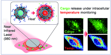
Control of cargo release from nanoscale carriers is an important technology for maximizing the benefits of nanoparticulate drug delivery systems. Herein, we attempt to trigger the release of cargo from liposomes by photothermal conversion of water with a 980 nm near-infrared (NIR) laser. This study examined liposomes of two types formulated by 1,2-dipalmitory-sn-glycero-3-phosphocholine (DPPC) or a mixture of DPPC/cholesterol with an anionic lipid and PEG-lipid as stabilizers encapsulating calcein as a cargo at different ionic strengths. Liposome formulation encapsulating a hypertonic solution with a lipid membrane shows that a gel to liquid-crystalline phase transition at around 40 °C effectively released the cargo from liposomes at temperature above 40 °C with NIR irradiation. Our proof of concept has been further demonstrated in a cancer cell with monitoring the actual “intracellular temperature” using a fluorescent thermosensor. Intracellular thermometry revealed that it was not until the intracellular temperature reached around 40 °C by NIR irradiation that the release of the cargo started gradually, showing good agreement with the result from the extracellular in vitro study. This targeted release of cargo from thermosensitive liposomes based on a photothermal effect using a NIR laser offers a potent nanoscale platform for the on-demand release of drugs in intracellular space with local hyperthermia. The intracellular thermometry facilitates the quantitative monitoring and control of the hyperthermia at the cellular level.


Introduction

Nanoscale carriers encapsulating drugs offer a promising approach to site-specific drug delivery and release control of the drugs, thereby enabling pinpoint cancer therapy with improved safety and efficacy over those of conventional chemotherapy.1–4 In a nanocarrier-based drug delivery system, the encapsulation structure shields the efficacy of drugs and toxicity to the normal healthy cells and tissues. These beneficial or undesirable activities of drugs appear once the drugs are released from the nanocarriers. Consequently, on-demand drug release from nanocarriers with appropriate timing in the targeted region is regarded as a key technology to maximize drug efficacy at the target site and to minimize effects on normal tissues.

Stimulus-responsive carriers that release the cargo by physicochemical stimulations such as temperature, pH, light, and magnetic field are attracting great interest in producing drug delivery systems that provide important benefits for drug release.2,3,5–8 Particularly, liposomes have been studied as widely applicable nano scale carriers that stably encapsulate drugs with biocompatible lipid bilayer membranes.9–11 Thermosensitive liposomes are widely applicable because stimulation by heating is technically possible in a wide range of bodies. Because the gel to liquid-crystalline phase transition in a lipid bilayer membrane at a specific temperature induces drastic changes of membrane fluidity, to control the phase transition of the lipid membrane has been considered as traditional approach to design thermosensitive liposomes. However, the thermosensitive drug release was slow and inefficient for the traditional thermosensitive liposomes.12 In order to achieve rapid and efficient drug release, the traditional thermosensitive liposomes have been expected to be improved further.

In addition to the development of advanced thermosensitive liposomes, the methodology for heating the target region to trigger the drug release from liposomes is important. For regional hyperthermia, the traditional method is heating with water baths heated to the designated temperature.13–15 This method is available as an option to heat the superficial tissues but a shortcoming of this method is the difficulty in heating the localized spot of the target. For localized hyperthermia, the use of microwaves, radio waves, and ultrasound have been investigated.16–18 Recently, magnetic resonance-guide focused ultrasound is attracting attention as a potent tool for imaging-guide thermal therapy.19,20 Laser is an effective source to focus the energy on the remote target portion. In addition to generating heating, the laser light can be converted to fluorescence for imaging-guide thermal treatment. Consequently, near infrared (NIR, 700–1100 nm wavelength) radiation is receiving increased attention as an energy source to produce localized heating by photothermal conversion.21–23 Several groups have applied NIR laser to trigger the release of drugs from drug carriers aimed at developing the controlled drug release system.24–26 Very recently, Li et al. reported liposomes with an embedded NIR dye in the lipid membrane causing the photothermal release of drugs with NIR irradiation.27 However, these conventional studies require special materials such as NIR dyes and gold nanoparticles to convert NIR light to heat, which are limited to use for practical clinical applications.

We proposed here a dye- or inorganic materials free system where hyperthermia to release cargo from improved traditional thermosensitive liposomes is achieved by heating the water with NIR irradiation. Recent years, it is proved that the irradiation of pulsed 980 nm NIR laser heats water molecules and then induces the release of cargos in polymeric particles without dyes.28 This kind of concept is also expected to be applicable for fully hydrated liposomes.

Our results show that the release properties of traditional thermosensitive liposomes can be improved by encapsulating hypertonic solution. We achieved on-demand release of cargo from the improved thermosensitive liposomes in cancer cells by the irradiation of 980 nm NIR laser. Importantly, the actual temperature rise inside the cell by NIR irradiation was measured using a fluorescent thermosensor in real-time, concurrent with monitoring of the release of the cargos. Despite of many reports regarding thermosensitive cargo release systems, this report is the first to describe cargo release observations during monitoring of the actual “intracellular temperature.”

Experimental

Materials and methods

Materials. 1,2-Dipalmitoyl-sn-glycero-3-phosphocholine (DPPC), cholesterol, and L-glutamic acid, N-(3-carboxy-1-oxopropyl)-, 1,5-dihexadecyl ester (SA) were purchased from Nippon Fine Chemical Co. Ltd. (Osaka, Japan). 1,2-Distearoyl-sn-glycero-3-phosphoethanolamine-N-[monomethoxy poly(ethylene glycol) (5000)] (PEG-DSPE) was purchased from NOF Corp. (Tokyo, Japan). Calcein, sodium gluconate, and phosphate buffered saline (PBS) were purchased from Sigma-Aldrich Corp. (St. Louis, MO).
Preparation and characterization of liposomes. Liposome samples were prepared using DPPC, cholesterol, SA, and PEG-DSPE. DPPC/SA/PEG-DSPE (9/1/0.06, molar ratio) were dissolved in t-butyl alcohol for DPPC-based liposomes (DPPC). DPPC/cholesterol/SA/PEG-DSPE (5/5/1/0.066, molar ratio) were dissolved in t-butyl alcohol for DPPC/cholesterol-based liposomes (DPPC/Chol). These solutions were freeze-dried to obtain mixed lipid powder. The mixed lipid powder was hydrated with calcein solution (50 mM calcein, 200 mM NaOH, and 0, 150 or 300 mM sodium gluconate). Then the dispersion was introduced into an extruder (Lipex Biomembrane Inc., Canada) and was extruded through membrane filters (0.22 μm final pore size, Durpore®; Millipore Corp., Tokyo, Japan) under pressure using nitrogen gas. The unencapsulated calcein was removed two times using gel filtration (PD10 column; GE Healthcare, Buckinghamshire, UK). The prepared liposomes encapsulating calcein were stored in a refrigerator under dark conditions. The diameters of the resulting liposomes were ascertained using a particle size analyzer based on dynamic light scattering (DLS) (Zeta-Sizer Nano ZS; Malvern Instruments, Ltd., Malvern, Worcestershire, UK). The average diameter ± standard deviation (SD) was calculated. The phospholipid concentration was determined using a choline oxidase method (Phospholipid C Test Wako; Wako Pure Chemical Industries Ltd., Tokyo).
Differential scanning calorimetry (DSC). Liposome dispersion ([lipids] = 77 mg mL−1, 50 μL) was put in an aluminum pan. The same volume of dispersant (PBS) was put in the reference pan. The DSC thermograms were recorded using a differential scanning calorimeter (DSC-60; Shimadzu Corp.) with a rate of temperature increase of 2 °C min−1.
Calcein release experiment. The dispersion of the liposomes encapsulating calcein (1 mL, [calcein] = 3 μM) was incubated at various temperatures (25–60 °C). Before heating, the blank fluorescence intensity of the liposome dispersion (Ipre) was measured with a fluorescence spectrophotometer (λex = 490 nm, λem = 515 nm, F-2700; Hitachi Ltd.). After heating, the fluorescence intensity of the liposome dispersion (Ipost) was measured again. For the fluorescence intensity of the 100% release of calcein from liposomes (I100%), the liposomes were solubilised using detergent (octyl-glucoside). The percentage of the calcein release from liposomes was calculated using eqn (1).
 
image file: c5ra19729e-t1.tif(1)
980 nm laser irradiation experiment. The diode-pumped solid state 980 nm CW laser was coupled with a flexible optical fibre terminated with a collimating lens (5 mm ϕ spot size). It was controlled using a VD-IIA DPSS laser driver. The actual laser power was measured using a power meter. The sample solution (1 mL) was put in a 10 mm × 10 mm square quartz cell. The 980 nm NIR laser was exposed to the sample from the side of the cell (10 mm light path length). The sample temperature was measured over time using a digital thermometer with a thermocouple (TES 1310 TYPE-K; TES Electrical Electronic Corp., Taipei, Taiwan). For the calcein release experiment, liposome-encapsulating calcein dispersion was put in a 10 mm × 10 mm square quartz cell. The 980 nm NIR laser was exposed for 10 min to the sample from the side of the cell (10 mm light path length).
Release of cargo from liposomes in cell culture. In a CO2 incubator, HeLa (ATCC CCL-2) cells were maintained in Dulbecco's modified Eagle's medium (DMEM) supplemented with fetal bovine serum (FBS, 10%) and penicillin–streptomycin (1%). The HeLa cells, seeded on the 35 mm glass-bottom dishes (50–70% confluence), were incubated with liposomes encapsulating calcein at 37 °C under 5% CO2 for 3 h, were replaced with the fresh DMEM with 10% FBS; and were then incubated with ER thermo yellow (f.c. 250 nM) at 37 °C for 30 min. Before imaging experiments, the cells were replaced with fresh basal medium (1 mL). Fluorescence imaging was done using a confocal microscope (FV 1000; Olympus Corp.) with an oil immersion objective lens (PLAPO, 60 × 1.45 NA). For excitation of calcein and ER thermo yellow, 488 nm and 543 nm lasers were used, respectively. Their fluorescence was recorded in the emission ranges of 500–530 nm and above 560 nm. Images were taken every 2 s. For heating HeLa cells transiently, the diode-pumped solid state 980 nm CW laser (described in the detail above) was conducted. All experiments were conducted using a temperature-controlled circulation chamber with the CO2 incubator.

Results and discussion

Thermotropic phase behaviour of liposomes

The target temperature to trigger the cargo release is an important factor affecting the design of thermosensitive liposome. For biomedical internal use such as drug delivery, the liposomes should retain the drugs stably below body temperature and release them after heated to a temperature higher than body temperature. Human cells are damaged when the surrounding temperature is increased to a temperature higher than 42.5 °C,29,30 which is regarded as an upper limit of medical hyperthermia. General hyperthermia for cancer therapy is performed at around 43 °C at a maximum. Consequently, the target temperature range for triggering drug release with heating at minimal damage to human cells is between the physiological body temperature (37 °C) and 43 °C. We attempted to demonstrate an on-demand release system in which the effective release of cargo from liposomes is triggered at around 40 °C by heating of water facilitated with the 980 nm NIR laser, as shown in Fig. 1A. For this purpose, the lipid composition of liposome membrane and osmotic pressure in inner aqueous phase was optimized in this study. In addition, the laser power and irradiation time to achieve heating the water to a higher temperature than 40 °C has been investigated.
image file: c5ra19729e-f1.tif
Fig. 1 NIR light triggers release of cargo from thermosensitive liposomes. (A) Schematic showing the concept of cargo release and (B) chemical structure of the lipid components.

Lipid formulation of two types, DPPC/SA/PEG-DSPE (DPPC; 9/1/0.06, molar ratio) and DPPC/cholesterol/SA/PEG-DSPE (DPPC/Chol; 5/5/1/0.066, molar ratio), were examined in this study. The chemical structure of the lipid is shown in Fig. 1B. These liposomes introduced the encapsulation of water-soluble fluorescent marker (calcein) with different ionic concentration, which varied by the concentration of sodium gluconate, as presented in Table 1. The gel to liquid-crystalline phase transition temperature (Tc) of the bilayer membrane consisting of pure DPPC is around 41 °C.31 Actually, SA has a negative charge on the hydrophilic head group, which increases the entrapment capacity of liposomes to reduce the lamellarity of the bilayer membrane because of the electrostatic repulsion force between bilayer membranes.32 Furthermore, the electrostatic repulsion force between liposomes prevents their aggregation. The high biocompatibility and unique biodistribution of liposomes containing SA served to increase the potential of the liposomes for use in medical applications such as drug delivery carriers.33,34 PEG-DSPE is used widely to stabilize the liposomes in circulation as well as in a dispersion state.

Table 1 Specification of liposome samples
Samples Lipid compositions (molar ratio) Encapsulating solution Size (μm)
DPPC(L) DPPC/SA/PEG-DSPE (9/1/0.06) 50 mM calcein, 200 mM NaOH 0.21 ± 0.10
DPPC/Chol(L) DPPC/Chol/SA/PEG-DSPE (5/5/1/0.066) 50 mM calcein, 200 mM NaOH 0.26 ± 0.12
DPPC(M) DPPC/SA/PEG-DSPE (9/1/0.06) 50 mM calcein, 200 mM NaOH, 150 mM sodium gluconate 0.21 ± 0.09
DPPC/Chol(M) DPPC/Chol/SA/PEG-DSPE (5/5/1/0.066) 50 mM calcein, 200 mM NaOH, 150 mM sodium gluconate 0.29 ± 0.14
DPPC(H) DPPC/SA/PEG-DSPE (9/1/0.06) 50 mM calcein, 200 mM NaOH, 300 mM sodium gluconate 0.24 ± 0.10
DPPC/Chol(H) DPPC/Chol/SA/PEG-DSPE (5/5/1/0.066) 50 mM calcein, 200 mM NaOH, 300 mM sodium gluconate 0.26 ± 0.12


The DSC thermogram showed that DPPC has clear endothermic peak of which Tc was 40.3 °C (Fig. 2A). This endothermic peak is regarded as the phase transition from ordered (gel) to a disordered (liquid-crystalline) lipid bilayer membrane with the melting of DPPC acyl chains (Fig. 2B).35,36 This gel to liquid-crystalline phase transition induces dynamic change in physical characteristics of DPPC membrane in which the area per lipid molecule changes from 47.9 Å2 (at 20 °C) to 64 Å2 (at 50 °C) and the membrane thickness changed from 5.5 nm (at 22 °C) to 3.6 nm (at 50 °C).35,36 The Tc of DPPC-liposomes was similar with that of DPPC itself, indicating that the incorporation of SA and PEG-DSPE had only a minimal effect on the thermotropic phase behaviour of the DPPC membrane. However, DPPC/Chol, containing cholesterol, showed no critical thermal peak at 25–55 °C. The incorporation of cholesterol changes the gel phase of the DPPC membrane to fluid phase, which does not transfer to other phase with critical temperature.37 Although the membrane fluidity is high in the DPPC/cholesterol membrane, the molecular packing in the fluid membrane is high in comparison with the fluid membrane of DPPC because the cholesterol fills the space among DPPC molecules (Fig. 2B).38 We examined liposomes of these two types, composed by membranes having different phase transition properties with temperature, to achieve the effective thermosensitive release of cargo from liposomes in an appropriate temperature range.


image file: c5ra19729e-f2.tif
Fig. 2 Thermotropic phase behaviour of lipid bilayer membrane: (A) DSC thermograms of DPPC (DPPC/SA/PEG-DSPE = 9/1/0.06, molar ratio) and DPPC/Chol (DPPC/Cholesterol/SA/PEG-DSPE = 5/5/1/0.066, molar ratio) and (B) schematic illustrations of different phase states on DPPC-membrane and DPPC/Chol-membrane.

Release of cargo from liposomes with thermal stimulation

The release of cargo from liposomes was evaluated using liposomes encapsulating calcein, as shown in Fig. 3. The percentage of released calcein was small in liposomes encapsulating solution of 50 mM calcein and 200 mM NaOH (Fig. 3A). Although the DPPC(L) exhibited increased release of calcein with increasing temperature higher than 40 °C, the percentage of released calcein was 12.4% after incubation at 50 °C for 10 min. The encapsulation of DPPC/Chol(L) was highly stable. As little as 0.4% of calcein was released from DPPC/Chol(L) after incubation at 50 °C for 10 min. DPPC/Chol(L) is available for stable encapsulation. DPPC(L) might be available for the slow release of cargo from liposomes with thermal stimulation.
image file: c5ra19729e-f3.tif
Fig. 3 Calcein release from liposomes at various temperatures. The percentage of calcein released from liposomes was measured after incubation in PBS at various temperatures for 10 min. Liposomes were prepared with the following conditions: (A) 50 mM calcein and 200 mM NaOH; (B) 50 mM calcein, 200 mM NaOH, and 150 mM sodium gluconate; (C) 50 mM calcein, 200 mM NaOH, and 300 mM sodium gluconate. (D) Calcein release from liposomes with time at 42 °C. The percentage of calcein released from liposomes was measured after incubation in PBS at 42 °C. Liposomes (lipid compositions: DPPC/SA/PEG-DSPE = 9/1/0.06, molar ratio) were prepared in 50 mM calcein, 200 mM NaOH, and 150 mM sodium gluconate solution for DPPC (M) or 50 mM, 200 mM NaOH, calcein and 300 mM sodium gluconate solution for DPPC (H).

In addition to the thermotropic phase behaviour of the lipid bilayer membrane, the osmotic pressure is an important factor affecting the stability of the liposome encapsulation structure.39 In physiological conditions, the osmotic pressure is 300 mOsmol L−1. Liposomes encapsulating a hypertonic solution higher than 300 mOsmol L−1 can be exposed to pressure from the inner aqueous phase because the lipid bilayer membrane is a semipermeable membrane. Once the pressure balance between surface tension and osmotic pressure is collapsed by the phase transition of lipid membrane, the encapsulated inner hypertonic solution is released from liposome to bulk phase. Based on this idea, we prepared liposomes encapsulating hypertonic solution adjusted with 50 mM calcein, 200 mM NaOH, and 150 mM or 300 mM sodium gluconate. As expected, the release of calcein was highly accelerated on liposomes encapsulating hypertonic solution. Particularly, the DPPC(M) showed significant release of calcein at temperatures higher than 40 °C, although negligibly small percentages of calcein were released on both DPPC(M) and DPPC/Chol(M) at 37 °C (Fig. 3B). Further increased osmotic pressure on DPPC(H) was effective for releasing most calcein above 40 °C (Fig. 3C). With high osmotic pressure on both DPPC(H) and DPPC/Chol(H), the calcein leaked sluggishly at temperatures lower than 37 °C. The release profiles of calcein on DPPC(M) and DPPC(H) at 42 °C indicated that the percentage of calcein release reached a plateau in 5 min (Fig. 3D).

Although the present liposomes are a class of traditional thermosensitive liposomes with thermosensitivity based on the phase transition of the lipid bilayer membrane, enhanced release of cargo from the liposomes could be achieved with encapsulation of a hypertonic solution. It is believed that the formation of grain boundaries between solid phase and liquid phase domains during the phase transition enhances the permeability of membrane on traditional thermosensitive liposomes.40 In this release mechanism, the rate and amount of drugs released from liposomes are generally small in the traditional thermosensitive liposomes.12 Anyarambhatla and Needham et al. achieved rapid drug release by modifying the lipid bilayer membrane of traditional thermosensitive liposomes with lysolipid, which is a phospholipid having a single acyl chain.41,42 It is hypothesized that the lysolipids accumulate at grain boundary between solid phase and liquid phase domains of liposome membrane to stabilize the boundary defect, resulting in rapid and enhanced drug release from liposomes.41 Oku et al. reported the enhanced release of cargo from traditional thermosensitive liposomes by controlling the encapsulated osmotic pressure.43 Because the lipid bilayer is a semipermeable membrane, difference in osmotic pressure between inner aqueous phase of liposomes and bulk dispersant exerts pressure stress on the membrane. The membrane of liposomes encapsulating a hypertonic solution is pressed to the outside by water from the inner aqueous phase. Once the grain boundary is formed by the phase transition upon heating, the inner aqueous solution is pushed out from liposomes. This pushing out is a possible mechanism for the enhanced release of cargo from thermosensitive liposomes encapsulating hypertonic solution. With increasing concentration of sodium gluconate, the release of cargo was increased. However, even if the release of cargo was enhanced when the concentration of sodium gluconate was increased to 300 mM in DPPC(H), the considerable release of cargo from liposomes was observed below physiological temperatures (37 °C). These results demonstrate that DPPC(M) is a potent formulation to retain the cargo in encapsulation at physiological temperature (37 °C) and release the cargo from liposomes with increasing the temperature above 40 °C for several minutes.

Release of cargo from liposomes by dye-free photothermal conversion with 980 nm near-infrared laser

Liquid water showed an absorption band at 980 nm, as shown in Fig. 4A. The bands at 980 nm have been assigned as a combination band (2ν1 + ν3; 10[thin space (1/6-em)]210 cm−1) where the ν1 is a frequency of symmetric stretching and ν3 is a frequency of asymmetric stretching.44 Water absorbs light of 980 nm and converts it into heat (photothermal conversion).28,45 The temperature of water was increased depending on the laser power and irradiation time. The water temperature reached a plateau within 20 min, as shown in Fig. 4B. As shown in Fig. 4C, the water temperature increased proportionally, concomitantly with increasing laser power. To confirm that the heat was produced by absorption of 980 nm laser energy by water in the present experiment settings, we performed irradiation of D2O using the same experimental settings. Because the atomic mass is different between hydrogen and deuterium, the frequencies of symmetric stretching and asymmetric stretching in D2O are shifted to a lower frequency compared with those of H2O. The combination band (2ν1 + ν3) of D2O has been assigned to 7470 cm−1.46 In this way, the absorption peak at 980 nm in H2O is shifted to at around 1300 nm in D2O, as shown in Fig. 4A. Therefore, no heating should be observed in D2O with 980 nm laser irradiation. As expected, the increase of temperature was not observed in D2O after irradiation of 980 nm laser for 10 min (Fig. 4C). These data support the notion that absorption of 980 nm laser light by water molecule (H2O) is effective for photothermal conversion. Heat production was controllable by the laser power and the water temperature was reached above 40 °C after laser irradiation for 10 min when the laser power was above 1015 mW. In addition, the temperature reached a plateau within 20 min at various temperatures depending on the laser power. This profile is expected to be suitable for mild hyperthermia in which the region is treated with a constant temperature at around 40–43 °C.
image file: c5ra19729e-f4.tif
Fig. 4 Release of cargo from liposomes by irradiation of 980 nm near infrared laser (CW): (A) absorption spectra of H2O (PBS) and D2O; (B) temperature profiles of dispersant (PBS) with time by continuous irradiation of 980 nm NIR laser at various power; (C) correlation between dispersant temperature and laser power after continuous irradiation of 980 nm NIR laser for 10 min; (D) percentage of calcein release from liposomes after continuous irradiation of 980 nm NIR laser for 10 min; (E) DLS size distributions of DPPC(M) before and after release of cargo by irradiation of 980 nm NIR laser with laser power 1015 mW for 10 min.

We applied this water heating system to trigger release of cargo from liposomes. We chose DPPC(M) which exhibited the best performance in retaining the cargo in encapsulation at physiological temperatures (37 °C) and released the cargo from liposomes above 40 °C for this experiment. Furthermore, we tested DPPC/Chol(M) as a reference liposomes which stably encapsulated cargo at temperatures even higher than 40 °C. As shown in Fig. 4D, the cargo release was triggered in DPPC(M) when the liposomes were irradiated with laser power 1015 and 1226 mW for 10 min. No release of cargo was observed with laser power 610 or 791 mW. Considering that the critical temperature to trigger the release of cargo on DPPC(M) was 40 °C (Fig. 3B) and the required laser power to heat water above 40 °C was more than 1015 mW (Fig. 4B and C), the release of cargo above 1015 mW is reasonable. No release of cargo from DPPC/Chol(M) was observed. According to the temperature-independent properties of DPPC/Chol(M), this result is reasonable. Consequently, the temperature to release the cargo from liposomes corresponds to the environmental temperature controlled by the 980 nm laser irradiation. After irradiation of the 980 nm NIR laser with laser power 1015 mW for 10 min, the size distribution of the liposomes was slightly shifted toward smaller one (0.18 ± 0.08 μm) compared with that of liposomes before irradiation (0.21 ± 0.09 μm) as shown in Fig. 4E. Such small change in size distribution suggested that the liposomes almost maintain their original structure after release of cargo with laser irradiation. The DPPC(M) encapsulating hypertonic solution might be swelled by the pressure from the inner aqueous phase. After release of cargo, the swelling is cancelled because the osmotic pressure in liposomes would become equal with the outside medium. The difference in swelling rate of liposomes is a possible reason for the slight shift to smaller size distribution after cargo release.

On-demand release of cargo from liposomes in living cells under intracellular temperature monitoring

The thermosensitive release of calcein from liposomes in living cells by irradiation of 980 nm NIR laser was examined in HeLa cells. The laser power was fixed to 1015 mW, which triggered the release of calcein by heating the water to temperature greater than 40 °C (Fig. 4D). The temperature change in HeLa cells induced by the irradiation of 980 nm NIR laser was monitored using a fluorescent thermosensor: ER thermo yellow. This sensor can stain the endoplasmic reticulum evenly and report the change in the intracellular temperature as a detectable fluorescence signal.47 Concurrent with real-time monitoring the temperature at the cellular level, the release of cargo was measured by the fluorescence increase of calcein released from liposomes because the self-quenching of calcein encapsulated in liposomes is eliminated.

First, HeLa cells were incubated with the liposome encapsulating hypertonic calcein solution and were then stained with ER thermo yellow. Dual imaging with ER thermo yellow and calcein during heating was done using a confocal microscope equipped with the 980 nm NIR laser (Fig. 5A). When the shutter of the NIR laser was open, the stained HeLa cells were exposed with heating. In response to the increase in temperature, ER thermo yellow exhibited a fluorescence decrease and a “square wave” pattern (Fig. 5C). The shutter being closed, the fluorescence returned to the basal level within about 20 s, indicating that our heating system creates a reversible temperature gradient at the microscopic level. Furthermore, a fluorescence of calcein in the cytoplasm was increased because of the heating (Fig. 5B and C). Fig. 5D, in which the axis of timescale at the very beginning of heating process is expanded, shows that the fluorescence of calcein remains silent, although the temperature increases gradually. When the normalized intensity of ER thermo yellow reached 0.72 ± 0.029, corresponding to 40.4 °C ± 0.7 (means ± standard deviation), fluorescence of calcein started to increase gradually. These results for live HeLa cells are compatible with the data from the in vitro study showing that the release of the cargo was observable at temperatures higher than 40 °C. Various temperature-sensitive cargo-release systems have been developed in recent year. In this research field, a thermocouple thermometer has been a common means of monitoring temperatures created by external devices. Nevertheless, it only provides the temperature in the medium. Therefore, previous studies have been unable to provide insight into how an individual cell senses the actual temperature change given by the device. Our evaluation system coupled with an intracellular thermosensor presents a standardized means for developing a thermosensitive release system. Here, we stress the importance of thermometry at cellular level again. For example, if the NIR laser irradiation causes uneven temperature variation in multi-cellular samples, some of cells do not reach the threshold temperature for the release.48 Even if it is only 1–2 °C below, the release of cargo based on the thermosensitive system is not allowed, leading to the poor therapeutic efficiency.


image file: c5ra19729e-f5.tif
Fig. 5 On-demand release of calcein from liposomes in HeLa cells by irradiation of 980 nm laser: (A) schematic image of a confocal microscope equipped with the NIR 980 nm laser; (B) representative fluorescence images of ER thermo yellow (upper panel) and calcein (lower panel) in HeLa cells during heating experiments. The elapsed time is shown at the top left of each image. (C) The square-wave in response to a laser-heating process (red, ER thermo yellow) and the release of cargo (green, calcein). Images were taken every 2 s. The normalized intensity of ER thermo yellow and calcein (F/F0) is shown versus time. The average of fluorescence intensity in a whole cell area was calculated. Then the average of cells 1–3 is shown. (D) To support (C), the x-axis was expanded and the actual temperature was calculated from the normalized intensity and the temperature sensitivity 3.9%/°C. Through the experiments, the base temperature was maintained at 33.3 °C.

Conclusions

Non-contact and remotely controllable laser technology is a powerful tool for use in biomedical applications. Irradiation of 980 nm NIR laser can be used to trigger the release of cargo from thermosensitive liposomes by mild hyperthermia based on the dye-free photothermal conversion from absorption of 980 nm NIR by water. This system is useful not only for on-demand release of drugs at target region but also for combination with local tumour hyperthermia and chemotherapy. Furthermore, in combination with the method of the intracellular temperature measurement, we propose “fluorescent intracellular thermometry”-guided heating system, which will pave the way for a novel concept of heat therapy at nanoscale.

Acknowledgements

This work was partly supported by a start-up research grant for Waseda-SP joint lab from Waseda University, SP R&D (TIEFA) grant number 198 from Singapore Polytechnic, and A*STAR-JST (Agency for Science, Technology and Research, The Japan Science and Technology Agency) joint grant.

Notes and references

  1. D. Peer, J. M. Karp, S. Hong, O. C. Farokhzad, R. Margalit and R. Langer, Nat. Nanotechnol., 2007, 2, 751–760 CrossRef CAS PubMed.
  2. S. Mura, J. Nicolas and P. Couvreur, Nat. Mater., 2013, 12, 991–1003 CrossRef CAS PubMed.
  3. V. P. Torchilin, Nat. Rev. Drug Discovery, 2014, 13, 813–827 CrossRef CAS PubMed.
  4. J. Y. Yhee, S. Lee and K. Kim, Nanoscale, 2014, 6, 13383–13390 RSC.
  5. T. Hoare, J. Santamaria, G. F. Goya, S. Irusta, D. Lin, S. Lau, R. Padera, R. Langer and D. S. Kohane, Nano Lett., 2009, 9, 3651–3657 CrossRef CAS PubMed.
  6. S. Pal, S. G. Roy and P. de, Polymer Chemistry, 2014, 5, 1275–1284 RSC.
  7. Y. Bae, S. Fukushima, A. Harada and K. Kataoka, Angew. Chem., Int. Ed. Engl., 2003, 42, 4640–4643 CrossRef CAS.
  8. N. Ž. Knežević and V. S.-Y. Lin, Nanoscale, 2013, 5, 1544–1551 RSC.
  9. D. Papahadjopoulos, T. M. Allen, A. Gabizon, E. Mayhew, K. M. Ii, S. K. Huang, K. Lee, M. C. Woodle, D. D. Lasic, C. Redemann and F. J. Martin, Proc. Natl. Acad. Sci. U. S. A., 1991, 88, 11460–11464 CrossRef CAS.
  10. G. Gregoriadis, Trends Biotechnol., 1995, 13, 21303–21306 CrossRef.
  11. B. S. Pattni, V. V Chupin and V. P. Torchilin, Chem. Rev., 2015, 115, 10938–10966 CrossRef CAS PubMed.
  12. M. B. Yatvin, J. N. Weinstein, W. H. Dennis and R. Blumenthal, Science, 1978, 202, 1290–1293 CAS.
  13. Q. Chen, S. Tong, M. Dewhirst and F. Yuan, Mol. Cancer Ther., 2004, 3, 1311–1318 CAS.
  14. G. Kong, R. Braun and M. Dewhirst, Cancer Res., 2001, 61, 3027–3032 CAS.
  15. J. May, M. Ernsting, E. Undzys and S. Li, Mol. Pharm., 2013, 10, 4499–4508 CrossRef CAS PubMed.
  16. M. L. Hauck, S. M. LaRue, W. P. Petros, J. M. Poulson, D. Yu, I. Spasojevic, A. F. Pruitt, A. Klein, B. Case, D. E. Thrall, D. Needham and M. W. Dewhirst, Clin. Cancer Res., 2006, 12, 4004–4010 CrossRef CAS PubMed.
  17. Y. Xu, A. Karmakar, W. E. Heberlein, T. Mustafa, A. R. Biris and A. S. Biris, Adv. Healthcare Mater., 2012, 1, 493–501 CrossRef CAS PubMed.
  18. S. Mitragotri, Nat. Rev. Drug Discovery, 2005, 4, 255–260 CrossRef CAS PubMed.
  19. M. de Smet, E. Heijman, S. Langereis, N. M. Hijnen and H. Grüll, J. Controlled Release, 2011, 150, 102–110 CrossRef CAS PubMed.
  20. N. Hijnen, S. Langereis and H. Grüll, Adv. Drug Delivery Rev., 2014, 72, 65–81 CrossRef CAS PubMed.
  21. K. Hribar, M. Lee, D. Lee and J. Burdick, ACS Nano, 2011, 5, 2948–2956 CrossRef CAS PubMed.
  22. S. Bhana, G. Lin, L. Wang, H. Starring, S. R. Mishra, G. Liu and X. Huang, ACS Appl. Mater. Interfaces, 2015, 7, 11637–11647 CAS.
  23. G. Yang, R. Lv, F. He, F. Qu, S. Gai, S. Du, Z. Wei and P. Yang, Nanoscale, 2015, 7, 13747–13758 RSC.
  24. A. K. Rengan, M. Jagtap, A. de, R. Banerjee and R. Srivastava, Nanoscale, 2014, 6, 916–923 RSC.
  25. A. Abhiruchi, M. A. Mackey, M. A. El-Sayed and R. V. Bellamkonda, ACS Nano, 2011, 5, 4919–4926 CrossRef PubMed.
  26. Z. Zhang, J. Wang and C. Chen, Adv. Mater., 2013, 25, 3869–3880 CrossRef CAS PubMed.
  27. M. Li, C. Teh, C. Y. Ang, S. Y. Tan, Z. Luo, Q. Qu, Y. Zhang, V. Korzh and Y. Zhao, Adv. Funct. Mater., 2015, 25, 5602–5610 CrossRef CAS.
  28. M. Viger, W. Sheng, K. Doré, A. Alhasan, C.-J. Carling, L. Jacques, C. de, G. Lux, G. Madeleine, R. Malinow and A. Adah, ACS Nano, 2014, 8, 4815–4826 CrossRef CAS PubMed.
  29. W. C. Dewey, Int. J. Hyperthermia, 2009, 25, 3–20 CrossRef CAS PubMed.
  30. A. Samali, C. I. Holmberg, L. Sistonen and S. Orrenius, FEBS Lett., 1999, 461, 306–310 CrossRef CAS PubMed.
  31. K. Jacobson and D. Papahadjopoulos, Biochemistry, 1975, 14, 152–161 CrossRef CAS PubMed.
  32. K. Sou, Chem. Phys. Lipids, 2011, 164, 211–215 CrossRef CAS PubMed.
  33. K. Sou and E. Tsuchida, Biochim. Biophys. Acta, 2008, 1778, 1035–1041 CrossRef CAS PubMed.
  34. K. Sou, B. Goins, S. Takeoka, E. Tsuchida and W. T. Phillips, Biomaterials, 2007, 28, 2655–2666 CrossRef CAS PubMed.
  35. J. F. Nagle and S. Tristram-Nagle, Biochim. Biophys. Acta, 2000, 1469, 159–195 CrossRef CAS.
  36. Z. V. Leonenko, E. Finot, H. Ma, T. E. S. Dahms and D. T. Cramb, Biophys. J., 2004, 86, 3783–3793 CrossRef CAS.
  37. S. Mabrey, P. Mateo and J. Sturtevant, Biochemistry, 1978, 17, 2464–2468 CrossRef CAS PubMed.
  38. M. Sankaram and T. Thompson, Proc. Natl. Acad. Sci. U. S. A., 1991, 88, 8686–8690 CrossRef CAS.
  39. M. Ohno, T. Hamada, K. Takiguchi and M. Homma, Langmuir, 2009, 25, 11680–11685 CrossRef CAS PubMed.
  40. C. D. Landon, J.-Y. Park, D. Needham and M. W. Dewhirst, Open Nanomed. J., 2011, 3, 38–64 Search PubMed.
  41. G. R. Anyarambhatla and D. Needham, J. Liposome Res., 1999, 9, 491–506 CrossRef CAS.
  42. D. Needham, G. Anyarambhatla, G. Kong and M. W. Dewhirst, Cancer Res., 2000, 60, 1197–1201 CAS.
  43. N. Oku, R. Naruse, K. Doi and S. Okada, Biochim. Biophys. Acta, 1994, 1191, 389–391 CrossRef CAS.
  44. M. Takeuchi, G. Martra, C. Salvatore and A. Masakazu, J. Phys. Chem. B, 2005, 109, 7387–7391 CrossRef CAS PubMed.
  45. B. Li, Y. Zhang, R. Zou, Q. Wang, B. Zhang, L. An, F. Yin, Y. Hua and J. Hu, Dalton Trans., 2014, 43, 6244–6250 RSC.
  46. J. G. Bayly, V. B. Kartha and W. H. Stevens, Infrared Phys., 1963, 3, 211–223 CrossRef CAS.
  47. S. Arai, S.-C. Lee, D. Zhai, M. Suzuki and Y.-T. Chang, Sci. Rep., 2014, 4, 6701 CrossRef CAS PubMed.
  48. S. Arai, M. Suzuki, S. J. Park, J. S. Yoo, L. Wang, N.-Y. Kang, H.-H. Ha and Y.-T. Chang, Chem. Commun., 2015, 51, 8044–8047 RSC.

This journal is © The Royal Society of Chemistry 2015