One-pot hydrothermal synthesis of carbonaceous nanocomposites for efficient decontamination of copper

Tian Longlong ab, Liu Danac, Huang Lingxina, Cao Shiweid, Qi Weia, Lin Jinga, Wu Qianga, Li Zhan*e and Wu Wangsuo*a
aLanzhou University, Lanzhou, Gansu, China 730000. E-mail: wuws@lzu.edu.zn
bSoochow University, Suzhou, Jiangsu, China 215000
cWuhan University, Wuhan, Hubei, China 430000
dInstitute of Modern Physics, Chinese Academy of Sciences, Lanzhou, 730000, China
eLanzhou Institute of Chemistry Physics, Chinese Academic of Science, Lanzhou, Gansu, China 730000. E-mail: lizhancg@licp.cas.cn

Received 15th August 2015 , Accepted 29th October 2015

First published on 3rd November 2015


Abstract

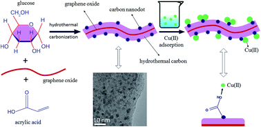
Environmental pollution associated with heavy metal ions has always been a serious environmental problem. To date, carbonaceous adsorbents are still the most promising candidates for the decontamination of these ions, but are limited by either cost or adsorption capacity. Here, carbonaceous nanocomposites containing numerous surface acidic groups were easily synthesized via one-pot hydrothermal carbonization of glucose in the presence of acrylic acid and a small amount of graphene oxide, and characterized in detail. Batch adsorption of Cu(II) on the carbonaceous nanocomposites from aqueous solution showed that these materials exhibited an excellent adsorption affinity for Cu(II) and the maximum adsorption capacity was as high as 146.1 mg g−1 at pH 5 and T = 298 K, which was much higher than any previous reports. The effect of the degree of functionality on adsorption behaviors, as well as the effects of pH, ionic strength, complex anions, temperature, and the presence of natural organic compounds (humic acid and fulvic acid) and organic pollutants (ionic liquid), were studied systematically to understand the adsorption mechanism. In addition, X-ray photoelectron spectroscopy was further used to confirm that surface complexation reactions played an important role in the adsorption process. This work would provide cheap nanocomposites which could be candidate materials for efficient decontamination of copper ions.


Introduction

Heavy metal ion pollution in industrial wastewater has attracted global attention due to its adverse effects on the environment and human health.1,2 Copper, an abundant and naturally occurring element present in municipal wastewaters,3 is one of the most widespread and common heavy metal contaminants in the environment.4 Many methods have been investigated to remove Cu(II) from wastewaters such as chemical precipitation, adsorption, ion exchange, membrane separation, reverse osmosis, electrolysis froth flotation and solvent extraction.5,6 Among these methods, adsorption has increasingly received more attention as it is simple and cost effective. Up to now, various adsorbents have been used to remove Cu(II), such as soil, clay mineral7 and resins.3,8,9 However, all things considered, these materials suffer from either high cost or low adsorption capacity, which limit their value for further study.

Carbonaceous materials are promising adsorbents as they are facile and environmentally friendly.10 But standard methods for the manufacture of activated carbons usually require very harsh conditions and have several drawbacks.11 What’s worse, the adsorption capacity of activated carbon needs to be further enhanced.12 Graphene oxide13 and carbon nanotubes14 have shown high adsorption capacities due to their large specific surface areas and numerous surface functional groups,15–17 while their application still remains at the theory stage.18

Here, a new type of carbonaceous nanocomposite was synthesized easily via a one-step aqueous hydrothermal carbonization route using cheap glucose in the presence of acrylic acid and a small amount of graphene oxide. The weight ratio of glucose to graphene oxide was fixed at 24 to lower the cost and acquire a large specific surface area.19 And acrylic acid added to the reaction could offer abundant functional groups and adsorption sites. The prepared materials, containing rich acidic groups, were not only inexpensive and easy to obtain, but also exhibited an excellent adsorption affinity and high maximum adsorption capacity for Cu(II). In order to assess the adsorption capacity of the as-prepared materials and understand the adsorption mechanism, batch experiments, including the effect of pH, ionic strength, complex anions and temperature, were performed and combined with X-ray photoelectron spectroscopy.20 Considering the presence of many natural organic compounds in the real environment, both humic acid and fulvic acid were evaluated.21 In addition, the presence of 1-butyl-3-methylimidazolium chloride ([Bmim][Cl]) was also studied, as it is the most used water soluble ionic liquid22 and also a potential contaminant.23

Experimental

Material preparation

Firstly, graphene oxide was prepared by the improved Hummers method.24,25 To obtain materials with a high degree of functionality, an acrylic acid monomer was added to the reaction mixture of glucose (3.6 g) and graphene oxide (0.15 g) at different amounts: 0, 3.6, 7.2 and 10.8 mL (mole ratios of acrylic acid to glucose were 0, 2.5, 5 and 7.5). Then deionized water was added to reach a total volume of 70 mL. And the final mixture was sealed into the Teflon inlet in an autoclave, and was hydrothermally treated at 198 °C for 16 h. Then, the materials were washed several times with deionized water and vacuum-dried overnight at 50 °C. The final products obtained were ultrasonically dispersed in deionized water as a stock suspension (1 mg mL−1).

Characterization

The prepared materials were collected and characterized by element content analysis (EC), scanning electron microscopy (SEM), transmission electron microscopy (TEM), X-ray photoelectron spectroscopy (XPS), Raman spectroscopy, Fourier transform infrared (FTIR) spectroscopy, powder X-ray diffraction (PXRD) and potentiometric titration. EC was performed with a Vario EL (ELEMENTAR). SEM was performed using a MIRA 3 XMU (Tecan). TEM was performed using a Tecnai-G2-F30 Field Emission Transmission Electron Microscope (FEI Corporation). Raman spectra from 500 to 4000 cm−1 were collected on an inVia-Reflex Raman scope using a 632.8 nm He–Ne laser (Renishaw). FTIR spectra were recorded from 400 to 4000 cm−1 on a NEXUS 6705-DX 170SX spectrometer (Nicolet Instrument Corporation). XPS spectra were obtained using an ESCALAB 250Xi (Thermo Fisher Scientific). The PXRD patterns were scanned from 5° to 60° on an X’Pert PRO X-ray diffractometer (Panalytical). The potentiometric acid–base titrations were conducted under argon using a DL50 Automatic Titrator (Mettler Toledo) in NaCl as a background electrolyte.

Adsorption experiments

Batch adsorption experiments were carried out at a 200 rpm equivalent shaking rate in 10 mL polyethylene test tubes for 24 h. The stock suspension of adsorbents and the stock solutions of Cu(II) were added in polyethylene test tubes to achieve the desired concentrations of the different components (solid-to-liquid ratio was fixed at 0.2 g L−1). In most experiments, the solution pH was adjusted to 2–10 as measured with a pH-meter (pHS-3C, Shanghai). For cases where the pH was adjusted, a 0.1 mol L−1 NaOH or HCl solution was used to change the initial pH value. A 5 mol L−1 NaCl (NaNO3 or NaClO4) background electrolyte solution was used to adjust ionic strength. After each adsorption equilibrium, the mixtures were centrifuged and the concentration of Cu(II) in the supernatant was determined using an Atomic Absorption Spectrometer (AAS, Perkin-Elmer). The adsorption amount was calculated according to the difference between the Cu(II) concentrations before and after adsorption. Adsorption isotherms were obtained by analysing different concentrations of Cu(II) at fixed temperature (298 K) and pH (5.0). At each condition, adsorption experiments were performed in triplicate and then the average was calculated.

Adsorption kinetics fitting model

The pseudo-second-order model26,27 general form was:
image file: c5ra16451f-t1.tif
where t (h) is the contact time, Qt (mg g−1) is the adsorption amount at time t, Qe (mol g−1) is the adsorption amount at the equilibrium time, and k (g mg−1 h−1)is the pseudo-second-order rate constant.

Adsorption isotherm fitting model

The General Langmuir Freundlich model28,29 was employed to fit the adsorption data. Its general form was:
image file: c5ra16451f-t2.tif
where Qe (mg g−1) is the amount of Cu(II) adsorbed on the adsorbent; Qm (mg g−1) is the maximum amount of Cu(II) adsorbed per unit mass of the adsorbent; Ce (mg L−1) is the adsorbate concentration at equilibrium; k (L mg−1) is the adsorption affinity coefficient; and α is an indicator of isotherm nonlinearity related to the heterogeneity of sorption sites.

Results and discussion

Characterization results

It has been reported that three-dimensional graphene-based porous materials were synthesized at the bulk scale via hydrothermal carbonization of glucose and graphene oxide.19 The reported materials showed large specific surface areas, but lacked surface functional groups for adsorption of metal ions. So we added acrylic acid as a comonomer to the reaction mixture of glucose and graphene oxide to provide more surface functional groups. For instance, GA0, GA1, GA2 and GA3 respectively represent 0, 3.6, 7.2 and 10.8 mL of acrylic acid used in the reaction. Fig. 1 shows the morphological images of the materials. Compared to the smooth surface of GA0 (Fig. 1a), large amounts of carbon nanodots (∼2.8 nm, ESI Fig. S1) were found to be deposited on the surfaces of GA1, GA2 and GA3 (Fig. 1b–d). And the SEM images (Fig. 1e and f) of the materials confirm that the surface of the materials became rough after adding acrylic acid. The morphological characterization results proved that adding acrylic acid successfully enhanced the surface functional groups.
image file: c5ra16451f-f1.tif
Fig. 1 TEM images of GA0 (a), GA1 (b), GA2 (c) and GA3 (d); SEM images of GA0 (e) and GA1 (f).

The element content analysis (Table 1) revealed that the oxygen content increased with increasing acrylic acid volume in the reaction. Compared with that of the precursors, an oxygen content of 38.6% seemed to be very high for the final product. The FTIR spectra of the products were recorded (Fig. 2a), and the following functional groups were identified in all samples: O–H stretching vibrations (3431 cm−1), C[double bond, length as m-dash]O stretching vibrations (1705 cm−1), C[double bond, length as m-dash]C from sp2 carbon (1627 cm−1), C–O vibrations (1406 cm−1), and O–H bending vibrations (1203 cm−1).25 XPS (Fig. S4) and Raman spectroscopy (Fig. 2b) were used to further analyze the carbon hybridization and oxygen-containing functional group state. The characteristic peaks of the D-band (1360 cm−1) and G-band (1601 cm−1)24 were clear in the Raman spectra. And the results that the ID/IG of GA0, GA1, GA2 and GA3 gradually reduced indicated that the sp2 carbon content increased with increasing acrylic acid volume in the reaction, because the carbon atoms in the acrylic acid molecule demonstrated sp2 hybridization. The powder X-ray diffraction (Fig. 2c) confirmed the broad (002) peak of carbon.

Table 1 Selected properties of the materials
Material Element content ID/IG O1s/C1s Hs (mmol g−1)
C (%) O (%) H (%) O/C
GA0 68.15 27.67 4.18 0.41 1.49 0.31 1.48
GA1 63.48 32.34 4.18 0.51 0.56 0.54 5.37
GA2 59.77 35.19 5.03 0.59 0.20 0.74 6.54
GA3 55.97 38.62 5.41 0.69 0.17 0.76 7.57


image file: c5ra16451f-f2.tif
Fig. 2 FTIR (a), Raman spectra (b), XRD (c) and surface charge densities (ΔQH) (d) of GA0, GA1, GA2 and GA3.

Potentiometric titrations were performed under argon atmosphere to characterize the surface charge densities (ΔQH, mol g−1) and the total concentration of surface acidic groups per solid weight (Hs, mmol g−1) of material.30 ΔQH was determined by subtracting the titration curve of the background electrolyte solution (blank) from that of the material suspension. In general, a suspension of material in 50 mL 0.1 mol L−1 NaCl solution was titrated with a standard NaOH solution (0.05 mol L−1) up to pH ∼ 10 at room temperature. Fig. 2d shows that the pHPZC (pH of point of zero charge) depended strongly on oxygen content.

(CaCb)susp = [H+] − [OH] + ΔQsolid + ΔQblank

(CaCb)blank = [H+] − [OH] + ΔQblank

ΔQH = V/m[(CaCb)susp − (CaCb)blank]
where Ca and Cb (mol L−1) are the concentrations of acid and base added, respectively; ΔQblank (mol L−1) represents consumption or release of H+ by side reactions; ΔQsolid (mol L−1) and ΔQH (mol g−1) represents the proton excess of the material in different units, respectively; V (L) is the volume of aqueous solution and m (g) is the mass of material.

Hs calculated from the two equivalence points on the Gran plot (Veb1 and Veb2, ESI S2 and Table S1) is defined by the following equation:

Hs = (Veb2Veb1)Cb/ms

The Hs values calculated from the titration curves are listed in Table 1. It was clear that the higher the oxygen content, the higher the Hs value, while the relationship was non-linear. These characterization results indicated that carbonaceous nanocomposites with different degrees of functionality were successfully synthesized (detailed data are summarized in Table 1). Then GA1 was chosen to systematically study the adsorption mechanism.

Kinetics

Fig. 3a shows the adsorption kinetics of Cu(II) on GA1. The adsorption amount increased quickly in the first 1 h, and then slowed down until the adsorption process reached equilibrium, which indicated that the adsorption mechanism was mainly chemical adsorption or surface complexation rather than physical sorption.3 After simulating several adsorption kinetics models, the pseudo-second-order model was found to be the most suitable (fitting parameter in Table S2) with the correlation coefficient (R2) of 0.998. Although the adsorption process was fast, the shaking time in the following batch experiments was selected as 24 h to achieve complete equilibrium.
image file: c5ra16451f-f3.tif
Fig. 3 Adsorption of Cu(II) on GA1 as a function of contact time. (a) Plot of Qt vs. t for Cu(II) adsorption on GA1. (b) Plot of t/Qt vs. t for the pseudo-second-order model. T = 298 K, pH = 5, initial CCu(II) = 0.2 mmol L−1 and INaCl = 0.1 mol L−1.

Effect of pH

The pH of a solution influences the surface chemical properties of a suspended material and the species of metal ions.31 So it is important to study the effect of pH on adsorption behaviors. Fig. 4a shows the adsorption of Cu(II) on GA1 as a function of pH. The sharp increase of the adsorption percentage over pH 4 was attributed to the deprotonation of the surface acidic groups which had a strong affinity for the positively charged Cu(II) species. According to the Cu(II) speciation distribution as a function of pH value (Fig. 4b), Cu(II) began to form a precipitate at pH 6 if no Cu(II) was adsorbed, while most Cu(II) was adsorbed by GA1 before pH 6, which proved that the adsorption of Cu(II) was not due to the surface precipitation of Cu(OH)2(s).21 In order to completely eliminate the influence of Cu(II) precipitation, the adsorption isotherms of Cu(II) on the materials were obtained at pH 5.0.
image file: c5ra16451f-f4.tif
Fig. 4 Adsorption of Cu(II) on GA1 as a function of pH (a), T = 298 K, CCu(II) initial = 0.2 mmol L−1 and INaCl = 0.1 mol L−1. Concentration of Cu(II) species as a function of pH value (b), T = 298 K CCu(II) = 0.2 mmol L−1 and INaCl = 0.1 mol L−1.

Effect of ionic strength and anion

The ionic strength and anion are important factors to study the adsorption mechanism, so the effect of NaCl, NaNO3 and NaClO4 concentration on the adsorption percentage of Cu(II) on GA1 is shown in Fig. 5a. The adsorption was inhibited gradually by NaCl due to strong coordination of Cl reducing the actual concentration of Cu(II); while the adsorption was promoted by NaNO3 and NaClO4, suggesting that the oxideacid anion took part in surface complexation. Comparing the adsorption isotherms of Cu(II) on GA1 in the presence of 0.1 mol L−1 NaCl, NaNO3 or NaClO4 (Fig. 5b, fitting parameters in Table S3), similar results were observed. The results suggested that the materials were favorable for use with wastewater containing the oxideacid anion.
image file: c5ra16451f-f5.tif
Fig. 5 The effect of NaCl, NaNO3 and NaClO4 concentration on the adsorption percentage of Cu(II) on GA1 at pH = 5 and T = 298 K (a). Adsorption isotherms of Cu(II) on GA1 in 0.1 mol L−1 NaCl, NaNO3 and NaClO4, pH = 5 and T = 298 K (b).

Effect of temperature

Temperature is also a key factor that affects ion adsorption. Fig. 6 shows the adsorption isotherms of Cu(II) on GA1 at 298, 313 and 328 K (fitting parameters in Table S4). Temperature showed a slightly positive influence on the adsorption behaviors, which indicated that the adsorption of Cu(II) on GA1 is an endothermic process. Some researchers reported that Cu(II) ions had to be denuded of their hydration sheaths to some extent, and this dehydration process needs energy.3,32 The materials showed stable adsorption behaviors against temperature.
image file: c5ra16451f-f6.tif
Fig. 6 Adsorption isotherms of Cu(II) on GA1 at pH = 5, INaCl = 0.1 mol L−1, T = 298, 313 and 328 K.

Effect of the presence of organic compounds

In the natural environment, there are many coexistent organic compounds. So the effect of the presence of humic acid, fulvic acid and [Bmim][Cl] were studied, and the results are shown in Fig. 7 (fitting parameters in Table S5). The results showed that the presence of humic acid and fulvic acid greatly promoted the adsorption capacity, while, on the contrary, the presence of [Bmim][Cl] slightly inhibited the adsorption behaviors. Those organic compounds were easily adsorbed by the materials through π–π interaction. And the negatively charged humic acid and fulvic acid21 were coordinated with the Cu(II), forming a Cu–O bond, and also further negatively charged the materials, which significantly improved the adsorption capacity of the positively charged Cu(II). However, although the adsorbed [Bmim]+ could form a strong Cu–N bond which promoted the adsorption behaviors, the positively charged [Bmim]+ changed the surface charge properties of the materials and inhibited further adsorption of positively charged Cu(II). The results clearly indicated that numerous natural organic compounds would promote the decontamination process.
image file: c5ra16451f-f7.tif
Fig. 7 Adsorption isotherms of Cu(II) on GA1 in the presence of 20 mg L−1 humic acid, fulvic acid and [Bmim][Cl] at pH = 5, T = 298 K and INaCl = 0.1 mol L−1.

Effect of degree of functionality

Four kinds of materials with different degrees of functionality were prepared. The adsorption isotherms of Cu(II) on GA0, GA1, GA2 and GA3 at pH 5, 0.1 mol L−1 NaCl and T = 298 K are shown in Fig. 8. The maximum adsorption amounts (Qm) of GA0, GA1, GA2 and GA3 were 10.2, 97.2, 122.6 and 146.1 mg g−1, respectively (fitting parameters in Table 2). And it seemed to be clear that the higher the degree of functionality, the higher the adsorption capacity. Modeling the relationship between Qm and oxygen content or Hs (Fig. 9 and S5) showed that the Qm was linearly dependent on Hs (R2 = 0.99995). The results indicated that the adsorption behavior was related to the surface acidic groups, not the oxygen content, because some inner oxygen atoms at the hydrothermal carbon layer couldn’t adsorb Cu(II). And previous reports also claimed that the adsorption of metal ions on a series of carbons was found to be related to the type and concentration of the surface functional groups.33
image file: c5ra16451f-f8.tif
Fig. 8 Adsorption isotherms of Cu(II) on GA0, GA1, GA2 and GA3 at pH = 5, T = 298 K and INaCl = 0.1 mol L−1.
Table 2 The fitting parameters for the general Langmuir Freundlich model of Cu(II) adsorption isotherms
Material Qm (mg g−1) k α R2
GA0 10.2 0.096 1.85 0.73
GA1 97.2 0.332 2.47 0.98
GA2 122.6 0.358 2.24 0.97
GA3 146.1 0.314 2.46 0.96


image file: c5ra16451f-f9.tif
Fig. 9 Relationship between Qm and Hs.

To further evaluate our result, XPS studies of GA1 before and after Cu(II) adsorption were conducted. The XPS results (Fig. 10) show that the relative content of C–O–C/C–OH was reduced, that of C[double bond, length as m-dash]O/COO increased, and the binding energy (B.E.) shifted after adsorption which suggested that oxygen atoms were the main adsorption sites and Cu2+–O coordinate bonds were formed in the adsorption process.5 And the Cu2p XPS also clearly manifested that Cu(II) was adsorbed on the surface of the material.


image file: c5ra16451f-f10.tif
Fig. 10 XPS of GA1 before adsorption (GA1) and after adsorption (GA1-Cu).

Compared to the adsorption capacities of other adsorbents (Table 3), such as modified activated carbon,12 graphene oxide15 and pecan nutshell,34 the adsorption capacity of the carbonaceous nanocomposites in this work was as high as 146.1 mg g−1, which was the highest.

Table 3 Comparison of the Cu(II) adsorption capacities of different adsorbents
Adsorbents Experimental conditions Qm (mg g−1) Reference
Graphene oxide aerogel pH = 6.3, T = 298 K 19.7 16
Oxidized multi-walled CNTs pH = 4, T = 298 K 29.7 14
Modified activated carbon pH = 5, T = 298 K 38 11
H2SO4/KMnO4-modified CNTs pH = 6, T = 300 K 38.6 8
CMCD-MNPs pH = 6, T = 298 K 47.2 5
Graphene oxide pH = 5, T = 308 K 74.9 15
Pecan nutshell pH = 5.5, T = 298 K 91.2 34
Carbonaceous nanocomposites pH = 5, T = 298 K 146.1 This work


Competitive adsorption

The competitive adsorption of multi-metal ions on the materials was studied to explore their potential for practical applications. The initial solution was prepared with 0.8 mmol L−1 of each of Cu(II), Sr(II), Zn(II), Mg(II), Ca(II), Cd(II), K(I) and Na(I). After adsorption to the carbonaceous nanocomposites, the concentrations in the supernatant were determined using ICP-AES, and the adsorption percentages are shown in Fig. 11. The adsorptions of Cu(II), Sr(II), and Zn(II) on the materials were higher than those of the other five ions, and the adsorption affinity (∼48%) of the materials for Cu(II) was the strongest when compared with Sr(II) (∼26%) and Zn(II) (∼14%), which meant that the carbonaceous nanocomposites had a high potentiality for selectively removing Cu(II).
image file: c5ra16451f-f11.tif
Fig. 11 The competitive adsorption of multi-metal ions, C[metal ions]initial = 0.8 mmol L−1, pH = 5, T = 298 K.

Conclusion

Carbonaceous nanocomposites with a high degree of functionality were synthesized via one-pot hydrothermal carbonization of cheap glucose in the presence of acrylic acid and a small amount of graphene oxide. The materials exhibited an excellent adsorption affinity for Cu(II) and the maximum adsorption capacity was as high as 146.1 mg g−1, which was much higher than any previous reports and was linearly dependent on surface acidic groups. The adsorption kinetics showed that the adsorption behavior was fast and well fitted with the pseudo-second-order kinetic model. And, more remarkably, the presence of the oxideacid anion and natural organic compounds could promote the adsorption behaviors. A schematic diagram elucidating the whole process is illustrated in Fig. 12. Overall, the cheap carbonaceous nanocomposites in this work exhibited a high potential for efficient decontamination of copper ions.
image file: c5ra16451f-f12.tif
Fig. 12 A schematic diagram elucidating the whole process. The materials were synthesized via one-pot hydrothermal carbonization. Then the materials were used to efficiently remove Cu(II) from aqueous solution.

Acknowledgements

This project is supported by National Science Foundation of China J1210001, 21327801 and 21405165. The authors thank Professor AFM Rahman and Doctor Zhang Rui for modification of the manuscript.

References

  1. J. Duruibe, M. Ogwuegbu and J. Egwurugwu, Int. J. Phys. Sci., 2007, 2, 112–118 Search PubMed.
  2. L. Järup, Br. Med. Bull., 2003, 68, 167–182 CrossRef.
  3. X. Wu, D. Zhao and S. Yang, Desalination, 2011, 269, 84–91 CrossRef CAS.
  4. C. Yuwei and W. Jianlong, Chem. Eng. J., 2011, 168, 286–292 CrossRef.
  5. A. Z. M. Badruddoza, A. S. H. Tay, P. Y. Tan, K. Hidajat and M. S. Uddin, J. Hazard. Mater., 2011, 185, 1177–1186 CrossRef CAS PubMed.
  6. F. Fu and Q. Wang, J. Environ. Manage., 2011, 92, 407–418 CrossRef CAS PubMed.
  7. R. Zhang, C. Chen, J. Li and X. Wang, Appl. Surf. Sci., 2015, 349, 129–137 CrossRef CAS.
  8. C.-Y. Kuo, Desalination, 2009, 249, 781–785 CrossRef CAS.
  9. J. He, Y. Lu and G. Luo, Chem. Eng. J., 2014, 244, 202–208 CrossRef CAS.
  10. X. Guo, C. Wang, M. Chen, J. Wang and J. Zheng, J. Power Sources, 2012, 214, 107–112 CrossRef CAS.
  11. R. Demir-Cakan, N. Baccile, M. Antonietti and M.-M. Titirici, Chem. Mater., 2009, 21, 484–490 CrossRef CAS.
  12. L. Monser and N. Adhoum, Sep. Purif. Technol., 2002, 26, 137–146 CrossRef CAS.
  13. R. K. Upadhyay, N. Soin and S. S. Roy, RSC Adv., 2014, 4, 3823–3851 RSC.
  14. J. Wang, Z. Li, S. Li, W. Qi, P. Liu, F. Liu, Y. Ye, L. Wu, L. Wang and W. Wu, PLoS One, 2013, 8, 1016–1020 Search PubMed.
  15. X. Ren, J. Li, X. Tan and X. Wang, Dalton Trans., 2013, 42, 5266–5274 RSC.
  16. G. Huang, W. Wang, X. Mi, W. Xie, Y. Liu and J. Gao, Carbon, 2012, 50, 4856–4864 CrossRef.
  17. M. Yusuf, F. M. Elfghi, S. A. Zaidi, E. C. Abdullah and M. A. Khan, RSC Adv., 2015, 5, 50392–50420 RSC.
  18. G. Zhao, T. Wen, X. Yang, S. Yang, J. Liao, J. Hu, D. Shao and X. Wang, Dalton Trans., 2012, 41, 6182–6188 RSC.
  19. L. Zhang, F. Zhang, X. Yang, G. Long, Y. Wu, T. Zhang, K. Leng, Y. Huang, Y. Ma and A. Yu, Sci. Rep., 2013, 3 Search PubMed.
  20. G. Sheng, Y. Li, X. Yang, X. Ren, S. Yang, J. Hu and X. Wang, RSC Adv., 2012, 2, 12400–12407 RSC.
  21. X. Tan, M. Fang, J. Li, Y. Lu and X. Wang, J. Hazard. Mater., 2009, 168, 458–465 CrossRef CAS PubMed.
  22. D. J. Gorman-Lewis and J. B. Fein, Environ. Sci. Technol., 2004, 38, 2491–2495 CrossRef CAS PubMed.
  23. B. Jastorff, K. Mölter, P. Behrend, U. Bottin-Weber, J. Filser, A. Heimers, B. Ondruschka, J. Ranke, M. Schaefer and H. Schröder, Green Chem., 2005, 7, 362–372 RSC.
  24. D. C. Marcano, D. V. Kosynkin, J. M. Berlin, A. Sinitskii, Z. Sun, A. Slesarev, L. B. Alemany, W. Lu and J. M. Tour, ACS Nano, 2010, 4, 4806–4814 CrossRef CAS PubMed.
  25. T. Longlong, Z. Xin, Q. Wei, L. Dan, J. Qiang, L. Jin, Y. Yuanlv, L. Zhan and W. Wangsuo, RSC Adv., 2014, 4, 58536–58545 RSC.
  26. L. Wang, C. Cheng, S. Tapas, J. Lei, M. Matsuoka, J. Zhang and F. Zhang, J. Mater. Chem. A, 2015, 3, 13357–13364 CAS.
  27. S. W. Zhang, J. X. Li, T. Wen, J. Z. Xu and X. K. Wang, RSC Adv., 2013, 3, 2754–2764 RSC.
  28. M. N. Carvalho, C. A. M. de Abreu, M. Benachour, D. C. S. Sales, O. S. Baraúna and M. A. D. Motta Sobrinho, Adsorpt. Sci. Technol., 2012, 30, 691–699 CrossRef CAS.
  29. G. P. Jeppu and T. P. Clement, J. Contam. Hydrol., 2012, 129–130, 46–53 CrossRef CAS PubMed.
  30. Q. H. Fan, P. Li, W. S. Wu, D. Q. Pan, K. F. Ding, Y. Lu, T. Yu and J. Xu, Sci. China: Chem., 2011, 54, 1138–1147 CrossRef.
  31. A. Schierz and H. Zänker, Environ. Pollut., 2009, 157, 1088–1094 CrossRef CAS PubMed.
  32. M. Wang, H. Jiang and Z. C. Wang, J. Therm. Anal. Calorim., 2006, 85, 751–754 CrossRef CAS.
  33. B. Xiao and K. M. Thomas, Langmuir, 2005, 21, 3892–3902 CrossRef CAS PubMed.
  34. J. C. P. Vaghetti, E. C. Lima, B. Royer, B. M. da Cunha, N. F. Cardoso, J. L. Brasil and S. L. P. Dias, J. Hazard. Mater., 2009, 162, 270–280 CrossRef CAS PubMed.

Footnotes

Electronic supplementary information (ESI) available. See DOI: 10.1039/c5ra16451f
These authors contributed equally to this work.

This journal is © The Royal Society of Chemistry 2015