Solute-induced perturbation of methanol–water association

B. Hemmateenejad*a, Z. Shojaeifarda, M. Shamsipurb, K. Neymeyrcd, M. Sawallcd and A. Mohajeria
aDepartment of Chemistry, Shiraz University, Shiraz, Iran. E-mail: hemmatb@shirazu.ac.ir
bDepartment of Chemistry, Razi University, Kermanshah, Iran
cInstitute of Mathematics, Rostock University, Rostock, Germany
dLeibniz Institute for Catalysis, Rostock, Germany

Received 10th July 2015 , Accepted 7th August 2015

First published on 7th August 2015


Abstract

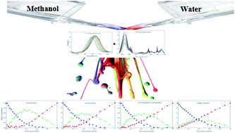
This article addresses experimental evidence of changes in the equilibrium constant of methanol–water association induced by solutes. The behavior of four solutes (solvatochromic dyes) in methanol–water binary mixtures is probed using augmentation of infrared and UV-Vis spectroscopy methods followed by multivariate analysis. In order to get more reliable concentration and spectral profiles, with fewer ambiguities, complementarity and coupling theorems have been utilized. A ternary system was resolved using multivariate analysis on the noted augmented data for each chemical probe. Resultant concentration profiles showed the unique behavior of each solute in the binary-solvent system. Therefore, an individual association constant for the formation of the methanol–water association was achieved for each solvatochromic probe. Indeed, different solutes can change the solvent–solvent interaction based on their hydrogen binding affinity. Diverse hydrogen binding affinities for different solutes were investigated using charge distribution analysis via molecular electrostatic potential (MEP) calculations as well as the atomic charge on the oxygen atoms. In addition, obtained spectral profiles of standard solvatochromic probes were used to calculate solvatochromic parameters of dipolarity/polarizability and hydrogen-bonding basicity of three resolved components (i.e. methanol, water and methanol–water complex surrounded probes).


Introduction

Solvent–solute and solvent–solvent interactions are of vital importance for studying chemical, environmental, industrial, and biological processes in the liquid phase. Solvent-induced changes of the dynamics and equilibria of such processes (known as preferential solvation) are well understood.1–5

Methanol–water binary solvents, as the widely used solvents in liquid chromatography, have been studied in the past using both theoretical and experimental methods. In this regard, multivariate curve resolution (MCR) as a powerful chemometric technique has been used to analyze the spectroscopic and physicochemical data of solvents alone or in the presence of solutes.6–9 It has now been accepted that, in the binary mixtures of methanol and water, a methanol–water association (or cluster) is formed.6–12 However, all of the previous studies ignored the solute effects on the structure of this association. This led to inconsistencies between the obtained results by different authors. There is no agreement between the stoichiometric ratio(s) of the associate(s)6–9,11–13 and also the reported equilibrium constants for even the same association model, e.g. a 1[thin space (1/6-em)]:[thin space (1/6-em)]1 molar ratio association, are largely different.9,12,13

Besides the pronounced effect of solvents on the structure and function of solutes, recently Ben-Amotz and coworkers discovered the effect of solutes on the structure of solvents.14–21 Using Raman spectroscopy and multivariate curve resolution, they could identify the changes in the structure and hydrogen-bonding network of water in the presence of different solutes including ions17,19 as well as polar and nonpolar molecules.15 However, nothing has been reported regarding solute effects on the structure of mixed solvents.

Here, we demonstrate the effect of solute on the methanol–water association in their binary mixtures. In the past, both native FTIR spectra of pure solvents as well as mixed solvents10–12 and the spectra of solvatochromic dyes in pure and mixed solvents2,6,22–24 have been used to monitor the methanol–water association. However, the changes in the absorbance spectra of the solvatochromic dyes can also provide some indirect measurement of the solvent–solvent association. Thus, in this work, we used both techniques simultaneously for following the solute effects on the dynamics parameters of binary solvents.

In this regard, the ATR-FTIR and visible electronic absorbance spectra of methanol–water mixtures in the presence of different solvatochromic dyes as probes have been analyzed using chemometrics methods.9 The results revealed changes in the equilibrium constant of the methanol–water association through perturbation of the hydrogen bonding interactions between the solvents.

Although the MCR method has been used in many methanol–water mixture studies,6–9,25 because of the existence of a range of feasible solutions (so-called rotational ambiguity) in this system, a unique and appropriate analysis could not be achieved.26

Here, we aim to use a more reliable method with fewer ambiguities to investigate solute effects. In this regard, complementarity and coupling theorems have been used as a soft model. In this method, any additional information on the system could be utilized to reduce ambiguity. Complementarity and coupling theory have a solid mathematical foundation and can be formulated in terms of linear and affine linear subspaces to which certain concentration profiles and spectra are restricted. This theory allows the formulation of restrictions on the feasible concentration profiles if information on the spectra is available and vice versa.27,28

Additionally, it has been shown that the dipolarity/polarizability and hydrogen acidity/basicity of solutes depend strongly on the composition and properties of the solvent mixture and mobile phases.6,8 Therefore, as well as the dynamics parameters, the resolved spectra are used to estimate the dipolarity/polarizability parameter (π*)29 and the hydrogen-bonding basicity (β)30 parameter for the pure solvent species and also for the solvent–water complexes.

Experimental

Chemicals and materials

N,N-Dimethyl-4-nitroaniline (DMA), 4-nitroanisol (NAS), and 4-nitroaniline (NAL) were purchased from Sigma-Aldrich. Merck supplied indigo carmine (ICM). Water (chromatography grade) and methanol (spectroscopy grade) were from Sigma Chemical Co. and were used directly without any further purification.

UV-Vis absorptions were studied using a Shimadzu 1601 UV-Vis spectrometer, using quartz analytical cells of 1 cm path length.

IR spectra were recorded on a FT-IR Perkin Elmer FTIR spectrometer-spectrum RX1 equipped with a Perkin Elmer ZnSe ATR sampler accessory.

Binary solvent mixtures of methanol and water of variable mole fractions (21 binary solvents) were prepared by mixing of the desired weights of the pure solvents. Also, the mole fraction compositions for IR spectroscopy were the same as for absorption spectroscopy.

The concentrations of all of the dyes in the solvent mixtures were kept constant at 2 × 10⁻⁵ M. For each solute, the IR and UV-Vis spectra of the resulting methanol–water binary solvent solutions of various compositions were recorded. The temperature was fixed at 25 °C. The collected spectra were converted to ASCII format and transferred to MATLAB (Mathwork, Inc.) environment. The data were analyzed with programs written in the MATLAB programming environment.

Data analysis method

Data matrices were constructed for each dye solvent mixture by collecting the digitized spectra of the dye in solvent mixtures at compositions ranging from 100% to 0% organic phase. For each solute and each spectroscopic method, an absorbance data matrix D of size (m × n) was obtained, where m is the number of different solvent compositions at which spectra are measured and n is the number of absorbance readings per spectrum.

According to the bilinear Lambert–Beer law, the absorbance data matrix D equals the product CA with C being the matrix of concentration profiles and A being the matrix of pure component spectra. Usually the concentration profiles are taken along the time axis, but here these values correspond to the different bulk solvent compositions.

The starting point is a singular value decomposition (SVD) of D with the form D = UΣVT. The number of significant singular values indicates the number of independent components. Then the first s columns of U (left singular vectors) and the first s columns of V (right singular vectors) are used for the expansion of an approximate factorization with C = UΣT−1 and A = TVT. Therein T is a regular s × s matrix. In general the matrix T is not unique, but many nonnegative factorizations exist. The term rotational ambiguity paraphrases the non-uniqueness of T. In order to extract the chemically most meaningful and hopefully correct solution, hard and soft models can be used as constraints on the factors C and A.31–33

Whenever additional information on the pure component factors is available, it should be used in order to reduce the rotational ambiguity; see e.g. the complementarity and coupling theory.27,28

Results and discussion

Factor numbers

Four standard solvatochromic indicators34 including N,N-dimethyl-4-nitroaniline (DMA), 4-nitroanisol (NAS), 4-nitroaniline (NAL) and indigo carmine (ICM) were used to probe methanol–water association. A fixed amount of each dye was dissolved in the methanol–water binary solvents and then the ATR-FTIR as well as the UV-Vis absorbance spectra of the resultant solutions were recorded. The resulting spectra are shown in the ESI (Fig. S1 and S2 (ESI)). To ignore the concentration effect, the same concentration of dye was used for all dyes. For each dye and for each spectroscopic method, an absorbance data matrix D, each row of which was the digitized absorbance spectrum at a specified methanol–water composition, was provided. Then, for each dye, the data matrices obtained from the two spectroscopic methods were augmented in a column-wise manner35 to monitor the manner of solutes in binary solvents using both IR and absorption spectroscopy techniques.

The first step in the analysis of methanol–water mixtures is the determination of the number of significant principal components in the mixture. Both singular value decomposition (SVD) and evolving factor analysis (EFA) were employed to determine the number of significant principal components, attributing to the number of solvated complexes in the systems. Eigen-value (EV) plots (obtained from SVD) and the results of EFA vs. number of principal components as a function of the eigenvalue numbers are shown in Fig. S3. In addition, we used reduced eigenvalue (REV), root mean square (RMS) and residual standard deviation (RSD) as mathematical criteria to determine the number of factors.36 The results of the SVD analysis are summarized in Table S1. In summary, principal component analysis (PCA)37 of the data matrices of all dyes revealed the presence of three significant chemical species (see the ESI, Fig. S3 and Table S1 for details), attributed to pure methanol, pure water and the methanol–water association.6–9 The same results were obtained when PCA was run on the extended data matrix obtained by stacking the data matrices of all dyes under each other (Table S1).

Pure contribution of species and stoichiometric analysis

The detection of three principal components using PCA suggested that when a dye is dissolved in a binary methanol–water solvent, it can be solvated by three solvent components and hence there are three different solvated states of each dye. According to the Beer–Lambert law of absorbance additives, the absorbance of each dye in the binary mixtures can be assumed as the summation of the absorbance of the dye in its three different states. So, the absorbance data matrix of each dye can be decomposed into the contribution of each solvated state of the dye:
 
D = Dm + Dw + Dmw = cmsm + cwsw + cmwsmw = [cmcwcmw] [sm; sw; smw] = CS (1)

The indices m, w and mw denote pure methanol, pure water and the methanol–water association, respectively. The column vectors cm, cw and cmw correspond to the concentration profile of each solvent species as a function of the bulk solvent composition. The row vectors sm, sw and smw denote the pure spectrum of the different states of the solvated dye. The same explanations apply for the FTIR spectra of the solvents. The recorded FTIR absorbance spectrum at a specified methanol–water mole fraction can be assumed as the summation of the absorbance of the three solvent species.

Generally multivariate curve resolution (MCR) as a soft-modeling method has been used to decompose the data matrix D into the pure contribution of each species (to obtain estimates of C and S as matrices of concentration profiles and pure spectra, respectively).38 Comparing the obtained concentration profiles of different solvent species in the presence of different solutes will reveal the effect of solutes on the methanol–water association. But, one major drawback inherent to all MCR methods is rotational ambiguity, which usually leads to obtaining a range of feasible solutions instead of a unique solution for C and S.26,39 Since the effect of solutes on solvent structure is very small, there might be an overlap between the ranges of solutions of different dyes and, hence, the comparison might not be meaningful. So, obtaining a unique solution is key in comparing the effects of solutes. The recent works conducted by Sawall and Neymeyr allow extraction of unique solutions for the given three component system.27,28 In this regard, the data analysis combines classical MCR techniques with the relatively new complementarity theory. The spectral data matrices for all our experiments indicate three-component systems. In all cases two of the three pure component spectra are available. By means of the complementarity theorem,27 this allows computation of the concentration profile of the third component. In order to supply the missing information for the remaining pure component spectra and concentration profiles, an additional soft constraint is used in the form of a mass balance for the three components. This constraint maximizes the smoothness of the concentration profiles of the two remaining components; mathematically the norm of the discretized second derivatives of these concentration profiles is minimized.27,28

The resultant concentration profiles for augmented matrices using complementarity and coupling theorems are given in Fig. 1. The starting point for these computations is the known pure component spectra of water and methanol. So the three component system has only two degrees of freedom, which is a result of the representation of the rotational ambiguity of the factorization for a three component system.32


image file: c5ra13514a-f1.tif
Fig. 1 Results of complementarity and coupling theorems: concentration profiles of the augmented spectra of UV-Vis and IR for dyes as solvatochromic probes. Dyes involved: 4-nitroanisol (A), 4-nitroaniline (B), N,N-dimethyl-4-nitroaniline (C) and indigo carmine (D) in a methanol–water mixture. For all experiments, water solvated solutes (blue, ●), methanol solvated solutes (red, ■) and methanol–water solvated solutes (green, ▼) are shown in the graphs.

In order to reduce the remaining two-parameter ambiguity, two soft-constraint functions (namely, on the mass balance and on the smoothness of the concentration profiles) have been used.31

The concentration profiles are similar for all studied dyes. All show a single association model. The methanol–water associated solvent reaches its maximum concentration value at a bulk molar ratio of about 0.5. This suggests a 1[thin space (1/6-em)]:[thin space (1/6-em)]1 stoichiometric ratio of methanol–water in the associated solvent.

Determination of the complex association constant

As shown in Fig. 1, the four dyes have a similar ternary contribution in the methanol–water mixed solvents. However, the concentrations of the associated solvent change as the dyes vary. The observed differences can be attributed to the change in the equilibrium constant of the association model with the form:
 
M + WMW (2)

The association constant can be estimated by nonlinear fitting of the concentration profiles to the association model of eqn (2). As observed from Table 1, there is a large difference in the estimated association constants for the different solutes. The largest association constant (2.32 M−1) was obtained in the presence of NAS whereas the smallest value (0.10 M−1) was found in the presence of ICM. The major difference between these compounds is their polarity and their ability for hydrogen-bonding. Indigo carmine is a large polyanionic compound capable of forming hydrogen bonds with both methanol and water, whereas 4-nitroanisole as a small molecule with fewer donor functional groups has a lower ability for forming hydrogen bonds.

Table 1 Association constant of methanol–water complexes using various dyes
Dye Association constant ssq
4-Nitroanisol (NAS) 2.32 ± 0.0003 1.60
4-Nitroaniline (NAL) 0.64 ± 0.0007 0.35
N,N-Dimethyl-4-nitroaniline (DMA) 0.54 ± 0.0013 0.82
Indigo carmine (ICM) 0.10 ± 0.0043 0.34


So, the lower instability of the methanol–water association in the presence of ICM can be attributed to the perturbation of methanol–water interaction (mostly because of hydrogen bonding) by the polar groups of ICM. On the other hand, the estimated association constants in the presence of DMA and NAL were of an intermediate level with association constants of 0.54 M−1 and 0.64 M−1, respectively. These compounds could be considered to be lower polarity relative to NAS and higher polarity relative to ICM. It should be noted that there are two reports on estimating the association constant of MW in the absence of solute. Using the bulk properties of binary mixtures such as volume changes on mixing, refractive indices and densities, an association constant of 5.04 has been suggested by Katz et al.40 In addition, by chemometric analysis of various solvent properties, Hemmateenejad reported an association constant of 13.7.9 Interestingly, both values are larger than those found in the presence of solutes. This confirms the instability of the methanol–water association in the presence of solutes.

In order to confirm the solutes’ preferential hydrogen binding in the methanol–water mixture, charge distributions and molecular electrostatic potential analysis have been performed in the following section.

Meanwhile, to be sure about the 1[thin space (1/6-em)]:[thin space (1/6-em)]1 association model, other association models like 2[thin space (1/6-em)]:[thin space (1/6-em)]1, 1[thin space (1/6-em)]:[thin space (1/6-em)]2 or 3[thin space (1/6-em)]:[thin space (1/6-em)]2 were also examined and the resultant concentration profiles were fitted to these models. The results revealed that none of these models could be fitted as good as the 1[thin space (1/6-em)]:[thin space (1/6-em)]1 model, i.e., the standard errors of fitting for these models were much larger than that for the 1[thin space (1/6-em)]:[thin space (1/6-em)]1 association model.

Charge distribution analysis

In order to get a deeper insight into the role of structural properties of solutes affecting the stability of the methanol–water association, we performed the charge distribution analysis via molecular electrostatic potential (MEP) calculations.41 The electrostatic potential that the nuclei and electrons of a molecule create in the surrounding space is well established as a guide to the interpretation and prediction of molecular reactive behavior. This quantity is a useful tool in studying both electrophilic and nucleophilic processes, including hydrogen-bonding interactions and, in particular, is well suited for studies that involve the identification of key features necessary for the “recognition” of one molecule by another.42,43 In this context, the structures of all solutes were optimized in the framework of density functional theory using the B3LYP method. All calculations were performed with the 6-31G+(d) basis set by means of the Gaussian 09 suite of programs.44 Natural population analysis was also employed to describe the charge distribution in each solute.45 The resulting electrostatic potential maps of all considered solutes are plotted in Fig. 2. Clearly, a negative electrostatic potential (red color) is present at the O end of the NO2 group whereas a positive one (blue color) is observed around the hydrogen atoms.
image file: c5ra13514a-f2.tif
Fig. 2 Electrostatic potential isosurfaces (0.025 au) of 4-nitroanisol (A), 4-nitroaniline (B), N,N-dimethyl-4-nitroaniline (C) and indigo carmine (D). The red and blue colors represent the negative and positive molecular electrostatic potentials; respectively. Natural charges on the oxygen atoms are shown below the figures.

Inspection of the MEP plots in Fig. 2 and the computed atomic charge on the oxygen atoms in different solutes reveal that the polarizability of the oxygen atoms and their tendency to participate in hydrogen bonding interactions exhibit the following order: ICM ≫ NAL > DMA > NAS. This trend is consistent with the association constant of the methanol–water complex in the presence of the mentioned solutes (Table 1). The only exception is the relative position of NAL and DMA. The association constants in the presence of these compounds are very close and the differences in the values of the association constants are in the range of experimental error.

Pure spectra of the species

Besides concentration profiles, complementarity and coupling theorems give the pure spectra of the species contributing to the system under study. We have two individual sets of spectral data; the UV/Vis absorbance spectra of the solutes and the FTIR spectra of the solvents.

The resolved FTIR spectral data are attributed to the solvents. Comparing the pure spectra obtained in the presence of different solutes gives information about the solute-induced perturbation of the solvent spectra (or, in other words, the solvent structure). The solute-induced changes of the spectral profiles of pure solvents have already been studied, But the effect of solute on the spectral characteristics of the methanol–water association has not been investigated yet. Therefore, an interesting point is to compare the pure spectrum of each solvent species in the presence of different solutes. Comparison of the resolved IR spectra (over the OH stretching frequencies) of each solvent species in the presence of different solutes clearly shows the perturbation of solvent spectra by the solutes.

This effect is minute for water and methanol (Fig. 3A and C). However, significant solute-induced perturbation of the IR spectra is observed for the methanol–water association (Fig. 3B).


image file: c5ra13514a-f3.tif
Fig. 3 Infrared spectra obtained with complementarity and coupling theorems for four solvent solvated dyes (solutes). Water solvated solutes (A), methanol–water solvated solutes (B) and methanol solvated solutes (C).

Solvatochromic parameter

The extracted UV/Vis pure spectral profiles are related to the solutes solvated by different solvent species (i.e. pure methanol, pure water and the methanol–water cluster) (Fig. 4). One can use these spectral profiles to calculate the solvatochromic parameters of the solvents.
image file: c5ra13514a-f4.tif
Fig. 4 Estimated spectral profiles obtained using complementarity and coupling theorems of UV-Vis spectra for four probes, 4-nitroanisol (A), 4-nitroaniline (B), N,N-dimethyl-4-nitroaniline (C) and indigo carmine (D) in methanol–water mixtures. For all experiments, water solvated solutes (blue, [dash dash, graph caption]), methanol solvated solutes (red, –··) and methanol–water solvated solutes (green, [thick line, graph caption]) are shown in the graphs.

Kamlet–Taft solvatochromic parameters are the most comprehensive and commonly used quantitative measurement of solvent properties. These parameters are the hydrogen bond basicity (β), the hydrogen bond acidity (α) and the dipolarity–polarizability (π*). Important information about the polarity and hydrogen bonding ability of the formed methanol–water complexes, allows prediction of solvent properties as a function of composition. This information can be useful in the analysis of retention models in chromatography.

The solvatochromic properties of pure methanol and pure water can be simply measured by recording the absorbance spectrum of the solute in the pure solvents. However, the spectral data of solutes in the methanol–water association could not be determined experimentally. An interesting application of complementarity and coupling theorems analysis is estimating the spectral profiles of the solutes in the methanol–water association. The calculated solvatochromic parameters of the methanol–water association and the pure solvents, using the resolved spectral profiles of the standard solutes, are listed in Table 2 and are compared with previous literature values.

Table 2 π* and β values of three species of dye in the methanol–water mixture, calculated using complementarity and coupling theorems
  π* Standard dye β Standard dye
N,N-Dimethyl-4-nitroaniline 4-Nitroanisol 4-Nitroaniline
Exp value Our theorem Exp value Our theorem Literature Exp value Our theorem Literature
Water 1.46 1.46 1.14 1.09 1.14 0.48 0.48 0.47 (ref. 2)
1.09 (ref. 47)
1.35 (ref. 6)
Methanol–water complex 1.18 0.97 1.21 (ref. 6) 0.81 1.09 (ref. 14)
Methanol 0.77 0.75 0.61 0.61 0.60 (ref. 47) 0.79 0.79 0.70 (ref. 47)
0.78 (ref. 6)


The estimated values of π* using the dyes N,N-dimethyl-4-nitroaniline and 4-nitroanisol (Fig. 4A and C) which were solvated with the methanol–water complex are 0.97 and 1.18, respectively.

The two probes confirm that dipolarity/polarizability is closer to water than methanol. The differences in the various π* scales are caused by the different mixtures of dipolarity and polarizability measured by the respective indicator.2 In this regard, the average value of 1.07 is suggested for π*.

The β parameter30 obtained from the estimated profiles has been compared with the experimental values in Table 2. The β value of 0.81 was obtained for the methanol–water complex. The solute 4-nitroaniline solvated by the methanol–water complex produces a larger bathochromic shift than water. Therefore, its β value for the methanol–water complex is higher than that of both pure methanol and water. The tendency of the methanol–water complex for hydrogen bond accepting is higher than pure solvents. It can be related to the enhancement of the hydrogen bonding basicity of the complex compared to the unmixed methanol and water. The results are in agreement with previous studies about the α parameter.6,46

As can be seen, a good agreement is observed between our results and the reported values. The polarity/polarizability index of the methanol–water association is between that of the pure solvents.

Conclusions

In conclusion, we illustrate the ability of different solutes in the perturbation of the methanol–water solvent association by applying a unique method, a complementarity and coupling theorem. Each solute gives an individual association constant for the methanol–water association. These constants can be related to the affinity of variant electron donor/acceptor functional groups toward hydrogen bonding.

Therefore, attributing a fixed and invariant association constant to binary solvent association is not acceptable and even the type of solute should be considered.

In addition, the spectral profiles of the three components have been used for determination of solvatochromic parameters. Dipolarity/polarizability (π*) and hydrogen-bonding basicity (β) have been defined separately for all three components of the methanol–water system. The average π* value for methanol–water is 1.07. The β parameter of 0.58 was achieved for the complex species.

Notes and references

  1. Y. Marcus, The Properties of Solvents, Wiley-Vch, 1998 Search PubMed.
  2. C. Reichardt and T. Welton, Solvents and Solvent Effects in Organic Chemistry, Wiley-Vch, 4th edn, 2011 Search PubMed.
  3. A. K. Laha, P. K. Das, D. Banerjee and S. Bagchi, J. Chem. Soc., Faraday Trans., 1996, 92, 1499–1502 RSC.
  4. P. Chatterjee and S. Bagchi, J. Chem. Soc., Faraday Trans., 1991, 87, 587–589 RSC.
  5. M. Roscs, C. Rafols, J. Ortega and E. Bosch, J. Chem. Soc., Perkin Trans. 1, 1995, 1607–1615 Search PubMed.
  6. S. Nigam, A. de Juan, R. Stubbs and S. Rutan, Anal. Chem., 2000, 72, 1956–1963 CrossRef CAS.
  7. C. A. Holden, S. S. Hunnicutt, R. Sanchez-Ponce, J. M. Craig and S. C. Rutan, Appl. Spectrosc., 2003, 57, 483–490 CrossRef CAS.
  8. S. Nigam, A. de Juan, V. Cui and S. C. Rutan, Anal. Chem., 1999, 71, 5225–5234 CrossRef CAS.
  9. B. Hemmateenejad, J. Chemom., 2005, 19, 657–667 CrossRef CAS PubMed.
  10. E. E. Fileti and S. Canuto, Int. J. Quantum Chem., 2005, 104, 808–815 CrossRef CAS PubMed.
  11. M. K. Alam and B. Callis, Anal. Chem., 1994, 66, 2293–2301 CrossRef CAS.
  12. Z. Zhao and E. R. Malinowski, Anal. Chem., 1999, 71, 602–608 CrossRef CAS PubMed.
  13. E. Bosch, P. Bou, H. Allemann and M. Roses, Anal. Chem., 1996, 68, 3651–3657 CrossRef CAS.
  14. P. Perera, M. Wyche, Y. Loethen and D. Ben-Amotz, J. Am. Chem. Soc., 2008, 130, 4576–4577 CrossRef CAS PubMed.
  15. D. Ben-Amotz and R. Underwood, Acc. Chem. Res., 2008, 41, 957–967 CrossRef CAS PubMed.
  16. B. M. Rankin, M. D. Hands, D. S. Wilcox, K. R. Fega, L. V. Slipchenko and D. Ben-Amotz, Faraday Discuss., 2013, 160, 255–270 RSC.
  17. P. N. Perera, B. Browder and D. Ben-Amotz, J. Phys. Chem. B, 2009, 113, 1805–1809 CrossRef CAS PubMed.
  18. J. Tomlinson-phillips, J. Davis, D. Ben-amotz, D. Spångberg, L. Pejov and K. Hermansson, J. Phys. Chem. A, 2011, 115, 6177–6183 CrossRef CAS PubMed.
  19. B. M. Rankin and D. Ben-Amotz, J. Am. Chem. Soc., 2013, 135, 8818–8821 CrossRef CAS PubMed.
  20. J. G. Davis, B. M. Rankin, K. P. Gierszal and D. Ben-Amotz, Nat. Chem., 2013, 5, 796–802 CrossRef CAS PubMed.
  21. J. G. Davis, K. P. Gierszal, P. Wang and D. Ben-Amotz, Nature, 2012, 491, 582–585 CrossRef CAS PubMed.
  22. C. Reichardt, Chem. Rev., 1994, 94, 2319–2358 CrossRef CAS.
  23. L. G. Nandi, F. Facin, V. G. Marini, L. M. Zimmermann, L. a. Giusti, R. da Silva, G. F. Caramori and V. G. Machado, J. Org. Chem., 2012, 77, 10668–10679 CrossRef CAS PubMed.
  24. P. G. Jessop, D. A. Jessop, D. Fu and L. Phan, Green Chem., 2012, 14, 1245–1259 RSC.
  25. M. López-Pastor, M. J. Ayora-Cañada, M. Valcárcel and B. Lendl, J. Phys. Chem. B, 2006, 110, 10896–10902 CrossRef PubMed.
  26. H. Abdollahi and R. Tauler, Chemom. Intell. Lab. Syst., 2011, 108, 100–111 CrossRef CAS PubMed.
  27. M. Sawall, C. Fischer, D. Heller and K. Neymeyr, J. Chemom., 2012, 26, 526–537 CrossRef CAS PubMed.
  28. M. Sawall and K. Neymeyr, Anal. Chim. Acta, 2014, 828, 17–26 CrossRef CAS PubMed.
  29. C. Laurence, P. Nicolet, M. T. Dalati, J. L. M. Abboud and R. Notario, J. Phys. Chem., 1994, 98, 5807–5816 CrossRef CAS.
  30. M. J. Kamlet and R. W. Taft, J. Am. Chem. Soc., 1975, 98, 377–383 CrossRef.
  31. K. Neymeyr, M. Sawall and D. Hess, J. Chemom., 2010, 24, 67–74 CAS.
  32. H. Haario and V. M. Taavitsainen, Chemom. Intell. Lab. Syst., 1998, 44, 77–98 CrossRef CAS.
  33. A. Juan, M. Maeder, M. Martinez and R. Tauler, Chemom. Intell. Lab. Syst., 2000, 54, 123–141 CrossRef.
  34. C. Reichardt, Solvents and Solvent Effects in Organic Chemistry, 2004 Search PubMed.
  35. R. Tauler, M. Maeder and A. de Juan, in Comprehensive Chemometrics, 2009, pp. 473–505 Search PubMed.
  36. A. Elbergali, J. Nygren and M. Kubista, Anal. Chim. Acta, 1999, 379, 143–158 CrossRef CAS.
  37. E. R. Malinowski, Factor Analysis in Chemistry, Wiley-VCH, 2002 Search PubMed.
  38. A. de Juan, J. Jaumot and R. Tauler, Anal. Methods, 2014, 6, 4964–4976 RSC.
  39. R. Tauler, A. Smilde and B. Kowalski, J. Chemom., 1995, 9, 31–58 CrossRef CAS PubMed.
  40. R. P. W. Scott, E. D. Katz and C. H. Lochmuller, Anal. Chem., 1989, 61, 349–355 CrossRef.
  41. K. C. Politzer and P. Daiker, In the Force Concept in Chemistry, Van Nostrand Reinhold, 1981 Search PubMed.
  42. Chemical Applications of Atomic and Molecular Electrostatic Potentials, ed. P. Politzer and D. G. Truhlar, Plenum Press, New York, 1981 Search PubMed.
  43. P. Politzer, P. R. Laurence, K. Jayasuriya and J. McKinney, Environ. Health Perspect., 1985, 61, 191–202 CrossRef CAS.
  44. M. J. Frisch, G. W. Trucks, H. B. Schlegel, G. E. Scuseria, M. A. Robb, J. R. Cheeseman, G. Scalmani, V. Barone, B. Mennucci, G. A. Petersson, et al., Gaussian 09, revision A.02, Gaussian, Inc., Wallingford, CT, 2009 Search PubMed.
  45. A. E. Reed, L. A. Curtiss and F. A. Weinhold, Chem. Rev., 1988, 88, 899–926 CrossRef CAS.
  46. U. Buhvestov, F. Rived, C. Rafols, E. Bosch and M. Roses, J. Phys. Org. Chem., 1998, 11, 185–192 CrossRef CAS.
  47. M. J. Kamlet, J. L. M. Abboud, M. H. Abraham and R. W. Taft, J. Org. Chem., 1983, 48, 2877–2887 CrossRef CAS.

Footnote

Electronic supplementary information (ESI) available. See DOI: 10.1039/c5ra13514a

This journal is © The Royal Society of Chemistry 2015