Guanidine-functionalized Fe3O4 magnetic nanoparticles as basic recyclable catalysts for biodiesel production

Evelyn C. S. Santosa, Thiago C. dos Santosa, Renato B. Guimarãesb, Lina Ishidac, Rafael S. Freitasc and Célia M. Ronconi*a
aInstituto de Química, Universidade Federal Fluminense, Campus do Valonguinho, CEP 24020-141, Niterói, Rio de Janeiro, Brazil. E-mail: cmronconi@id.uff.br
bInstituto de Física, Universidade Federal Fluminense, Campus da Praia Vermelha, Niterói, RJ, Brazil
cInstituto de Física, Universidade de São Paulo, CP 66318, 05314-970 São Paulo, SP, Brazil

Received 22nd April 2015 , Accepted 19th May 2015

First published on 19th May 2015


Abstract

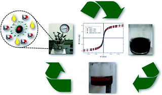
Two organic superbases, 1,5,7-triazabicyclo[4,4,0]dec-5-ene (TBD) and 1,1,3,3-tetramethylguanidine (TMG), were anchored onto silica-coated and uncoated iron oxide nanoparticles, resulting in three (MNP–TBD, MNP–TMG and MNP@SiO2–TBD) recoverable basic nanocatalysts. The nanocatalysts were fully characterized by elemental analysis, infrared and Raman spectroscopies, X-ray diffraction, transmission electron microscopy, N2 adsorption/desorption isotherms, thermogravimetric analysis and magnetic measurements. X-ray diffraction indicated the presence of a spinel-structured iron oxide, and Raman spectroscopy revealed both magnetite and maghemite phases in the prepared nanocatalysts. The average particle sizes of the nanocatalysts were in the range of 11 to 12 nm, and they exhibited superparamagnetic behaviour at 300 K. Infrared spectroscopy indicated the presence of the superbases (TBD and TMG) on the surface of the silica-coated and uncoated iron oxide nanoparticles. The performance of the nanocatalysts was tested in the methanolysis reaction of soybean oil under different conditions. At the end of each reaction, the nanocatalysts were magnetically recovered from the medium, and the product was analysed and quantified by high-performance liquid chromatography (HPLC). MNP–TBD exhibited the best catalytic performance in the first cycle (96% biodiesel conversion); however, MNP@SiO2–TBD exhibited the best reusability.


Introduction

Petroleum is a non-renewable source of energy; therefore, the development of technologies to generate energy from renewable feedstocks remains a major societal need.1 Biodiesel is garnering attention as an alternative fuel derived from vegetable oils or animal fats. This fuel is considered to be safer, more biodegradable and causes less pollution than traditional diesel.2–4 Biodiesel is produced by the transesterification process catalyzed by acids, bases or enzymes in the presence of an alcohol. Industrial production is carried out under homogeneous basic catalysis using hydroxides and alkoxides of alkaline-earth metals to achieve high yields.5,6 Catalyst recovery, however, is one of the limitations of the process.7,8 In addition, the remaining catalyst should be removed during water washings; however, to our knowledge, the maximum residual concentration of catalysts in biodiesel is not specified by the American Society for Testing and Materials (ASTM). Excess residual basic or acid catalysts can cause fuel tank corrosion and other engine problems. To overcome this limitation, immobilization of the catalysts onto various solid supports facilitates their recovery. Hydrotalcites,9,10 modified zeolites11,12 and metal oxides13,14 are among the solids investigated to date. Furthermore, catalytic compounds such as guanidines have been supported on mesoporous silica (MCM-41)15–18 and subsequently used in transesterification reactions.19–22 Previously, we have demonstrated 99% biodiesel conversion using the guanidine 1,5,7-triazabicyclo[4,4,0]dec-5-ene (TBD) anchored onto MCM-41.20 However, the biodiesel conversion rate decreases drastically to 0.8% after the fifth cycle, likely because of neutralization of basic sites on the guanidine by free fatty acids contained in soybean oil.

In the field of catalysis, magnetic nanoparticles (MNPs) are interesting and versatile materials because of their high surface-area-to-volume ratio, which facilitates surface modification.23 Thus, MNPs are an alternative support material because of their easy synthesis and functionalization, low cost, and low toxicity.24 The greatest advantage of MNPs is their easy recovery from the reaction medium under an applied external magnetic field, eliminating steps such as centrifugation, filtration, and extraction.25–28 To protect MNPs from external attack and avoid agglomeration, a thin coat of silica can be deposited onto their surface, giving rise to a core–shell structure.29 Subsequently, this silica shell can be functionalized with a coupling agent such as 3-(chloropropyl)triethoxysilane (CPTES) to enable further surface modifications.

In this work, we successfully anchored two organic superbases, TBD and 1,1,3,3-tetramethylguanidine (TMG), onto silica-coated and uncoated magnetic supports, resulting in three (MNP–TBD, MNP–TMG and MNP@SiO2–TBD) recoverable basic nanocatalysts (Scheme 1). The catalysts were prepared in three steps: (i) synthesis of Fe3O4 nanoparticles via co-precipitation of Fe(II) and Fe(III) ions from basic solution; (ii) surface modification with chloropropyl groups; and (iii) functionalization with guanidine bases through SN2 nucleophilic substitution of the chloro-functionalized Fe3O4 nanoparticles. For the preparation of the silica-coated magnetic nanocatalyst, tetraethylorthosilicate (TEOS) was added to the reaction medium containing Fe3O4 nanoparticles and guanidine functionalization was performed as previously described (Scheme 1). The characterization results for the three materials and the results of an evaluation of their catalytic properties towards biodiesel production are presented. We also discuss the differences in performance among the three materials.


image file: c5ra07331f-s1.tif
Scheme 1 Synthesis of magnetic silica-coated and uncoated catalysts (MNP–TBD, MNP–TMG and MNP@SiO2–TBD).

Results and discussion

Characterization of the catalysts

On the basis of CNH elemental analysis, the amount of guanidine base present on each magnetic nanocatalyst was 0.20, 0.16 and 0.3 mmol g−1 of MNP–TBD, MNP–TMG MNP@SiO2–TBD, respectively.

Fig. 1 shows the FT-IR spectra of the magnetic nanocatalysts and their precursors. The IR spectrum of the Fe3O4 MNPs showed a broad band at 3414 cm−1 associated with the stretching vibration of O–H groups on their surface.30 An intense band due to Fe–O bond vibrations was split into two peaks at 635 and 570 cm−1. This band splitting has been associated with the nanometric size of Fe3O4 MNPs and was observed for all of the prepared catalysts and their precursors.31 Grafting of 3-(chloropropyl)triethoxysilane onto MNP–Cl was confirmed by the bands at 998 cm−1, 1124 cm−1 and 2861 to 2938 cm−1, which are assigned to the Fe–O–Si,32 Si–O–Si33 and C–H stretching vibrations,34 respectively. The band at 998 cm−1 was not observed in the spectrum of the silica-coated material because of the presence of a strong and broad stretching vibration band of the siloxane groups (Si–O) at 1007 cm−1. The bands at 1440 cm−1 and 1625 cm−1 present in the spectra of guanidine-functionalized magnetic nanocatalysts correspond to C–N and C[double bond, length as m-dash]N stretching vibrations of the guanidines, respectively.35,36 These latter bands indicate that the guanidine bases were grafted onto silica-coated and uncoated magnetic supports.


image file: c5ra07331f-f1.tif
Fig. 1 FTIR spectra of the magnetic nanocatalysts (MNP–TBD, MNP–TMG and MNP@SiO2–TBD) and their precursors.

X-ray diffraction (XRD) patterns of the MNPs and the nanocatalysts (Fig. 2) indicate the presence of a spinel-structured oxide identified as either as magnetite (Fe3O4) or maghemite (γ-Fe2O3).37 The XRD pattern of MNP@SiO2–TBD did not show peaks related to the silica, probably because of the thinness of the shell.


image file: c5ra07331f-f2.tif
Fig. 2 XRD patterns of the Fe3O4 MNPs and the magnetic nanocatalysts.

Raman spectroscopy can be used to distinguish both phases of iron oxides.38 A confocal Raman microscope was used to collect the spectra of all of the nanocatalysts (Fig. 3 and S1, see ESI). Spectra were collected at several regions of each sample. All of the spectra exhibit a main band at approximately 666 cm−1 that is assigned to the symmetric stretch of oxygen atoms along Fe–O bonds (A1g) of magnetite; the bands at approximately 717 (A1g), 500 (T2g) and 357 (Eg) cm−1 are associated with maghemite.39 Thus, all of the prepared catalysts are composed of both the magnetite and maghemite phases of iron oxide.


image file: c5ra07331f-f3.tif
Fig. 3 Raman spectra of the Fe3O4 MNPs and the magnetic nanocatalysts.

Transmission electron microscopy (TEM) of MMP–TBD, MNP–TMG and MNP@SiO2–TBD show spherical particles (Fig. 4). The particle-size distribution curves (Fig. 4) indicate that the average particle diameters of MNP–TBD, MNP–TMG and MNP@SiO2–TBD are 12, 12.3 and 11 nm, respectively. The average sizes of Fe3O4 nanoparticles obtained by the co-precipitation method, in which the base is rapidly injected, are typically smaller and more uniform than those obtained by the conventional co-precipitation method.40 Fast injection of the base (NH4OH) results in a fast nucleation process, yielding small particles. TEM images of MNP@SiO2–TBD show a uniform and continuous silica shell around the surface of Fe3O4 particles (Fig. S2, see ESI).


image file: c5ra07331f-f4.tif
Fig. 4 TEM images and particle-size distributions (PSDs) of (a) MNP–TBD, (b) MNP–TMG and (c) MNP@SiO2–TBD.

N2 adsorption/desorption isotherms of the nanocatalysts (Fig. S3, see ESI) are of type IV in the IUPAC classification, which is typical of mesoporous materials. The presumed mesoporosity of the nanocatalysts is probably related to spaces between the nanoparticles arising from their aggregation,36,41 as evident in the TEM images. The Brunauer–Emmett–Teller (BET) specific surface areas are 62 m2 g−1, 53 m2 g−1 and 84 m2 g−1 for MNP–TBD, MNP–TMG and MNP@SiO2–TBD, respectively. The BET surface area of the MNP@SiO2–TBD catalyst was greater than those of the other catalysts because of the presence of the silica shell.

Fig. 5 shows the field dependence of the magnetization for all three nanocatalysts at room temperature. The absence of hysteresis indicates the superparamagnetic nature of the nanocatalysts at T = 300 K. The saturation value of the magnetization at this temperature and at a magnetic field of 70 kOe corresponds to approximately 70 emu g−1 of nanocatalyst.


image file: c5ra07331f-f5.tif
Fig. 5 Field dependence of the magnetization measured at 300 K and 2 K (inset) for MNP–TBD, MNP–TMG and MNP@SiO2–TBD.

Fig. 6 shows the temperature dependence of the zero-field-cooled (ZFC) and field-cooled (FC) magnetization of the three nanocatalysts. The blocking temperature TB of the magnetic moment is indicated by the maximum in the ZFC curve. The silica-coated nanocatalyst MNP@SiO2–TBD exhibits a lower blocking temperature as well as a lower coercivity at low temperatures (inset of Fig. 5) compared to those of the other nanocatalysts. This result is commonly explained as a consequence of the surface anisotropy reduction due to the silica coating.42


image file: c5ra07331f-f6.tif
Fig. 6 Field-cooled (closed symbols) and zero-field-cooled (open symbols) magnetization of the nanocatalysts measured at 100 Oe.

Thermogravimetric analysis (TGA) of all of the prepared nanocatalysts (Fig. S4, see ESI) was conducted to verify their thermal stabilities and to establish the appropriate temperature for the catalytic reactions. The TGA curves show two main decomposition steps. The initial weight loss of approximately 1% is likely due to the presence physisorbed water or solvent molecules on the surface of the nanocatalysts. The data suggest that the second step, at temperatures ranging from 200 to 600 °C, comprises two consecutive weight losses. Both are related to the decomposition of the organosilane and the guanidine bases grafted onto the MNPs. The weight loss during this step is approximately 8.6, 8.0, and 9.0 wt% for MNP–TBD, MNP–TMG and MNP@SiO2–TBD, respectively. These results corroborate the relative quantities of guanidine estimated by elemental analysis for each nanocatalyst. In addition, the nanocatalysts were demonstrated to be thermally stable at the temperature used for the transesterification reaction (120 °C).

Catalytic activities of the MNP–TBD, MNP–TMG and MNP@SiO2–TBD catalysts towards biodiesel production

To evaluate the catalytic activities of the guanidine-functionalized Fe3O4 superparamagnetic nanoparticles and to determine the most appropriate reaction conditions, the transesterification reaction of soybean oil with methanol was investigated (Table 1).
Table 1 Methanolysis of soybean catalyzed by MNP–TBD, MNP–TMG and MNP@SiO2–TBD at 120 °C, using 10% for each catalyst
Entry Catalyst MeOH/oil Time (h) Biodiesel yield (%)
1 None 30[thin space (1/6-em)]:[thin space (1/6-em)]1 24 0
2 MNP–TBD 30[thin space (1/6-em)]:[thin space (1/6-em)]1 24 96
3 MNP–TMG 30[thin space (1/6-em)]:[thin space (1/6-em)]1 24 11
4 MNP–TBD 30[thin space (1/6-em)]:[thin space (1/6-em)]1 12 78
5 MNP–TBD 30[thin space (1/6-em)]:[thin space (1/6-em)]1 6 60
6 MNP–TMG 30[thin space (1/6-em)]:[thin space (1/6-em)]1 6 9
7 MNP@SiO2–TBD 30[thin space (1/6-em)]:[thin space (1/6-em)]1 24 80


The results in Table 1 show that the MNP–TBD was an efficient nanocatalyst in the transesterification reaction of soybean oil at 120 °C and at a 30[thin space (1/6-em)]:[thin space (1/6-em)]1 methanol-to-oil molar ratio. The biodiesel yield was 60% after 6 h (entry 5) and 96% after 24 h (entry 2). Thus, the biodiesel yield increases with increasing reaction time. Using the MNP@SiO2–TBD nanocatalyst resulted in an 80% biodiesel yield after 24 h (entry 7), which is close to the yield obtained using the MNP–TBD catalyst. MNP–TMG nanocatalyst was the least efficient, yielding only 11% biodiesel at the same reaction time (entry 3). Control experiments showed that, in the absence of the nanocatalysts, no biodiesel conversion was observed under the same reaction conditions (Table 1, entry 1).

Table 2 shows the turnover number (TON), which is expressed as yield per mmol of basic sites, and the turnover frequency (TOF), which is expressed as TON per hour during the experiments. Because the kinetics of the reactions were not investigated, the TON values calculated in this work provide only a trend for the activity of the nanocatalysts.

Table 2 Activity, expressed in TON and TOF, and basicity, expressed in terms of the pKa, of the catalysts
Catalyst TONa TOFb
a (% biodiesel per mmol of basic sites).b (TON per h).
MNP–TBD 480 20
MNP–TMG 69 3
MNP@SiO2–TBD 267 11


The MNP–TBD nanocatalyst exhibits the highest TON and TOF, followed by the MNP@SiO2–TBD and the MNP–TMG nanocatalysts. The higher TON and TOF for the MNP–TBD and MNP@SiO2–TBD compared to those for the MNP–TMG are attributed to the intrinsic basicity of the guanidines. TMG and TBD are considered organic superbases because of the resonance stability of their conjugated acids.43 The pKa of TBD is 25.9 (CH3CN), whereas that of TMG is 23.3 (CH3CN). The six-membered ring of TBD increases its basicity compared to the acyclic TMG. The transesterification reaction homogeneously catalyzed by TMG (10%) under reflux conditions produced a biodiesel yield of 75% biodiesel after 1 h of reaction, whereas TBD yielded 97%. However, heterogenization of TMG drastically decreased its catalytic performance. Both superbases were anchored to the magnetic nanoparticles modified with 3-(chloropropyl)triethoxysilane by the amine nitrogen, leaving the N imino atoms available to carry out methanol deprotonation (Fig. S5, see ESI). Compared to the N imino atoms of TBD in a heterogeneous phase, those of TMG are likely much less accessible to the methanol molecules because of steric effects.

Reusability and recovery of the MNP–TBD and MNP@SiO2–TBD nanocatalysts

The reusability and recovery of MNP–TBD and MNP@SiO2–TBD were investigated. Nanocatalyst recovery after the reactions was very simple and easy, as shown in Fig. 7.
image file: c5ra07331f-f7.tif
Fig. 7 Images showing the crude products obtained after the transesterification reaction before and after an external magnetic field was applied: (a) MNP–TBD and (b) MNP@SiO2–TBD.

The reusability of the MNP–TBD and MNP@SiO2–TBD catalysts was evaluated under the conditions described in Table 1 (entry 2 for MNP–TBD and entry 7 for MNP@SiO2–TBD). After each reaction cycle, the nanocatalysts were separated from the reaction mixture using a magnet, recovered, washed with methanol, and then washed with 0.1 mol L−1 NaOH aqueous solution and deionized water until the nanoparticle suspension reached a pH of 7. The nanocatalysts were air-dried before being reused in the next cycle. Washings with aqueous NaOH were carried out because neutralization of the basic sites of guanidine by the free fatty acids present in the soybean oil can occur, deactivating the catalysts.20

Fig. 8 shows the biodiesel yield upon four consecutive cycles using MNP–TBD and MNP@SiO2–TBD. The activity of the MNP–TBD nanocatalyst was almost negligible after the fourth cycle. To understand why this deactivation occurred, elemental analysis of the MNP–TBD nanocatalyst was carried out after the fourth reaction cycle; the results indicated that only 0.03 mmol g−1 of TBD remained on the magnetic support. Therefore, approximately 85% of the organic base was leached from the surface of the magnetic nanoparticles. Conversely, in the case of the MNP@SiO2–TBD nanocatalyst, much better reusability was observed; elemental analysis of this nanocatalyst after the forth cycle revealed that approximately 23% of the TBD was leached (0.23 mmol g−1 of TBD remained after the fourth cycle). Thus, the leaching process was more pronounced in the case of the magnetic nanocatalysts not coated with silica. To explain the difference in the leaching process between the two nanocatalysts, particle-size distribution (PSD) curves were obtained from TEM micrographs of MNP–TBD and MNP@SiO2–TBD after the fourth cycle (Fig. 9).


image file: c5ra07331f-f8.tif
Fig. 8 Reusability of the MNP–TBD and MNP@SiO2–TBD nanocatalysts.

image file: c5ra07331f-f9.tif
Fig. 9 TEM microscopies and PSDs of (a) MNP–TBD and (b) MNP@SiO2–TBD nanocatalysts after the forth cycle.

Fig. 9a shows that the PSD curve is shifted to smaller values compared to the PSD before the catalysis (Fig. 4a). In contrast, the PSD of MNP@SiO2–TBD did not significantly change. These results indicate that the guanidine (TBD) leaching from the surface of MNP–TBD is associated with a partial dissolution of the iron oxide nanoparticles. Iron oxide nanoparticles can dissolve by different mechanisms in the presence of organic or inorganic acids.44,45 At high temperatures (>90 °C), the rate of iron oxide dissolution can increase. Because soybean oil is composed of a mixture of fatty acids, including linoleic and oleic acids,46 the dissolution process can occur via the adsorption of these compounds onto the surface of the MNP–TBD nanocatalysts. Indeed Fig. 7 shows that the colour of the solution appears brown, indicating partial dissolution of iron oxide in the presence of fatty acids and also possible oxidation of Fe3O4 to Fe2O3. The silica coating protected the iron oxide nanoparticles from dissolution; consequently, MNP@SiO2–TBD exhibited better reusability and structural stability than the MNP–TBD nanocatalyst under the conditions used in this work.

Several efforts have been carried out to obtain structurally stable catalysts in addition to good recyclability. Zhang et al.47 prepared Pd/Fe3O4 binary superstructures for use in catalyzing dye degradation. Although the material exhibited good catalytic performance, the structural stability of the superstructures was quite poor. The low stability of the Pd/Fe3O4 binary superstructure was attributed to van der Waals week interactions between the particles that are short range in action. Coating the Pd/Fe3O4 binary particles with polypyrrole shell prevented the deconstruction of the superstructures and consequently, improved structural stability and reusability of the catalysts.47 In the case of the MNP–TBD nanocatalyst investigated in this work, we believe that the poor stability of this material might be related to dissolution process due to the presence of fatty acids in the soybean oil. Thus, protecting iron oxide nanoparticles with polymers or oxides seems to be the best strategy to improve structural stability and recyclability of the magnetic catalysts.

Conclusions

Three new recoverable magnetic nanocatalysts for biodiesel production were successfully prepared. All of the nanocatalysts exhibited superparamagnetic behaviour at room temperature and were observed to support methanolysis of soybean oil. The MNP–TBD nanocatalyst exhibited the best performance, with a biodiesel yield of 96% at 120 °C and 24 h of reaction time in the first cycle. The MNP–TMG nanocatalyst was the least efficient, most likely because of steric effects caused by its methyl groups. The nanocatalysts were easily recovered using an external magnetic field. The reusability of MNP@SiO2–TBD was better than that of MNP–TBD because the silica coating protected the iron oxide nanoparticles from dissolution caused by the free fatty acids present in the soybean oil. The results of the present study demonstrated the advantages of using magnetic nanoparticles as catalytic supports instead of conventional supports; the magnetic supports are useful for the clean and economical production of biodiesel. These nanocatalysts can also be used for other reactions and might exhibit better reusability under neutral or basic reaction conditions.

Experimental

Materials and methods

The following reagents were used without any prior treatment: anhydrous iron(II) chloride, iron(III) chloride hexahydrate, (3-chloropropyl)trimethoxysilane (CPTES) 95%, tetraethyl orthosilicate (TEOS) 98%, 1,1,3,3-tetramethylguanidine (TMG), 1,5,7-triazabicyclo[4.4.0]dec-5-ene (TBD) from Sigma-Aldrich and ammonium hydroxide, triethylamine and ethanol from Vetec. Toluene (Vetec) was dried under sodium and subsequently distilled. Methanol (Vetec) and commercial soybean oil (LIZA) were used in the transesterification reaction without further purification. Fourier-transform infrared (FT-IR) spectra were recorded in the range from 400 to 4000 cm−1 on a Varian 660-IR FT-IR spectrophotometer using the KBr pellet technique. The TGA of the samples was performed under flowing nitrogen (50 cm3 min−1) on a TG Instruments system, model Shimadzu DTG-60/60H; the samples were heated from 30 °C to 600 °C at a heating rate of 5 °C min−1. Elemental analyses (CHN) were carried out using in a Perkin-Elmer CHN 240 C analyser at the Central Analítica of the Instituto de Química, Universidade de São Paulo, Brazil. Powder XRD patterns were collected on a Bruker D8 Advance X-ray diffractometer equipped with a LynxEye detector and a Co-Kα1 (1.78901 Å) and Co-Kα2 (1.7929 Å) radiation source. The following databases were used: PDF 082-1533 for magnetite and PDF 039-1346 for maghemite, both from ICCD, 2003. Raman spectra were recorded with a Witec Alpha 300 confocal Raman imaging microscope. The experiments were performed at 25 °C using an Nd:YAG green laser with a wavelength of 532 nm and a small aperture (730 CCD cts for SiO2) to avoid decomposition or phase changes in the sample during the analysis. The laser light was focused onto the sample using a 100× objective lens (NA = 0.95); the integration time was 0.5 s, and the number of scans was 600. Two different points were focused, allowing better characterization of the samples. TEM images were obtained using a JEOL JEM-1011. For these analyses, 1.0 mg of the samples was dispersed in 1 mL of ethanol by sonication for 10 min. A drop of this dispersion was transferred into 50 mL of deionized water, and a carbon-coated copper grid was subsequently immersed into this dispersion. This grid was stored in a desiccator for one week to slowly remove the solvent. The magnetic measurements were carried out in a magnetometer with a SQUID sensor (MPMS-XL, Quantum Design). The specific surface areas of the nanocatalysts were characterized by isotherms of N2, which were obtained using an ASAP 2020 V304-4 serial 1200 apparatus. Prior to the measurements, the samples were degassed under vacuum at 100 °C for 24 h.

Preparation of the magnetic nanoparticles (MNPs)

Fe3O4 nanoparticles were prepared by a chemical coprecipitation method.32 FeCl3·6H2O (48.64 g, 180 mmol) and FeCl2 (11.34 g, 89 mmol) were dissolved in 1 L of deionized water. The solution was degassed by bubbling Ar for 30 min at room temperature under mechanical stirring (400 rpm). Then, 100 mL of 25% NH4OH was quickly added in one portion to the solution. The reaction mixture remained under mechanical stirring and Ar atmosphere at RT for 1 h. The black precipitate was separated from the solution using magnetic decantation and was subsequently washed with deionized water (10×). The resulting black solid was dried under vacuum at RT for 5 h, yielding a compound labelled MNPs (21.00 g).

Grafting of 3-(chloropropyl)triethoxysilane onto MNPs

MNPs (20 g) were dispersed in 400 mL of EtOH[thin space (1/6-em)]:[thin space (1/6-em)]H2O (1[thin space (1/6-em)]:[thin space (1/6-em)]1) by sonication for 1 h. The dispersion was then transferred to a round-bottom flask and heated to 40 °C under Ar atmosphere and mechanical stirring (300 rpm) for 30 min. After this period, 8 mL of CPTES (95%) was added. The reaction mixture remained under stirring for 3 h, after which five aliquots of 2 mL of CPTES were added during an interval of 6 h. The reaction mixture was left to stir for another 16 h, and the solid was collected by centrifugation (3000 rpm for 10 min) and washed with EtOH (3×). The final product was dried under vacuum at RT for 3 h and labelled as MNP–Cl.

Preparation of guanidine-functionalized magnetic nanocatalysts

The MNP–Cl (8.0 g) was dispersed in dry toluene (50 mL) by being sonicated for 1 h and placed to a 250 mL round-bottom flask. The TBD (5.0 g, 36 mmol) in triethylamine (11 mL, 79 mmol) was added to the dispersion under mechanical stirring (300 rpm), and the reaction mixture was refluxed for 24 h. The final product was separated by magnetic decantation and washed sequentially (4×) with CH2Cl2, EtOH and CH2Cl2. The product was then dried under vacuum at RT for 2 h and labelled as MNP–TBD. The same procedure was followed for the synthesis of MNP–TMG except that, in this case, TMG (5 mL, 40 mmol) was used.

Preparation of silica-coated MNPs

A one-step synthesis was used to obtain silica-coated Fe3O4 nanoparticles.48 Tetraethylorthosilicate (TEOS) (2.7 mL) in EtOH (60 mL) was added to the reaction mixture of Fe3O4 nanoparticles. The reaction was continued for 24 h at RT. The resulting silica-coated MNPs were separated by magnetic decantation and alternately washed (9×) with H2O and EtOH. The MNPs were then dried under vacuum at RT for 8 h, affording MNP@SiO2.

Grafting of 3-(chloropropyl)triethoxysilane onto silica-coated MNP

MNP@SiO2 (8.00 g) was dispersed in 200 mL of dry toluene by sonication for 1 h. The dispersion was subsequently refluxed under Ar atmosphere and mechanical stirring (800 rpm). After this period, 8 mL of CPTES was added and the reaction mixture remained under stirring for 6 h. Then, five 2 mL aliquots of CPTES were added during an interval of 3 h. The reaction mixture was left to stir for an additional 16 h. The solid was collected by magnetic decantation and washed with EtOH (6×). The final product was dried under vacuum at RT for 12 h, affording MNP@SiO2–Cl.

Guanidine functionalization of silica-coated magnetic nanocatalysts

MNP@SiO2–Cl (7.0 g) was dispersed in dry toluene (100 mL) by sonication for 1 h. Subsequently, TBD (4.0 g, 29 mmol) in triethylamine (5 mL, 36 mmol) was added to the dispersion under mechanical stirring (300 rpm) and the reaction mixture was refluxed for 24 h. The final product was separated by magnetic decantation, washed sequentially (7×) with CH2Cl2, EtOH and CH2Cl2, and dried under vacuum at RT for 8 h, affording MNP@SiO2–TBD.

Catalytic reactions

The transesterification reactions were carried out in a Parr 5500 HP (100 mL) reactor under magnetic stirring and at a controlled temperature. The reactor was loaded with soybean oil and a methanolic suspension of the catalysts (10% w/w), which provided a 30[thin space (1/6-em)]:[thin space (1/6-em)]1 methanol-to-oil molar ratio. Prior to the reaction, the catalysts were dispersed in methanol by sonication for 1 h. The system was closed and heated to 120 °C for 24 h under an autogenous pressure. After this period, the reactor was cooled to RT and the catalysts were magnetically separated from the reaction mixture. The method used to analyse and quantify the biodiesel formed in the reaction is described elsewhere.20 Briefly, we diluted (1% w/v) the lower phase (alkyl ester) in isopropyl alcohol and analysed it (in duplicate) using an Agilent 1200 high-performance liquid chromatograph. The products were separated using a 250 mm long column with 4.6 mm internal diameter; the column was packed with C18-covered silica with a particle size of 5 μm. A pre-column 2 cm in length was fitted between the injector and the separation column. The solvent gradient elution was performed using mobile phases A (methanol), B (n-hexane) and C (isopropanol). The phases were filtered through a membrane of expanded polytetrafluoroethylene (PTFE) with a diameter of 47 mm. Analyses were carried out at 40 °C and at a flow rate of 1 mL min−1 using spectrophotometric detection at 205 nm in the UV region. The sequence of the gradient was: at 0 min – 98% A, 1% B and 1% C; at 20 min – changed to 2% A, 49% B and 49% C; at 22 min – 98% A, 1% B and 1% C. The peaks were identified by comparison of the retention times of the samples with those of standard compounds (triglyceride and fatty acid methyl esters). The chromatograms, which were analysed and integrated with the aid of the Agilent ChemStation software, were used to quantify the amount of methyl fatty esters (biodiesel) formed in the reactions on the basis of the response factor obtained from a previously constructed calibration curve.

Reusability of the catalysts

The catalysts used in the first cycle of the transesterification reactions were separated from the reaction solution using a magnet, recovered, extensively washed with methanol, and then washed with 0.1 mol L−1 NaOH aqueous solution and deionized water until the pH of the suspension was 7. The catalysts were air-dried before reuse in the next cycle. Each catalyst was then used in a new reaction cycle under the previously described conditions.

Acknowledgements

The authors gratefully acknowledge FAPERJ (JCNE), CNPq 45 (Jovens Pesquisadores em Nanotecnologia grant number 550572/2012-0 and ECSS fellowship), CAPES (T.C.S. fellowship) and FINEP (0113031500) for financial support. C.M.R. is recipient of a CNPq research fellowship. We thank Prof. Cláudio J. A. Mota and Ana L. Lima (IQ-UFRJ, Brazil) for assistance with the catalytic experiments. We also thank the Multiuser Laboratory of Material Characterization (http://www.uff.br/lamate/), Multiuser Laboratory of Spectroscopy (http://www.uff.br/lame/), Multiuser Laboratory of Microscopy (Rômulo Custódio dos Santos) and Dr Pedro Pablo Florez Rodrigues for confocal Raman measurements.

Notes and references

  1. N. F. Nasir, W. R. W. Daud, S. K. Kamarudin and Z. Yaakob, Renewable Sustainable Energy Rev., 2013, 22, 631 CrossRef CAS.
  2. G. Knothe, J. Gerpen and J. Krahl, The biodiesel handbook, AOCS Press, Urbana, Illinois, 2005 Search PubMed.
  3. A. Islam, Y. H. Taufiq-Yap, C.-M. Chu, E.-S. Chan and P. Ravindra, Process Saf. Environ. Prot., 2013, 91, 131 CrossRef CAS.
  4. K. Ramachandran, T. Suganya, N. N. Gandhi and S. Renganathan, Renewable Sustainable Energy Rev., 2013, 22, 410 CrossRef CAS.
  5. U. Schuchardt, R. Sercheli and R. M. Vargas, J. Braz. Chem. Soc., 1998, 9, 199 CrossRef CAS.
  6. I. M. Atadashi, M. K. Aroua, A. R. A. Aziz and N. Sulaiman, J. Ind. Eng. Chem., 2013, 19, 14 CrossRef CAS.
  7. D.-W. Lee, Y.-M. Park and K.-Y. Lee, Catal. Surv. Asia, 2009, 13, 63 CrossRef CAS.
  8. E. Santacesaria, G. M. Vicente, M. Di Serio and R. Tesser, Catal. Today, 2012, 195, 2 CrossRef CAS.
  9. K. G. Georgogianni, A. P. Katsoulidis, P. J. Pomonis and M. G. Kontominas, Fuel Process. Technol., 2009, 90, 671 CrossRef CAS.
  10. J. Shen, J. Kobe, Y. Chen and J. Dumesic, Langmuir, 1994, 10, 3902 CrossRef CAS.
  11. G. J. Suppes, M. A. Dasari, E. J. Doskocil, P. J. Mankidy and M. J. Goff, Appl. Catal., A, 2004, 257, 213 CrossRef CAS.
  12. S. K. Karmee and A. Chadha, Bioresour. Technol., 2005, 96, 1425 CrossRef CAS PubMed.
  13. M. Di Serio, R. Tesser, L. Pengmei and E. Santacesaria, Energy Fuels, 2008, 22, 207 CrossRef CAS.
  14. Y. B. Cho and G. Seo, Bioresour. Technol., 2010, 101, 8515 CrossRef CAS PubMed.
  15. M. R. Mello, D. Phanon, G. Q. Silveira, P. L. Llewellyn and C. M. Ronconi, Microporous Mesoporous Mater., 2011, 143, 174 CrossRef CAS.
  16. T. C. Santos, S. Bourrelly, P. L. Llewellyn, J. W. d. M. Carneiro and C. M. Ronconi, Phys. Chem. Chem. Phys., 2015, 17, 11095 RSC.
  17. G. Q. Silveira, M. D. Vargas and C. M. Ronconi, J. Mater. Chem., 2011, 21, 6034 RSC.
  18. G. Q. Silveira, R. S. da Silva, L. P. Franco, M. D. Vargas and C. M. Ronconi, Microporous Mesoporous Mater., 2015, 206, 226 CrossRef CAS.
  19. J. M. Balbino, E. W. Menezes, E. V. Benvenutti, R. Cataluña, G. Ebeling and J. Dupont, Green Chem., 2011, 13, 3111 RSC.
  20. A. L. Lima, A. Mbengue, R. San Gil, C. M. Ronconi and C. J. A. Mota, Catal. Today, 2014, 226, 210 CrossRef.
  21. D. Meloni, R. Monaci, Z. Zedde, M. G. Cutrufello, S. Fiorilli and I. Ferino, Appl. Catal., B, 2011, 102, 505 CrossRef CAS.
  22. R. Sercheli, R. Vargas and U. Schuchardt, J. Am. Oil Chem. Soc., 1999, 76, 1207 CrossRef CAS.
  23. S. Paria and R. Chaudhuri, Chem. Rev., 2012, 112, 2373 CrossRef PubMed.
  24. H.-J. Xu, X. Wan, Y. Geng and X.-L. Xu, Curr. Org. Chem., 2013, 17, 1034 CrossRef CAS.
  25. L. M. Rossi, N. J. S. Costa, F. P. Silva and R. Wojcieszak, Green Chem., 2014, 16, 2906 RSC.
  26. Q. Zhang, X. Han and B. Tang, RSC Adv., 2013, 3, 9924 RSC.
  27. Z. Chen, W. Xu, L. Jin, J. Zha, T. Tao, Y. Lin and Z. Wang, J. Mater. Chem A, 2014, 2, 18339 CAS.
  28. N. Basavegowda, K. Mishra and Y. R. Lee, RSC Adv., 2014, 4, 61660 RSC.
  29. C. G. S. Souza, W. Beck and L. C. Varanda, J. Nanopart. Res., 2013, 15, 1545 CrossRef.
  30. A. Rostami, B. Atashkar and D. Moradia, Appl. Catal., A, 2013, 467, 7 CrossRef CAS.
  31. M. Ma, Y. Zhang, W. Yu, H.-Y. Shen, H.-Q. Zhang and N. Gu, Colloids Surf., A, 2003, 212, 219 CrossRef CAS.
  32. B. Atashkar, A. Rostami and B. Tahmasbi, Catal. Sci. Technol., 2013, 3, 2140 CAS.
  33. F. Galeotti, F. Bertini, G. Scavia and A. Bolognesi, J. Colloid Interface Sci., 2011, 360, 540 CrossRef CAS PubMed.
  34. R. A. Bini, R. F. C. Marques, F. J. Santos, J. A. Chaker and M. Jafelicci Jr, J. Magn. Magn. Mater., 2012, 324, 534 CrossRef CAS.
  35. F. Adam and M. S. Batagarawa, Appl. Catal., A, 2013, 454, 164 CrossRef CAS.
  36. A. C. Blanc, D. J. Macquarrie, S. Valle, G. Renard, C. R. Quinn and D. Brunel, Green Chem., 2000, 2, 283 RSC.
  37. L. Signorini, L. Pasquini, L. Savini, R. Carboni, F. Boscherini, E. Bonetti, A. Giglia, M. Pedio, N. Mahne and S. Nannarone, Phys. Rev. B: Condens. Matter Mater. Phys., 2003, 68, 195423 CrossRef.
  38. N. Pinna, S. Grancharov, P. Beato, P. Bonville, M. Antonietti and M. Niederberger, Chem. Mater., 2005, 17, 3044 CrossRef CAS.
  39. Raman Spectroscopy for Nanomaterials Characterization, ed. C. S. S. Kumar, Springer-Verlag, London, Berlin, Heidelberg, 2012 Search PubMed.
  40. M. C. Mascolo, Y. Pei and T. A. Ring, Materials, 2013, 6, 5549 CrossRef CAS.
  41. F. Márquez, T. Campo, M. Cotto, R. Polanco, R. Roque, P. Fierro, J. M. Sanz, E. Elizalde and C. Morant, Soft Nanosci. Lett., 2011, 1, 25 CrossRef.
  42. U. Gradmann, J. Magn. Magn. Mater., 1991, 100, 481 CrossRef CAS.
  43. Superbases for organic synthesis: guanidines, amidines and phosphazenes and related organocatalysts, ed. T. Ishikawa, John Wiley & Sons Ltd, West Sussex, 2009 Search PubMed.
  44. D. Panias, M. Taxiarchou, I. Paspaliaris and A. Kontopoulos, Hydrometallurgy, 1996, 42, 257 CrossRef CAS.
  45. B. Kalska-Szostkoa, U. Wykowska, K. Piekut and D. Satuła, Colloids Surf., A, 2014, 450, 15 CrossRef.
  46. S. P. Singh and D. Singh, Renewable Sustainable Energy Rev., 2010, 14, 200 CrossRef CAS.
  47. X. Zhang, M. Lin, X. Lin, C. Zhang, H. Wei, H. Zhang and B. Yang, ACS Appl. Mater. Interfaces, 2014, 6, 450 CAS.
  48. N. Chekiva, D. Horák, P. Jendelová, M. Trchová, M. Benes, M. Hrubý, V. Herynek, K. Turnovcová and E. Syková, J. Mater. Chem., 2011, 21, 7630 RSC.

Footnote

Electronic supplementary information (ESI) available. See DOI: 10.1039/c5ra07331f

This journal is © The Royal Society of Chemistry 2015