Highly stable oil-in-water emulsions with a gemini amphiphilic pseudopeptide

Ahmed H. Lotfallaha, M. Isabel Burguetea, Ignacio Alfonso*b and Santiago V. Luis*a
aDepartamento de Química Inorgánica y Orgánica, Universidad Jaume I, Avenida Sos Baynat, s/n, Castellón, Spain. E-mail: luiss@uji.es; Fax: +34 964728214; Tel: +34 964728239
bDepartamento de Química Biológica y Modelización Molecular, IQAC-CSIC, Jordi Girona 18-26, Barcelona, Spain. E-mail: ignacio.alfonso@iqac.csic.es; Fax: +34 932045904; Tel: +34 934006100

Received 23rd March 2015 , Accepted 16th April 2015

First published on 16th April 2015


Abstract

A gemini amphiphilic pseudopeptide promotes the spontaneous formation of an oil-in-water emulsion with a high thermal, mechanical and acid-medium stability. The micro-droplets thus formed are disassembled by strong bases or after the action of an enzyme, showing a potential for stimulus-responsive material formulations.
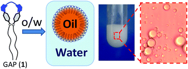

The preparation of stable emulsions has recently attracted attention in chemical research,1 especially after understanding some of the basis for the design of new self-assembling entities.2 Surfactant-based emulsions display a wide range of technological and industrial applications in pharmaceutics,3 food4 and cosmetic5 formulations or catalysis.6 However, some of the conventional surfactant agents have some limitations for long-term stability,7 availability and chemical modularity for tailor-made structural modifications.8 In this regard, the use of amino acid-based amphiphilic molecules for phase separation and emulsion stabilization will offer a tremendous advantage in terms of synthetic and structural versatility.9 Recently, we reported on the synthesis and study of a family of new Gemini Amphiphilic Pseudopeptides (GAP)10 that were able to self-assemble into different nanostructures depending on the environment, and even to respond to simple stimuli, like polarity11 or pH changes of the medium.12 The delicate tuning of the chemical structures allowed us to modulate the amphiphilic nature for optimizing their self-assembling abilities at the organic–aqueous or air–water interphases.13 Here we exploit that knowledge for the efficient preparation of emulsions with a high mechanical, thermal and chemical stability. Our pseudopeptidic systems show advantages in terms of biocompatibility and synthetic modularity, making them highly appealing for future biotechnological applications.

The designed GAP (1) is a flexible C2 symmetric pseudopeptide with a central polar linker (Fig. 1, in blue) and two lipophilic tails. The Val derivative was selected because the corresponding pseudopeptides bearing this side chain have shown specific self-assembling advantages.14 This molecule can be readily synthesized in relatively large scale from commercially available or easily accessible precursors (see ESI for the synthetic procedure). Its specific structural design favours the corresponding self-assembling of 1 in aqueous environments, with a critical aggregation concentration of 4–25 μM, depending on the pH and ionic strength of the medium. Actually, Scanning Electron Microscopy (SEM), Atomic Force Microscopy (AFM) and Transmission Electron Microscopy (TEM) images of dried samples of 1, obtained from acidic aqueous methanolic media showed the formation of soft spherical microstructures (Fig. 1). The three microscopy techniques consistently showed a spherical morphology of the microstructures, with a diameter in the range of 1–5 μm. The shape and size of the microstructures were not dramatically affected by the different procedures used for the preparation of the sample for each technique. This observation is quite remarkable considering the different nature of the surfaces used to prepare the samples for each technique: aluminium for SEM, mica for AFM and carbon for TEM. The careful analysis of the width and the height of the observed microstructures using AFM rendered that they flattened over the surface. This observation supported their soft nature. The observed size and softness suggested that they must be hollow vesicles.


image file: c5ra05121e-f1.tif
Fig. 1 (A) Chemical structure of the Gemini Amphiphilic Pseudopetide (GAP) 1, (B) SEM, (C) AFM and (D) TEM micrographs of dried samples of 1 grown from 1[thin space (1/6-em)]:[thin space (1/6-em)]1 water[thin space (1/6-em)]:[thin space (1/6-em)]MeOH solvent mixture at pH 2.

Most likely, these microstructures occurred through the self-assembling of a folded conformation of 1 where the polar linker is exposed to the aqueous medium and the hydrophobic tails are buried in a lipid phase packed by van der Waals interactions. Accordingly, this property could be exploited to use 1 for the stabilization of oil microdroplets in aqueous media toward the preparation of stable oil-in-water (o/w) emulsions (Fig. 2A). The o/w emulsion can be easily prepared by stirring an aqueous suspension of isopropylpalmitate as a model oily phase (<1% oil in water by volume), containing a small quantity (∼1 mg mL−1) of 1 (Fig. 2B and C). The typical white and opaque appearance of the sample was obtained after 30–90 minutes of vigorous magnetic stirring (Fig. 2C). Optical microscopy clearly showed the emulsifying effect of the GAP. While the sample prepared in the absence of 1 showed the oil–water phase separation in heterogeneous and amorphous drops (Fig. 2B), the presence of 1 led to the spontaneous formation of regular spherical microdroplets, most likely containing the organic phase (Fig. 2C) and surrounded by the aqueous solution. The emulsion thus formed showed to be perfectly stable for several months.


image file: c5ra05121e-f2.tif
Fig. 2 (A) Schematic representation of the self-assembling of the GAP (1) toward the formation of an o/w emulsion. (B and C) Macroscopic and microscopic pictures of the oil–water mixtures (O + W) alone (B) and after the addition of the GAP and 30–90 minutes of vigorous stirring (C).

In order to monitor the ability to incorporate organic molecules within the droplets of the emulsion, different fluorescent probes were assayed. For instance, the dansyl derivative (N,N-diethyldansylamide, 2) prepared by us revealed to be very appropriate for the visualization of the droplets formed in the emulsion under fluorescence optical microscopy (Fig. 3A and B). The images clearly showed the fluorescent microdroplets of the organic phase immersed in the aqueous medium. Interestingly, the microstructures were stable and emissive for at least eight weeks (Fig. 3C). Additionally, the emulsion preparation was studied with a highly hydrophobic molecule, 9,10-dimethylantracene (DMA), a well-known fluorescent probe for labelling membranes and lipid phases.15 The emission spectra of DMA in water and in the emulsion preparation are shown superimposed in Fig. 3D. Several changes in both the intensity and position of representative bands are remarkable. First of all, the normalized emission spectrum of DMA in the emulsion is fourfold more intense than in water. Besides, a slight red-shift of the I3 band from 450 nm (in water) to 456 nm (in the emulsion) was observed. More importantly, the ratio of the corresponding intensity of I1/I3 bands changed from 1.90 in water to 2.54 in the emulsion. These changes are consistent with the localization of the DMA probe in a highly hydrophobic microenvironment for the emulsion sample. Actually, control experiments showed that the emission spectrum of the DMA dissolved in pure isopropylpalmitate was very similar (although with lower intensity) to that obtained from DMA within the emulsion. These results suggested that the DMA probe in the emulsion was accumulated in the organic phase by passive diffusion into the microdroplets. Overall, these fluorescence experiments demonstrated the potential of our preparation for the stable long-term encapsulation of hydrophobic drugs and functional molecules.


image file: c5ra05121e-f3.tif
Fig. 3 (A) Schematic representation of the labelling of the microdroplets with the N,N-diethyldansylamide 2. (B and C) Fluorescence microscopy images of the emulsion loaded with 2 after one (B) and eight (C) weeks. (D) Normalized fluorescence emission spectra (λ excitation = 358 nm) of 9,10-dimethylantracene (DMA) acquired in water (black) or in GAP-stabilised o/w emulsion (red).

As previously stated, one of the drawbacks of some naturally occurring surfactants is their low long-term stability, specially under non-natural stress inputs, such as mechanical action, high temperatures or extreme pH values. We decided to test the stability of the o/w emulsion stabilized by 1 under different conditions. First, it was checked that the emulsion was perfectly stable for at least four months at 45 °C. On the other hand, the mechanical stability was studied by the centrifugation of the sample and checking both the macroscopic and the microscopic appearance (Fig. 4A and B). The emulsion showed a very good mechanical stability, being unaltered after 30 min of centrifugation at 3000 rpm. If longer mechanical stress was applied, the emulsion started to disassemble but, very interestingly, it was able to spontaneously recover back by simply standing for a couple of hours. This unexpected self-correction behaviour underscores the dynamic nature of the self-assembling process.


image file: c5ra05121e-f4.tif
Fig. 4 Optical microscopy of o/w emulsions formed by GAP 1: before (A) and after (B) centrifugation; at acidic (C) or basic pH (D), and after incubation with thermolysin (E). The corresponding macroscopic appearance are shown in (A and B).

The effect of the pH was also systematically studied by addition of either HCl or NaOH to the pre-formed emulsion. The emulsion remained stable at pH values from neutrality to highly acidic media (up to pH ∼ 0, Fig. 4C and ESI). However, the addition of NaOH (pH ∼ 12) rapidly destroyed the emulsion leading to the disruption of the microdroplets and the concomitant partial separation of the aqueous and organic phases (Fig. 4D). In this case, a white solid precipitate was observed that corresponded to the pure unaltered GAP (1), as confirmed by ESI mass spectrometry. These experimental observations suggested that the surfactant action of the GAP is efficient only when the amino groups of the polar moiety are partially or totally protonated, while the free base (obtained at high pH) is not able to stabilize the emulsion. Therefore, the surface of the microdroplets must be positively charged presenting a good scaffold for the attachment of anionic biomolecules such as nucleic acids.

Finally, the responsiveness of the emulsion toward a biochemically meaningful stimulus was also studied. After adding 200 μL of a solution of thermolysin (1.5 mg mL−1) to our preparation, we observed that this protease was also able to destroy the emulsion leading to the collapse of the microdroplets into larger, morphologically undefined, separated phases (Fig. 4E). Also in this case, a white precipitate was observed. However, ESI-MS analysis of the solid thus formed indicated that it mainly contained fragments of the initial GAP, suggesting that the enzyme was able to irreversibly cleave the molecule.

In conclusion, a simple and easy to synthesize Gemini Amphiphilic Pseudopeptide self-assembles into microspheres that allow stabilizing oil-in-water emulsions. This preparation showed high stability to mechanical and thermal stress, as well as to acidic media, although it disassembled in strongly basic medium or upon the action of an enzyme. The stability and responsiveness of this emulsion foresees potential applications as stabilizing and delivering vehicle for organic molecules in aqueous environments.

Acknowledgements

This work was supported by the Spanish Ministry of Economy and Competitiveness (MINECO, CTQ2012-38543-C03 project) and the GV (GV-PROMETEO/2012/020). Personal financial support for A. H. L. (GV, Grisolía fellowship 2011/036) is also gratefully acknowledged as well as technical support from the SCIC of the UJI for instrumental techniques.

Notes and references

  1. S. U. Pickering, J. Chem. Soc., 1907, 91, 2001–2021 RSC ; M. Gradzielski, Curr. Opin. Colloid Interface Sci., 2008, 13, 263–269 CrossRef CAS PubMed .
  2. G. M. Whitesides and B. Grzybowski, Science, 2002, 295, 2418–2421 CrossRef CAS PubMed ; J. M. Lehn, Science, 2002, 295, 2400–2403 CrossRef PubMed ; E. Gazit, Chem. Soc. Rev., 2007, 36, 1263 RSC ; S. Ghosh and S. Verma, Tetrahedron, 2008, 64, 6202–6208 CrossRef PubMed ; S. Cavalli, F. Albericio and A. Kros, Chem. Soc. Rev., 2010, 39, 241–263 RSC ; R. J. Brea, C. Reiriz and J. R. Granja, Chem. Soc. Rev., 2010, 39, 1448–1456 RSC .
  3. M. J. Lawrencea and G. D. Rees, Adv. Drug Delivery Rev., 2000, 45, 89–121 CrossRef CAS ; A. A. Date and M. S. Nagarsenker, Int. J. Pharm., 2008, 355, 19–30 CrossRef PubMed ; C.-X. He, Z.-G. He and J.-Q. Gao, Expert Opin. Drug Delivery, 2010, 7, 445–460 CrossRef PubMed ; Y.-L. Chiu, H. F. Chan, K. K. L. Phua, Y. Zhang, S. Juul, B. R. Knudsen, Y.-P. Ho and K. W. Leong, ACS Nano, 2014, 8, 3913–3920 CrossRef PubMed ; M. Windbergs, Y. Zhao, J. Heyman and D. A. Weitz, J. Am. Chem. Soc., 2013, 135, 7933–7937 CrossRef PubMed .
  4. D. J. Mcclements, E. A. Decker and J. Weiss, J. Food Sci., 2007, 72, R109–R124 CrossRef CAS PubMed .
  5. P. L. Lam and R. Gambari, J. Controlled Release, 2014, 178, 25–45 CrossRef CAS PubMed ; R. Y. Lochhead, W. J. Hemker and J. Y. Castaneda, Soap, Cosmet., Chem. Spec., 1987, 63, 84 Search PubMed .
  6. S. Wiese, A. C. Spiess and W. Richtering, Angew. Chem., Int. Ed., 2013, 52, 576–579 CrossRef CAS PubMed ; J. Duschmalé, S. Kohrt and H. Wennemers, Chem. Commun., 2014, 50, 8109–8112 RSC ; S. Crossley, J. Faria, M. Shen and D. E. Resasco, Science, 2010, 327, 68–72 CrossRef PubMed .
  7. S. Bai, C. Pappas, S. Debnath, P. W. J. M. Frederix, J. Leckie, S. Fleming and R. V. Ulijn, ACS Nano, 2014, 8, 7005–7013 CrossRef CAS PubMed .
  8. R. Adjonu, G. Doran, P. Torley and S. Agboola, J. Food Eng., 2014, 122, 15–27 CrossRef CAS PubMed ; N. Varghese, G. S. Shetye, D. Bandyopadhyay, N. Gobalasingham, J. Seo, J.-H. Wang, B. Theiler and Y.-Y. Luk, Langmuir, 2012, 28, 10797–10807 CrossRef PubMed ; Y. X. Liu, P. G. Jessop, M. Cunningham, C. A. Eckert and C. L. Liotta, Science, 2006, 313, 958–960 CrossRef PubMed .
  9. Y. Cheng, J. Chen and Y. L. Xiong, J. Agric. Food Chem., 2014, 62, 11575–11581 CrossRef CAS PubMed ; L. Pérez, A. Pinazo, R. Pons and M. R. Infante, Adv. Colloid Interface Sci., 2014, 205, 134–155 CrossRef PubMed ; J. M. Fletcher, R. L. Harniman, F. R. H. Barnes, A. L. Boyle, A. Collins, J. Mantell, T. H. Sharp, M. Antognozzi, P. J. Booth, N. Linden, M. J. Miles, R. B. Sessions, P. Verkade and D. N. Woolfson, Science, 2013, 340, 595–599 CrossRef PubMed ; I. W. Hamley, Soft Matter, 2011, 7, 4122–4138 RSC .
  10. J. Rubio, I. Alfonso, M. Bru, M. I. Burguete and S. V. Luis, Tetrahedron Lett., 2010, 51, 5861–5867 CrossRef CAS PubMed .
  11. J. Rubio, I. Alfonso, M. I. Burguete and S. V. Luis, Soft Matter, 2011, 7, 10737–10748 RSC .
  12. J. Rubio, I. Alfonso, M. I. Burguete and S. V. Luis, Chem. Commun., 2012, 48, 2210–2212 RSC .
  13. J. Rubio-Magnieto, S. V. Luis, M. Orlof, B. Korchowiec, G. Sautrey and E. Rogalska, Colloids Surf., B, 2013, 102, 659–666 CrossRef CAS PubMed .
  14. E. Faggi, S. V. Luis and I. Alfonso, RSC Adv., 2013, 3, 11556–11565 RSC ; I. Alfonso, M. Bru, M. I. Burguete, E. García-Verdugo and S. V. Luis, Chem.–Eur. J., 2010, 16, 1246–1255 CrossRef CAS PubMed .
  15. S. B. Dror, I. Bronshtein, Y. Garini, W. G. O'Neal, P. A. Jacobi and B. Ehrenberg, Photochem. Photobiol. Sci., 2009, 8, 354–361 Search PubMed .

Footnote

Electronic supplementary information (ESI) available: Synthetic procedures, experimental details, additional microscopy images. See DOI: 10.1039/c5ra05121e

This journal is © The Royal Society of Chemistry 2015