Electrospun composite nanofibres of PVA loaded with nanoencapsulated n-octadecane

Sidhharth Sirohi, Dhirendra Singh, Ratyakshi Nain, Dambarudhar Parida, Ashwini K. Agrawal* and Manjeet Jassal*
SMITA Research Lab, Department of Textile Technology, Indian Institute of Technology, Hauz Khas, New Delhi 110016, India. E-mail: manjeet.jassal@smita-iitd.com; ashwini@smita-iitd.com; Tel: +91-11-26591426/26591415

Received 24th December 2014 , Accepted 8th April 2015

First published on 8th April 2015


Abstract

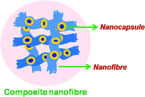
In this study composite nanofibres of PVA consisting of polystyrene/n-octadecane nanocapsules were prepared. The n-octadecane present as a core of the nanocapsules acts as a phase change material (PCM) for thermal storage properties. Controlled size nanoencapsulated PCMs were prepared by surfactant free RAFT miniemulsion polymerization of styrene. These nanocapsules were incorporated into nanofibres by electrospinning a mixture of nanocapsule latex containing octadecane as a core and polystyrene (PS) as a shell material with an aqueous solution of poly(vinyl alcohol). The morphology and thermal properties of the resultant PVA nanofibres were studied by field emission scanning microscopy (FE-SEM), scanning transmission electron microscopy (STEM), high resolution transmission electron microscopy (HRTEM) and differential scanning calorimetry (DSC). The composite nanofibres could show a latent heat storage capacity of approx. 4–5 J g−1. This method offers the opportunity to produce low diameter composite nanofibres with uniform dispersion of encapsulated material for various applications.
1 Introduction

Electrospinning provides a simple, straightforward, cheap and unique approach for the production of polymeric nanofibres in the range of 1000 nm or less.1–5 The advantages of electrospinning include production of ultrathin fibres with large surface area and superior properties.6–9 These outstanding properties have made electrospun webs promising candidates for various applications. These include filtration,10 fuel cells,11 nanowires,12 catalyst supports,13,14 chemical and biological protection,15 drug delivery devices,16 nanodevices,17 nanocomposites,18 and tissue scaffolds.19 The electrospinning of polymer solutions with particles,20 nanotubes or proteins embedded in them has been reported in the literature.21,22 The electrospinning of composite nanofibres loaded with capsules have also been reported.23,24 However, the diameter of these composite nanofibres was observed to increase to micron range due to the large size of the loaded capsules and inhomogeneity in their dispersions.25 Such systems are important for applications where handling of encapsulated sensitive materials such as biomolecules and catalysts is critical. The preparation of uniform size nanocapsules and their uniform dispersion for their optimum loading in nanofibres remain as some of the major challenges and require further investigation.

In this study, we have used electrospinning technique to produce composite PVA nanofibres loaded with controlled size nanoencapsulated PCMs. The PCMs of 97 nm size were prepared in our laboratory by surfactant free RAFT miniemulsion polymerization using co-oligomer of styrene and maleic anhydride (SM-RAFT) as amphi-RAFT agent. SM-RAFT was synthesized by RAFT polymerization using phenylethyl dithioactetate (PEPDA) as the RAFT agent. The SM co-oligomer acted as an emulsifier and RAFT agent for controlling size of nanocapsules in miniemulsion. The influence of controlled size of NCs on the final diameter of composite nanofibres was investigated. Experiments were also conducted to optimize loading of PCM nanocapsules (NCs) in PVA nanofibres.

2 Experimental work

2.1 Materials

Styrene (Merck, Germany) was distilled prior to its use and azobisisobutyronitrile (AIBN) was recrystallized twice in methanol. Maleic anhydride (Aldrich, USA), potassium per sulphate (KPS) (Merck, India), n-octadecane, (90%, Spectrochem, India), poly(vinyl alcohol) (cold, avg. mol. wt. 124[thin space (1/6-em)]000, degree of alcoholysis 80%) (Aldrich, USA), n-hexane (Specrtochem, India), and ammonia solution (25% w/v) (Merck, India) were used as obtained. The n-octadecane was used as phase change material with transition temperature of 298 K and latent heat of 215 J g−1.

2.2 Preparation of polystyrene/n-octadecane nanocapsules

The PS/n-octadecane NCs were prepared by surfactant free RAFT miniemulsion polymerization using amphiphilic co-oligomer of styrene and maleic anhydride (amphi-RAFT agent) (Scheme 1). The co-oligomer was synthesized using phenylethyl phenyl dithioactetate (PEPDA) as RAFT agent and AIBN as initiator. For this, Sty[thin space (1/6-em)]:[thin space (1/6-em)]MAn[thin space (1/6-em)]:[thin space (1/6-em)]PEPDA[thin space (1/6-em)]:[thin space (1/6-em)]AIBN ratio of 225[thin space (1/6-em)]:[thin space (1/6-em)]25[thin space (1/6-em)]:[thin space (1/6-em)]5[thin space (1/6-em)]:[thin space (1/6-em)]1 was taken and the reaction was carried out at 60 °C for 130 minutes. The co-oligomer was obtained by precipitation of reaction mixture in methanol which was further dried at 50 °C in vacuum oven overnight (characteristics shown in Table 1).
image file: c4ra16988c-s1.tif
Scheme 1 Procedure of PS/OD NCs preparation.
Table 1 Characteristics of SM co-oligomer
Co-oligomer Mn (theo.) Mn (cal.) Mw/Mn Tg (°C) CMC (mg ml−1)
SM-84 2073 1998 1.3 111 0.1584


For preparation of polystyrene/n-octadecane NCs, aqueous solution of ammonia (0.5% w/v) was added to mixture of styrene, macroRAFT agent (amphi-RAFT agent), n-octadecane and deionized water. The mixture was initially stirred for 20 minutes at 60 °C then subjected to ultrasonication for 10 minutes at 50 °C The mixture was further stirred at 60 °C for 60 minutes and finally sonicated for 15 minutes to complete the hydrolysis of anhydride units. The obtained miniemulsion was transferred to 25 ml two necked round bottom flask and kept in N2 purging for 15 minutes. The polymerization was initiated by injecting aqueous solution of potassium per sulphate. The reaction was continued for 5 hours at 68 °C. The nanocapsule latex prepared by RAFT miniemulsion polymerization was used for preparation of composite nanofibres using electrospinning.

2.3 Electrospinning

A 14% (w/w) homogeneous dope solution of PVA in DI water was prepared by stirring required quantity of polymer at 80 °C for 4 hours. Polystyrene nanocapsule latex containing octadecane as core material was added to 14% PVA solution so as to adjust concentration of octadecane to 10%, 15% and 20% on the weight of PVA. The dopes were stirred for 2 hours prior to electrospinning. Nanofibres were electrospun at minimum electrospinning voltage (MEV). MEV is defined3 as the minimum electrospinning voltage required for complete conversion (or maximum possible conversion for dilute solutions). MEV was determined as per the reported method.5 The electrospinning was carried out using an infusion syringe pump (KDS 100 from KD Scientific, Massachusetts USA) with a flat tip needle (inner diameter of 0.84 mm) as the spinning head and 40 kV high voltage supply (DES 40 PN from Gamma high voltage, Florida, USA). The distance from the tip of the needle to the collector was kept as 15 cm and a flow rate of 0.25 ml h−1 was used. A stainless steel disc was used as collector and aluminium foil was used as a substrate (Scheme 2). The applied needle voltage for electrospinning was 10–13 kV.
image file: c4ra16988c-s2.tif
Scheme 2 Schematic of electrospinning set up.

2.4 Characterization

1H NMR of co-oligomer. 1H NMR was performed on a 400 MHz (JEOL, Tokyo Japan) spectrometer at 25 °C using CDCl3 and tetramethylsilane as an internal standard. Experiments were conducted using relaxation delay time of 5 s.
Electron microscopy. The surface morphology of NCs and their composites with PVA were characterized by FE-SEM Quanta 200F (Eindhoven, The Netherlands) and HRTEM, JEOL (Tokyo, Japan). The diameter of the nanofibres was measured using ImageJ software (NIH, USA). Diameter of 100 fibres was measured and an average value with standard deviation has been reported for all the samples.
Fourier transform infrared spectroscopy (FT-IR). FT-IR analysis of dried PVA nanofibres webs was performed on FT-IR spectroscope model spectrum BX from Perkin Elmer (Waltham, USA). All spectra were collected with a 2 cm−1 wave number resolution.
Differential scanning calorimetry (DSC). The capsules were dried by lyophilizer (Labconco, Kansas USA) and washed with hot water to remove physically adhering OD before analysis. The loaded nanofibre webs were washed with n-hexane to remove physically adhering capsules on the web and dried under vacuum at 50 °C prior to DSC analysis, which was performed on Q200 DSC from TA Instruments (New Castle, USA). The samples were equilibrated at −20 °C then heated to 50 °C at rate of 10 °C min−1 under nitrogen atmosphere.

3 Results & discussion

3.1 SM-RAFT co-oligomer

The 1H NMR spectrum of co-oligomer is shown in Fig. 1. The chemical shift appeared at 6–7.5 ppm represents aromatic hydrogens of styrene while broad peak at 1–4.0 ppm shows presence of methine protons of maleic anhydride (δ = 2.5–4.0 ppm) and methine/methylene protons of styrene units (δ = 1.0–2.5 ppm) of co-oligomer.
image file: c4ra16988c-f1.tif
Fig. 1 1H NMR spectrum of co-oligomer.

FT-IR spectrum co-oligomer is shown in Fig. 2. The broad peak between 3000–3100 cm−1 corresponds to aromatic C–H stretching in co-oligomer and the peak between 2850–2975 cm−1 corresponds to aliphatic C–H stretching while peaks at 1775 and 1853 cm−1 were due to anhydride group stretching of maleic anhydride present in the co-oligomer.


image file: c4ra16988c-f2.tif
Fig. 2 FT-IR spectrum of co-oligomer.

3.2 Morphological study and thermal analysis of NCs

HRTEM and FE-SEM micrographs of NCs, shown in Fig. 3, revealed both filled NCs and unfilled polystyrene particles in the latex NCs. The TEM micrographs of NCs latex confirmed the presence of large number of octadecane (OD) filled polystyrene NCs consisting of well defined core shell structures with average diameter 97 ± 33 nm. The OD is seen as lighter density core surrounded by dense polymeric wall. The average diameter of core of NCs was 31 nm and shell thickness was found to be 17 nm. The encapsulation efficiency was 80%.
image file: c4ra16988c-f3.tif
Fig. 3 FE-SEM and HRTEM micrographs of PS/OD NCs.

The DSC thermograph is shown in Fig. 4. The absorption (endothermic transition) and release of heat (exothermic transition) of the NCs loaded with n-octadecane were observed at 27.0 and 23.0 °C, respectively. The latent heats of these transitions were nearly same at 44.0 and 43.0 J g−1, respectively. From the latent heat values, the core content of the capsules was calculated to be ∼20%, which is high considering the small size of the NCs.


image file: c4ra16988c-f4.tif
Fig. 4 DSC thermograph of PS/OD NCs.

3.3 Electrospinning of composite nanofibres loaded with PS/OD nanocapsules

The electrospinning of control PVA (14% w/w) was smooth and continuous. Our group has recently reported the influence of polymer elasticity and other electrospinning parameters such as spinning distance and flow rate on morphology of nanofibres based on PAN/DMF3,5,26,27 and PVA/water systems.4 It was found that when PVA was electrospun at MEV, the diameter of the nanofibres did not change significantly with either the spinning distance or the flow rate. However, the polymer rheology had a great influence because of the change in elasticity of the system. The concentration of PVA, which influences the solution rheology, changes the diameter of the nanofibres. The addition of nanocapsule latex into PVA solution resulted in dilution of PVA from 14 wt% solution. Because of this dilution, the diameters of nanofibres were found to change and are reported in Table 2. NCs equivalent to 10 to 20 wt% of OD on the weight of PVA were incorporated during electrospinning. A resultant PVA solution of 8.3 wt% was obtained after incorporation of 20 wt% OD. However, the diluted solution had sufficient viscosity for continuous spinning. 10 and 15 wt% of OD loading did not affect the spinning behaviour of PVA and the spinning was still continuous and yielded good quality fibres. However on increasing the OD loading to 20 wt%, the spinning was found to be intermittent. The nanofibre webs were washed with n-hexane to remove physically adhering NCs present on the surface of the nanofibres before further characterization.
Table 2 Properties of PS/OD nanocapsule-loaded PVA nanofibres
OD wt% (on the weight of PVA) Avg. dia. of composite nanofibres (nm) PVA conc. (w/w%) Avg. dia. of control PVA (nm) OD Tm (°C) Latent heat of melting of composite nanofibres (J g−1) Latent heat of freezing of composite nanofibres (J g−1)
Bulged portion Smooth portion
14.0 403 ± 40
10 515 ± 94 323 ± 55 10.4 361 ± 84 25.0 2.2 −2.7
15 438 ± 100 292 ± 49 9.2 230 ± 22 25.3 4.3 −4.8
20 425 ± 93 264 ± 35 8.3 204 ± 34 24.8 3.3 −4.0


The FT-IR spectra shown in Fig. 5 confirmed the presence of styrene, n-octadecane and PVA in PVA nanofibres loaded with PS/n-octadecane NCs. The peaks for aliphatic C–H stretching related to OD appeared at 2920 and 2852 cm−1 while aromatic C–H stretching of styrene was observed at 3000–3100 cm−1. The peaks at 1460 and 1375 cm−1 were associated with C–H bending vibrations of methylene bridge of n-octadecane and the peak at 720 cm−1 was associated with in-plane rocking vibration of CH2 group of n-octadecane. The vibrations of aromatic C–H stretch were absent in n-octadecane and PVA nanofibres.


image file: c4ra16988c-f5.tif
Fig. 5 FT-IR spectra of PVA nanofibres (PVA NF), octadecane (OD), octadecane nanocapsule latex (ODNC), PVA–octadecane latex (PVA–ODNC) at 10%, 15% and 20% loading.

The SEM micrographs of control PVA (without NCs) and loaded PVA nanowebs are shown in Fig. 6. The average diameter of control PVA nanofibres obtained from 14 wt% solution was 403 ± 40 nm. It was observed that loaded PVA nanofibres had larger diameter. The diameter was found to vary considerably along the length because of the presence of NCs inside them. However, the diameters were still within the submicron range.


image file: c4ra16988c-f6.tif
Fig. 6 Surface morphology of electrospun PVA nanofibres at (a) 0%, (b) 10%, (c) 15% and (d) 20% OD loading on weight of PVA.

The average diameter of nanofibres in loaded region was 515 ± 94 nm for 10 wt% OD loading. This decreased to 425 ± 93 nm as the OD loading was increased from 10 to 20 wt% (Table 2). The presence of NCs and their association with PVA polymeric chains is likely to increase the elasticity of the composite solutions, and is thus, primarily responsible for the increase in diameter of nanofibres even at lower concentration of PVA in comparison to the control PVA.

The diameter of the smooth portions was found to be a little lower for nanofibres with 10 wt% loading compared to that of control PVA. This may be attributed to the fact that the smooth portions of these fibres have none or fewer small sized NCs, compared to the bulged portions, which tend to incorporate more polymer in the bulged region. However, on increasing the loading to 15 and 20 wt%, several NCs and unfilled PS nanoparticles were also found to be present in the smooth portions, resulting in higher elasticity of the polymer solution, and hence, the higher diameter compared to the control PVA nanofibres.

Further, it was observed that as the loading was increased from 10 to 15 wt%, the frequency of occurrence of bulged portions also increased, which indicated increased concentration of NCs inside the nanofibres. On increasing the loading of OD to 20 wt%, the number of bulged portions further increased. However, the distribution of the portions was not even, which indicated that NCs could not be incorporated properly possibly because of the very low concentration of PVA at such high loading of OD.

The distribution of NCs inside the nanofibres was analysed using STEM and HRTEM imaging. The micrographs (shown in Fig. 7) of PCM loaded nanofibres revealed that NCs are present in both smooth and bulged portions. Smooth portions have fewer and smaller sized NCs, whereas the bulged portions have either bigger sized or larger number of NCs. At 10% loading, the nanofibres had good dispersion of NCs, though only a few, in the smooth portions. On increasing the loading of OD from 10 wt% to 15 wt%, the concentration of NCs in the nanofibres increased resulting in an increase of the number of bulged regions. The dispersion of NCs was still uniform in the smooth portions and the average distance among NCs had decreased. A small number of polystyrene particles were also observed inside nanofibres.


image file: c4ra16988c-f7.tif
Fig. 7 (i) STEM (ii) HRTEM micrographs of PCM loaded PVA nanofibres (a) 10%, (b) 15%, (c & d) 20% OD loading on the weight of PVA. Arrows show the presence of NCs.

However, on further increasing the loading to 20 wt%, some of the NCs seemed to have moved towards the periphery of the nanofibres compared to that with 15 wt% OD. Moreover, a few NCs were also present on the surface and the dispersion was poorer with aggregated NCs resulting in nanofibres with distorted shapes. At 10% OD loading, 1.5 ± 0.5 NCs were incorporated per unit length (i.e. 1 μm) of nanofibre, while for 15% OD loading, the NCs increased substantially to 2.9 ± 0.4 NC per μm. Further, on 20% loading, the occurrence of NCs inside the fibre could increase only marginally to 3.1 ± 0.2 NCs per μm, while a large number of NCs, 1.5 ± 0.5 NC per μm, were found adhering on the surface of nanofibres. In these fibres, the incorporated NCs were present in aggregated form. Therefore, 15 wt% was found to be the optimum loading concentration of OD in the PVA nanofibres.

DSC thermographs of n-hexane washed NCs loaded PVA nanowebs are shown in Fig. 8. In all the three cases, the phase change transition occurred at 25 ± 0.5 °C. The latent heat of fusion was maximum at 4.3 J g−1 for fibres with 15 wt% OD loading indicating that about 65% of the added NCs could be incorporated in the nanofibres. The results further confirmed that NCs could be incorporated inside the nanofibres and are well distributed. The latent of fusion was found to be decreased for 20 wt% loading, which further indicated that such a high concentration of NCs could not be properly incorporated in the nanofibres.


image file: c4ra16988c-f8.tif
Fig. 8 DSC thermographs of PVA nanofibres at 10, 15 and 20% OD loading.

4 Conclusions

Uniform sized PS/OD NCs were synthesized using SM-RAFT miniemulsion polymerization technique. These PS/OD NCs could be successfully incorporated inside PVA nanofibres using electrospinning technique to form composite nanofibres. The presence of NCs inside nanofibres was confirmed by FT-IR analysis. Morphological studies on FE-SEM and STEM/HRTEM showed that the NCs were dispersed within nanofibres. The dispersion and distribution of NCs was better at 15% OD loading and the average diameter of loaded and unloaded regions was in submicron range at 438 nm and 292 nm, respectively. The PS/OD loaded PVA nanofibres absorbed 4.3 J g−1 heat at 25.3 °C and released 4.8 J g−1 at 9.2 °C, indicating that about 65% of the added NCs were incorporated. The technique offers a method of making composite nanofibres loaded with encapsulated materials suitable for various applications.

Acknowledgements

The authors acknowledge partial financial support provided by Department of Science and Technology, Govt. of India under various research grants. One of the authors (S. Sirohi) would like to thank Bhaskaracharya College of Sciences (University of Delhi) for providing opportunity to pursue Ph.D. under which this work was carried out.

Notes and references

  1. D. Li and Y. Xia, Adv. Mater., 2004, 16, 1151–1170 CrossRef CAS PubMed.
  2. G. C. Rutledge, J. Phys.: Conf. Ser., 2008, 142, 1–5 CrossRef.
  3. S. Basu, A. K. Agrawal and M. Jassal, J. Appl. Polym. Sci., 2011, 122, 856–866 CrossRef CAS PubMed.
  4. D. Gupta, M. Jassal and A. K. Agrawal, Ind. Eng. Chem. Res., 2015, 54, 1547–1554 CrossRef CAS.
  5. S. Basu, M. Jassal and A. K. Agrawal, J. Text. Inst., 2013, 104, 158–163 CrossRef.
  6. J. Bai, Y. Li, S. Yang, J. Du, S. Wang, J. Zheng, Y. Wang, Q. Yang, X. Chen and X. Jing, Solid State Commun., 2007, 141, 292–295 CrossRef CAS PubMed.
  7. A. Greiner and J. H. Wendorff, Angew. Chem., Int. Ed., 2007, 46, 5670–5703 CrossRef CAS PubMed.
  8. S. Tan, X. Huang and B. Wu, Polym. Int., 2007, 56, 1330–1339 CrossRef CAS PubMed.
  9. Y.-H. Yu, C.-C. Chan, Y.-C. Lai, Y.-Y. Lin, Y.-C. Huang, W.-F. Chi, C.-W. Kuo, H.-M. Lin and P.-C. Chen, RSC Adv., 2014, 4, 56373–56384 RSC.
  10. A. Daneleviciute-Vaisniene, J. Katunskis, G. Buika and G. Buika, Fibres Text. East. Eur., 2009, 17, 40–43 CAS.
  11. C. A. Bessel, K. Laubernds, N. M. Rodriguez and R. T. K. Baker, J. Phys. Chem. B, 2001, 105, 1115–1118 CrossRef CAS.
  12. H. Ye, N. Titchenal, Y. Gogotsi and F. Ko, Adv. Mater., 2005, 17, 1531–1535 CrossRef CAS PubMed.
  13. G. L. Bezemer, J. H. Bitter, H. P. C. E. Kuipers, H. Oosterbeek, J. E. Holewijn, X. Xu, F. Kapteijn, A. J. van Dillen and K. P. de Jong, J. Am. Chem. Soc., 2006, 128, 3956–3964 CrossRef CAS PubMed.
  14. N. M. Rodriguez, M.-S. Kim and R. T. K. Baker, J. Phys. Chem., 1994, 98, 13108–13111 CrossRef CAS.
  15. S. Agarwal, J. H. Wendorff and A. Greiner, Polymer, 2008, 49, 5603–5621 CrossRef CAS PubMed.
  16. D.-G. Yu, L.-M. Zhu, K. White and C. Branford-White, Health, 2009, 1, 67–75 CrossRef.
  17. X. Xing, Y. Wang and B. Li, Opt. Express, 2008, 16, 10815–10822 CrossRef CAS.
  18. R. Nain, M. Jassal and A. K. Agrawal, Compos. Sci. Technol., 2013, 86, 9–17 CrossRef CAS PubMed.
  19. H. Yoshimoto, Y. M. Shin, H. Terai and J. P. Vacanti, Biomaterials, 2003, 24, 2077–2082 CrossRef CAS.
  20. I. Uslu, T. Tunc, M. Öztürk and A. Aytimur, Polym.-Plast. Technol. Eng., 2012, 51, 257–262 CrossRef CAS.
  21. A. L. Yarin, E. Zussman, J. Wendorff and A. Greiner, J. Mater. Chem., 2007, 17, 2585–2599 RSC.
  22. W. Salalha, Y. Dror, R. L. Khalfin, Y. Cohen, A. L. Yarin and E. Zussman, Langmuir, 2004, 20, 9852–9855 CrossRef CAS PubMed.
  23. S. Alay, F. Göde and C. Alkan, Fibers Polym., 2010, 11, 1089–1093 CrossRef CAS PubMed.
  24. R. Faridi-Majidi, M. Madani, N. Sharifi-Sanjani, S. Khoee and A. Fotouhi, Polym.-Plast. Technol. Eng., 2012, 51, 364–368 CrossRef CAS.
  25. M. Barari, R. Faridi-Majidi, M. Madani, N. Sharifi Sanjan and M. A. Oghabian, J. Nanosci. Nanotechnol., 2009, 9, 4348–4352 CrossRef CAS PubMed.
  26. S. Basu, N. Gogoi, S. Sharma, M. Jassal and A. K. Agrawal, Fibers Polym., 2013, 14, 950–956 CrossRef CAS.
  27. S. Basu, M. Jassal and A. K. Agrawal, J. Text. Inst., 2013, 104, 1071–1079 CrossRef.

Footnote

Electronic supplementary information (ESI) available: Recipe of PS/OD NCs, latex photograph, FE-SEM micrographs of composite PVA nanofibres at 20% OD loading, control PVA nanofibres and details of calculations. See DOI: 10.1039/c4ra16988c

This journal is © The Royal Society of Chemistry 2015