Photocatalytic organic degradation over W-rich and Cu-rich CuWO4 under UV and visible light

Haihang Chen and Yiming Xu*
State Key Laboratory of Silicon Materials and Department of Chemistry, Zhejiang University, Hangzhou, 310027, China. E-mail: xuym@zju.edu.cn

Received 6th November 2014 , Accepted 22nd December 2014

First published on 23rd December 2014


Abstract

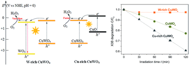
A hydrothermal reaction between Cu(NO3)2 and Na2WO4 at 170 °C, followed by sintering at 500 °C, resulted in the formation of stoichiometric CuWO4 at pH 5.2, while the reactions below and above pH 5.2 gave a mixture of CuWO4 with WO3 and CuO, respectively. For organic degradation in water under visible light, W-rich and Cu-rich samples were less and more active than CuWO4, respectively. A maximum activity was observed with the sample prepared at pH 8.5. Furthermore, this Cu-rich sample shown an activity greatly changing with its sintering temperature, and reaching a maximum at 600 °C. A possible mechanism responsible for the observed activity difference among the samples is proposed, involving the interfacial charge transfer between CuWO4 and WO3 or CuO.


Introduction

Heterogeneous photocatalysis for environmental remediation has been studied for many years.1–3 Among the various semiconductors, TiO2 has been widely studied, due to its low cost and high activity. However, TiO2 is active only in UV light, which would limit the utilization of solar energy. Recently, CuWO4 as a visible-light-driven photocatalyst has received increasing attention. This semiconductor can absorb light at wavelengths shorter than 540 nm.4 Under stimulated solar illumination, the CuWO4 films, fabricated by spin-coating, reactive cosputtering, spray pyrolysis and electrodeposition, can be used as the photoanodes for water oxidation to O2 at an applied potential bias.5–10 In addition, CuWO4 powders prepared by a co-precipitation method are also active for organic degradation in aerated aqueous solution under visible light.11–13 However, the observed photocatalytic activity of CuWO4 for organic degradation in water is very low, mainly due to fast recombination of the photogenerated charge carriers. In general, the photocatalytic activity of a semiconductor can be improved by increasing its crystallinity and surface area, and/or through surface modification with suitable co-catalysts. We have recently shown that deposition of CuWO4 with 1.8 wt% of CuO can increase the activity by 9 times under UV light, and 5 times under visible light for phenol degradation in water.13

In this article, we report on the hydrothermal synthesis of CuWO4, whose chemical composition, crystalline structure and photocatalytic activity are greatly influenced by the solution pH and sintering temperature used for the solid synthesis. Samples were prepared in aqueous solution at 170 °C with Cu(NO3)2 and Na2WO4 as precursors. Solids were characterized with several routine techniques, and their photocatalytic activities for organic degradation in water were measured under either UV or visible light. Furthermore, a possible mechanism responsible for the observed activity difference among the samples is discussed.

Experimental section

Reagents

Tungstate(VI) sodium dihydrate was purchased from Sinopharm Chemical Reagent Co., Ltd., and a textile azo-dye X3B (Reactive Orange 86) from Jining dye manufacture of China (pKa = 4.5). Copper(II) nitrate trihydrate, phenol and other chemicals in analytical grade were purchased from Shanghai Chemicals Inc., China, and used as received without further purification. The solution pH was adjusted with a dilute solution of NaOH or HCl. Milli-Q water was used throughout this work.
image file: c4ra13952f-u1.tif

Synthesis

Typically, the aqueous solutions of 0.10 M Na2WO4·2H2O and 0.10 M Cu(NO3)2·3H2O were first prepared. Then the former was added dropwise to the latter under magnetic stirring, followed by pH adjustment to a given value. After further stirring for 30 min, and sonication for 30 min, the suspension was transferred into a stainless autoclave, and heated at 170 °C for 20 h. After the reactor cooled down, the particles was collected by centrifuge, thoroughly washed with water, and dried at 60 °C overnight. Finally, the solid was sintered in air at a given temperature for 3 h.

Characterization

X-ray powder diffraction (XRD) pattern was recorded on a D/max-2550/PC diffractometer (Rigaku). According to the (100) diffraction, the crystal size of CuWO4 was calculated by using Scherrer equation. Adsorption isotherm of N2 on solid was measured at 77 K on a Micromeritics ASAP2020 apparatus, from which the Brunauer–Emmett–Teller (BET) surface area (SBET) and cumulative pore volume (Vp) were calculated. Scanning electron microscope (SEM) image was recorded on a Hitachi S-4800, attached with EDS (energy-dispersive X-ray spectroscopy). X-ray photoelectron spectroscopy (XPS) data were measured with Kratos AXIS UItra DLD spectrometer, and calibrated with C(1s) at 284.8 eV. Diffuse reflectance spectra were recorded on a Shimadzu UV-2550 with BaSO4 as a reference. The reflectance (R) is transformed to the Kubelka–Munk unit, F(R) = (1 − R)2/2R.

Photocatalysis

Reactions were carried out in a Pyrex-glass reactor, thermostated at 25 °C through a recycle system. Light sources were a 300 W high pressure mercury lamp (Shanghai Mengya) for UV light, and a 150 W Xenon lamp (USHIO) equipped with a 420 nm cut-off filter for visible light. The light intensity reaching the external surface of the reactor was 1.41, and 5.50 mW cm−2 for Hg and Xe lamps, respectively, measured with an irradiance meter (Beijing Normal University, China). The suspension (50 mL) containing 1.7 g L−1 catalyst and 0.066 mM X3B or 0.22 mM phenol was first stirred in the dark for 12 h, and then irradiated with UV or visible light. At given intervals, small aliquots were withdrawn, and filtered through a membrane. Dye concentration was analyzed at 533 nm on an Agilent 8451 spectrometer. Phenol concentration was measured with high performance liquid chromatography on a Dionex Ultimate 3000, equipped with Apollo C18 reverse column, and with 50% CH3OH aqueous solution as an eluent.

Results and discussion

Effect of synthesis pH

The solids were prepared from the reaction between Cu(NO3)2 and Na2WO4 in aqueous solution at a fixed temperature (170 °C), but at different pHs (1.1–11.6). Because of that, the samples were different not only in the chemical composition, but also in the crystal structures and optical properties. Element analysis by EDS showed that the mole ratio of Cu to W increased with the solution pH, the value of which was 0.05, 0.24, 0.25, 1.04, 1.86 and 3.00 for the samples prepared at pH 1.1, 2.4, 3.9, 5.2, 8.5 and 11.6, respectively. The relevant EDS spectra of the samples were showed in Fig. S1 (ESI). These results indicate that only the CuWO4 sample prepared at pH 5.2 was stoichiometric. Below and above this pH, the samples were W-rich and Cu-rich CuWO4, respectively. It is highly possible that the W-rich and Cu-rich sample are a mixture of CuWO4 with WO3 and CuO, respectively.

In order to confirm this hypothesis, the crystalline phase of the sample was analyzed by XRD. Fig. 1A shows the XRD patterns of the as-prepared samples. The solid obtained at pH 1.1 exhibited series of diffraction peaks mainly due to hexagonal WO3 (PDF # 33-1387), and partially due to orthorhombic WO3·0.33H2O (PDF # 35-0270). When the solid was prepared at pH 5.2, the peak intensities of WO3 and WO3·0.33H2O were greatly reduced, together with the formation of new crystalline phases of CuWO4·2H2O (PDF # 33-0503), and Cu2WO4(OH)2 (PDF# 34-1297).


image file: c4ra13952f-f1.tif
Fig. 1 XRD patterns of (A) the as-prepared solids at initial pHs of (a) 1.1, (b) 2.4, (c) 3.9, (d) 5.2, (e) 8.5, and (f) 11.6, and (B) the corresponding solids sintered at 500 °C. Note that h = hexagonal WO3, m = monoclinic WO3, t = triclinic CuWO4, (*) = WO3·0.33H2O, (○) = CuWO4·2H2O, (+) = tetragonal Cu2WO4(OH)2, and (#) = monoclinic CuO.

When the synthesis pH was further increased to 11.6, additional peaks at 2θ = 38.86 and 48.70° also appeared, due to the formation of monoclinic CuO (PDF # 65-2309). These observations indicate that in an acidic solution, WO42− prefers to react with H+ to produce H2WO4, followed by partial and complete dehydration to WO3·0.33H2O and WO3. In a basic solution, WO42− turns to react with Cu2+ to produce Cu2WO4(OH)2 and CuWO4·2H2O. In this case, Cu(OH)2 might be also formed, followed by dehydration to CuO.

After the above samples were sintered at 500 °C, there were great changes in the crystal structures and phase composition (Fig. 1B). With the sample prepared at pH 1.1, the original phases of hexagonal WO3 and WO3·0.33H2O completely disappeared, together with the formation of new phases of monoclinic WO3 (PDF # 43-1035) and triclinic CuWO4 (PDF # 21-0307). This observation indicates that there is a solid-state reaction between tungstate and cupric species present in the un-sintered samples. As the synthesis pH increased, the phase contents of WO3 and CuWO4 decreased and increased, respectively. Moreover, with the sample prepared at pH 11.6, the diffraction peaks due to crystalline CuO were also visible. Since the XRD technique has a certain detection limit, it follows that the phase contents of WO3 and CuO in the mixtures decrease and increase with the synthesis pH, respectively.

The solid composition that changes with the synthesis pH is also suggested by the diffuse reflectance spectra (Fig. 2). As the synthesis pH increased, not only the spectral onset of the sample shifted gradually from 480 nm toward a longer wavelength, but also the absorbance at given wavelength increased. In comparison, the spectral onset of pure WO3 and CuWO4 were located at 480 and 540 nm, respectively, while the absorption spectrum of black CuO expanded in the whole wavelength region (data not shown here). It becomes clear that increasing the synthesis pH leads to a gradual phase transition from WO3 to CuWO4 to CuO, the trend of which is in good agreement with that observed from XRD.


image file: c4ra13952f-f2.tif
Fig. 2 Diffuse reflectance spectra of the powders sintered at 500 °C. Samples were hydrothermally synthesized at different pHs of (a) 1.1, (b) 2.4, (c) 3.9, (d) 5.2, (e) 8.5, and (f) 11.6, respectively.

Photocatalytic activity of the sintered sample was evaluated under visible light by using X3B degradation as a model reaction, and the result is shown in Fig. 3. This azo-dye was stable against photolysis. In the presence of catalyst, the concentration of X3B in solution decreased with the irradiation time, indicative of the dye degradation. However, different catalysts showed different activities. A maximum rate of X3B degradation was observed with the catalyst prepared at pH 8.5. On the other hand, these catalysts were also different in their sorption capacities toward X3B in water. The amount of X3B adsorption (qe), measured in the dark, was negligible with the samples prepared at pH < 5.2, while the values of qe with the samples prepared at pH 5.2, 8.5, and 11.6 were 1.0, 2.4 and 8.1 μmol g−1, respectively. Since the trend in qe among the catalysts does not match that in the rate of dye degradation, it can concluded that W-rich and Cu-rich CuWO4 are less and more photoactive than CuWO4, respectively.


image file: c4ra13952f-f3.tif
Fig. 3 Photocatalytic degradation of X3B under visible light in aerated aqueous suspensions. Catalysts were prepared at different pHs of (a) 1.1, (b) 2.4, (c) 3.9, (d) 5.2, (e) 8.5, and (f) 11.6, followed by sintering at 500 °C in air for 3 h.

Effect of sintering temperature

Since Cu-rich sample is superior to W-rich sample, a further study has been made with the Cu-rich CuWO4 prepared at pH 8.5. Fig. 4 shows the XRD patterns of the samples sintered in air at different temperatures (Ts) for 3 h. At 300 °C, the sample was a mixture of CuWO4·2H2O and Cu2WO4(OH)2, similar to the un-sintered sample (curve e, Fig. 2A). However, at 400 °C, these phases of CuWO4·2H2O and Cu2WO4(OH)2 completely disappeared, together with the formation of CuWO4. As Ts further increased, the diffraction intensity of CuWO4 increased, indicative of the crystal growth. Moreover, at 800 °C, the crystalline phase of CuO was clearly observed. By using the Scherrer equation, the average crystallite size (dXRD) of CuWO4 for each sample was estimated, and the result is tabulated in Table 1. There is indeed a progressive increase in the primary crystallite size as Ts increases.
image file: c4ra13952f-f4.tif
Fig. 4 XRD patterns of the sample prepared at pH 8.5, and sintered in air for 3 h at different temperatures of (a) 300, (b) 400, (c) 500, (d) 600, (e) 700, and (f) 800 °C. The captions are the same as those in Fig. 1.
Table 1 Physical parameters of Cu-rich CuWO4 at different temperaturesa
Ts (°C) dXRD (nm) dSEM (nm) SBET (m2 g−1) Vp (cm3 g−1)
a n.d, not detectable; dXRD, crystallite size by XRD; dSEM, particle size by SEM; SBET, BET specific surface area; Vp, total pore volume.
170 n.d n.d 72.2 0.29
300 n.d n.d 53.0 0.27
400 25.4 28.1 25.3 0.23
500 30.3 34.1 17.3 0.14
600 38.2 43.7 12.2 0.06
700 >100 406.4 0.10 n.d
800 >100 3260 × 940 n.d n.d


The surface area (SBET) and pore volume (Vp) of the solid were measured by N2 adsorption at 77 K. As Ts increased, both the values of SBET and Vp decreased. Since N2 largely adsorbs onto the solid external surface, it follows that the particle size of the whole mixture also increases with Ts. To distinguish the crystal and particle growth, the SEM images were recorded with all the samples (Fig. 5). At low Ts, the particles were highly aggregated and opaque in appearance. As Ts increased, the contour between the particles became more and more clear. At the same time, the average grain size was significantly increased, from 28.1 nm at 400 °C, to 406.4 nm at 700 °C (Table 1). At 800 °C, the rod-like particles were produced, with an average size of 3.26 μm in length and 0.94 μm in width. These observations suggest that the primary crystallites of CuWO4 grow to big crystals on the thermal treatment. Moreover, there were also some small particles on the large particles (Fig. 5d). According to the crystallite size of CuWO4, the observed small and large particles are presumably assigned to CuO, and CuWO4, respectively.


image file: c4ra13952f-f5.tif
Fig. 5 SEM images of the sample prepared at pH 8.5, and sintered at (a) 400, (b) 500, (c) 600, (d) 700, and (e) 800 °C.

Fig. 6 shows the XPS spectra of W, O and Cu for the sample sintered at 600 °C. The binding energies of W(4f7/2), W(4f5/2) and W(5p3/2) at 35.4, 37.6 and 41.2 eV were in good agreement with the standard data for W6+.14 By using a Lorentzian–Gaussian function, the peak of O(1s) at 530.4 eV were deconvoluted into three peaks: W–O (530.1 eV), Cu–O (530.6 eV), and surface OH (531.6 eV).15–18 The peaks of Cu(2p3/2) and Cu(2p1/2) at 934.3 and 954.3 eV could be also deconvoluted into two components: 934.2, and 935.8 eV for Cu(2p3/2); 954.2, and 955.3 eV for Cu(2p1/2). The binding energies of Cu(2p3/2) at 934.2 eV and Cu(2p1/2) at 954.2 eV were similar to those reported with CuO,19–24 while the remaining peaks are due to Cu(II) in the lattice sites of CuWO4. In addition, there were two shakeup satellite peaks at 943 and 963 eV, typical of Cu(II) in cupric compounds.25 These observations indicate that this sample is a mixture of CuWO4 and CuO, and that only the chemical states of Cu(II) and W(VI) are present.


image file: c4ra13952f-f6.tif
Fig. 6 X-ray photoelectron spectra of W(4f), O(1s) and Cu(2p) for the sample prepared at pH 8.5, and sintered at 600 °C for 3 h. The colored curves correspond to deconvolution with a Lorentzian–Gaussian function.

Fig. 7A shows the results of X3B degradation in aerated aqueous suspension under visible light. With each catalyst, the concentration of X3B in solution decreased with the irradiation time. Among the catalysts, the rates of dye degradation were different. A maximum rate of dye degradation was observed with the sample sintered at 400 °C. However, these catalysts had different values of qe. Since the rate of dye degradation increase with its surface concentration, the observed rate should be normalized with qe for evaluation of the catalyst activity. This specific rate reached a maximum with the catalyst sintered at 600 °C. However, X3B is red in color, and its degradation may occur through dye sensitization, other than photocatalytic pathway.26 Because of that, it is better to use a colorless compound as a model substrate such as phenol. Fig. 7B shows the result of phenol degradation in water, measured under UV light for a fast data collection. Interestingly, the relative activity among the catalysts observed from phenol degradation under UV light was similar to that observed from dye degradation under visible light (Fig. 7A). A maximum activity was also obtained with the sample sintered at 600 °C. Such Ts-dependent activity observed with Cu-rich CuWO4 is similar to those reported with other semiconductor photocatalysts.2 With the increase of Ts, the increase of crystallinity (Fig. 5) and the decrease of surface area (Table 1) would benefit and disbenefit the photocatalytic reaction, respectively. As a result, the optimum Ts of 600 °C is observed with the Cu-rich CuWO4 (Fig. 7). As a reference, the commercial TiO2 of Degussa P25 was also used as a photocatalyst. Under visible light, this TiO2 was active for X3B degradation, due to the dye sensitization. But this activity of TiO2 is much lower than that of the Cu-rich CuWO4 sintered at 400–600 °C. Under UV light for phenol degradation, TiO2 was always more active than that obtained with any of Cu-rich CuWO4 samples.


image file: c4ra13952f-f7.tif
Fig. 7 (A) Rate constant (kobs) and qe-normalized kobs for X3B degradation in aqueous solution under visible light. (B) Rate constant of phenol degradation in aqueous solution under UV light. The catalysts were prepared at pH 8.5, followed by sintering in air for 3 h at (a) 100, (b) 300, (c) 400, (d) 500, (e) 600, (f) 700, (g) 800 °C, and (h) commercial P25.

Possible mechanism

It has been reported that the conduction band edge potentials (ECB) for CuO, CuWO4, and WO3 in water at pH 0, are −0.03, +0.20, and +0.30 V versus normal hydrogen electrode (NHE), respectively.13,27 All these values of ECB are more positive than the one-electron reduction potential of O2 (−0.05 V vs. NHE). Then, in thermodynamics, the one-electron reduction of O2 would be not allowed, and consequently organic degradation on these photocatalysts was inactive, as observed with WO3 and CuO.13,28 However, in the aerated aqueous suspension of CuWO4, organic degradation could occur under UV or visible light.11–13 In this case, the photocatalytic generation of H2O2 is also observed, ascribed to the two-electron reduction of O2 over the irradiated CuWO4.13

In thermodynamics, the electron transfer from CuWO4 to WO3, and the electron transfer from CuO to CuWO4 are both possible (Scheme 1). However, the transferred electron on WO3 hardly reacts with O2 to form HO2 or H2O2.28,29 This may explain the observation that W-rich CuWO4 is less active than CuWO4 for organic degradation in an aerated aqueous suspension (Fig. 3). On the contrary, the transferred electron on CuWO4 can be captured by O2 to form H2O2. As a result, the efficiency of charge separation is improved, and the rate of organic degradation is enhanced. However, at a high Cu/W ratio of 3.0, Cu-rich CuWO4 becomes to be less active. Since black CuO is nearly not active, the decreased activity of Cu-rich CuWO4 is due to the decreased concentration of net CuWO4 in the reactor, and due to the reduced number of photons reaching CuWO4, and/or both.


image file: c4ra13952f-s1.tif
Scheme 1 Band structures of WO3, CuO and CuWO4 in aerated aqueous suspension and possible interfacial transfers between two semiconductors.

Conclusion

In this work, CuWO4 has been synthesized in aqueous medium at 170 °C through a hydrothermal reaction between Cu2+ and WO42−. However, due to the pH-dependent hydrolysis of precursors, this material is easily contaminated by WO3 or CuO. The optimal pH for the synthesis of stoichiometric CuWO4 is around 5.2. On the other hand, these composite materials have different activities for organic degradation in aerated aqueous solution under UV and visible light. Comparatively, W-rich and Cu-rich CuWO4 are less and more active than CuWO4, respectively. Furthermore, the photocatalytic activity of Cu-rich CuWO4 is also a function of its sintering temperature, mainly ascribed to the combined effects of crystallinity and surface area. The present result would be useful to further development of the CuWO4-based photocatalysts for environmental use, and for water splitting as well.

Acknowledgements

This work was supported by the 973 program of China (no. 2011CB936003) and NSFC (no. 21377110).

Notes and references

  1. M. R. Hoffmann, S. T. Martin, W. Choi and D. W. Bahnemann, Chem. Rev., 1995, 95, 69 CrossRef CAS.
  2. O. Carp, C. L. Huisman and A. Reller, Prog. Solid State Chem., 2004, 32, 33 CrossRef CAS PubMed.
  3. T. L. Thompson and J. T. Yates, Chem. Rev., 2006, 106, 4428 CrossRef CAS PubMed.
  4. R. Lacomba-Perales, J. Ruiz-Fuertes, D. Errandonea, D. Martínez-García and A. Segura, EPL, 2008, 83, 37002 CrossRef.
  5. J. E. Yourey, K. J. Pyper, J. B. Kurtz and B. M. Bartlett, J. Phys. Chem. C, 2013, 117, 8708 CAS.
  6. J. C. Hill and K. S. Choi, J. Mater. Chem. A, 2013, 1, 5006 CAS.
  7. J. E. Yourey and B. M. Bartlett, J. Mater. Chem., 2011, 21, 7651 RSC.
  8. J. E. Yourey, J. B. Kurtz and B. M. Bartlett, J. Phys. Chem. C, 2012, 116, 3200 CAS.
  9. Y. Chang, A. Braun, A. Deangelis, J. Kaneshiro and N. Gaillard, J. Phys. Chem. C, 2011, 115, 25490 CAS.
  10. N. Gaillard, Y. Chang, A. DeAngelis, S. Higgins and A. Braun, Int. J. Hydrogen Energy, 2013, 38, 3166 CrossRef CAS PubMed.
  11. T. Montini, V. Gombac, A. Hameed, L. Felisari, G. Adami and P. Fornasiero, Chem. Phys. Lett., 2010, 498, 113 CrossRef CAS PubMed.
  12. U. M. García-Péreza, A. Martínez-de la Cruz and J. Peral, Electrochim. Acta, 2012, 81, 227 CrossRef PubMed.
  13. H. Chen and Y. Xu, J. Phys. Chem. C, 2014, 118, 9982 CAS.
  14. S. Penner, X. Liu, B. Klotzer, F. Klauser, B. Jenewein and E. Bertel, Thin Solid Films, 2008, 516, 2829 CrossRef CAS PubMed.
  15. J. H. Pan and W. I. Lee, Chem. Mater., 2006, 18, 847 CrossRef CAS.
  16. M. R. Bayati, F. Golestani-Fard, A. Moshfegh and Z. R. Molaei, Mater. Chem. Phys., 2011, 128, 427 CrossRef CAS PubMed.
  17. S. Poulston, P. M. Parlett, P. Stone and M. Bowker, Surf. Interface Anal., 1996, 24, 811 CrossRef CAS.
  18. M. Takeuchi, Y. Shimizu, H. Yamagawa, T. Nakamuro and M. Anpo, Appl. Catal., B, 2011, 110, 1 CrossRef CAS PubMed.
  19. G. Moretti, G. Fierro, M. L. Jacono and P. Porta, Surf. Interface Anal., 1989, 14, 325 CrossRef.
  20. G. Deroubaix and P. Marcus, Surf. Interface Anal., 1992, 18, 39 CrossRef CAS.
  21. T. Robert, M. Bartel and G. Offergeld, Surf. Sci., 1972, 33, 123 CrossRef CAS.
  22. B. R. Strohmeier, D. E. Leyden, R. S. Field and D. M. Hercules, J. Catal., 1985, 94, 514 CrossRef CAS.
  23. F. Parmigiani, G. Pacchioni, F. Illas and P. S. Bagus, J. Electron Spectrosc. Relat. Phenom., 1992, 59, 255 CrossRef CAS.
  24. J. G. Jolley, G. G. Geesey, M. R. Hankins, R. B. Wright and P. L. Wichlacz, Appl. Surf. Sci., 1989, 37, 469 CrossRef CAS.
  25. T. L. Hsiung, H. P. Wang, Y. Lu and M. C. Hsiao, Radiat. Phys. Chem., 2006, 75, 2054 CrossRef CAS PubMed.
  26. K. Lv and Y. Xu, J. Phys. Chem. B, 2006, 110, 6204 CrossRef CAS PubMed.
  27. J. R. Darwent and A. Mills, J. Chem. Soc., Faraday Trans., 1982, 78, 359 RSC.
  28. J. Kim, C. W. Lee and W. Choi, Environ. Sci. Technol., 2010, 44, 6849 CrossRef CAS PubMed.
  29. J. Sheng, X. Li and Y. Xu, ACS Catal., 2014, 4, 732 CrossRef CAS.

Footnote

Electronic supplementary information (ESI) available: Details of EDS spectra. See DOI: 10.1039/c4ra13952f

This journal is © The Royal Society of Chemistry 2015