The art in science of MicroTAS: the 2014 issue†
Darwin R.
Reyes
National Institute of Standards and Technology (NIST), Gaithersburg, MD, USA. E-mail: darwin.reyes@nist.gov
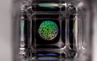 | ||
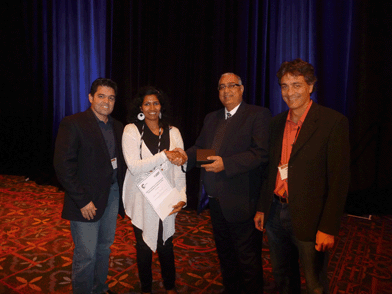
| Fig. 2 MicroTAS Art in Science 2014 award winner “The Sphere”, submitted by David Castro of the Department of Electrical Engineering, KAUST (Saudi Arabia). | ||
Since the establishment of the Art in Science Award 7 years ago, the Lab on a Chip journal has portrayed on its cover some of the most impressive images from the MicroTAS conference. The images shown on the cover and in the editorials that announce the winners are considered by many to have great artistic value. It seems as though in the same way that microfluidics have found a scientific niche at a dimensional scale where different than the usual laws dominate, the art that is found at the microfluidic “scale” can produce artistic features that are exclusively seen at this scale. One common example of a microfluidic feature is the common laminar flow. This type of flow can be tweaked in many different ways so that many images of great artistic value can be generated with a single device. This, and the constant pursuit of new types of devices by the scientific community makes the microTAS Art in Science award competition more challenging each year and at the same time an invaluable source of art.
The selection committee for the 2014 Art in Science award included representatives from industry, academia, government and scientific publishers. This year's committee was composed of Omar Jina from Dolomite, Prof. Albert Folch from the University of Washington, Darwin Reyes from NIST and Harpal Minhas from LOC/Royal Society of Chemistry. Several criteria were taken into account when considering each of the submissions. Specifically, the submissions were judged for their originality, scientific merit, visual appeal, and suitability for a Lab on a Chip front cover. As in previous years, the selection committee had a difficult task when scrutinizing the images to select the finalists and the winner of the award. The judges first agreed on the top four submissions, followed by deliberations and a final vote that ultimately decided the selection of the winner. The top 3 runners-up for the 2014 Art in Science award are as follows:
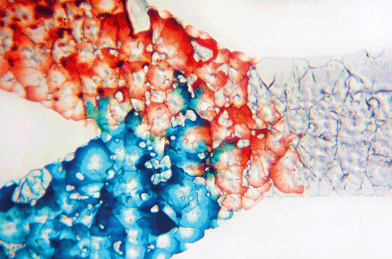
1st runner up - “Wicking glass channels” by Manuel Ochoa of Purdue University (Fig. 3).
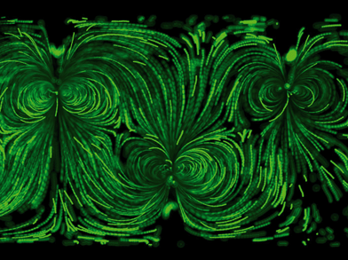
2nd runner up - “Acoustic Streaming Effects” by Po-Hsun Huang of Pennsylvania State University (Fig. 4).
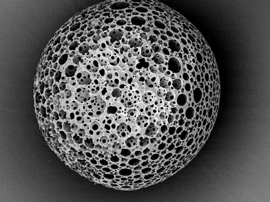
3rd runner up - “Scanning electron micrograph of a highly porous polymer” by Florian Lapierre of Royal Melbourne Institute of Technology (RMIT) (Fig. 5).
Acknowledgements
The Art in Science award is sponsored and supported by MicroTAS, the Chemical and Biological Microsystems Society (CBMS), the Lab on a Chip journal, and NIST. The award consists of a monetary prize ($2,500), an award certificate, and the coveted front cover of the Lab on a Chip journal. Please check the MicroTAS 2015 conference website for further details regarding the submission of images for the next MicroTAS Conference in Gyeongju, Korea.Reference
- S. Sivashankar, D. Castro, U. Buttner and I. G. Foulds, Real-Time Agglutination within a Microdroplet in a Three Phase Fluidic Well for Detection of Biomarkers, MicroTAS, 2014, 2097–2100 Search PubMed.
Footnote |
| † Any opinions or views expressed in the following article are entirely those of the author and do not represent the views of the journal, Lab on a Chip, or The Royal Society of Chemistry. |
| This journal is © The Royal Society of Chemistry 2015 |

