Sourav Ghosha,
Rituparna Dasa,
Ipsita Hazra Chowdhurya,
Piyali Bhanjab and
Milan Kanti Naskar*a
aSol-Gel Division, CSIR-Central Glass and Ceramic Research Institute, Kolkata 700 032, India. E-mail: milan@cgcri.res.in; Fax: +91 33 24730957
bIndian Association for the Cultivation of Science, Kolkata 700 032, India
First published on 13th November 2015
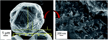
A hierarchical copper nanoassembly was synthesized by one-pot solvothermal treatment at 150 °C for 2 h in the presence of copper nitrate, formamide and water. The product exhibited phase pure hierarchical Cu microspheroids (2–7 μm) comprising a nanorod (50–100 nm) assembly. The Cu microspheroids showed excellent air-stability, antioxidative properties and catalytic reduction of p-nitrophenol.
It is well-known that in several catalytic processes Cu2O and/or CuO are the active species.29,30 However, there are few reports on Cu nanoparticles as catalysts due to their susceptibility to oxidation.13 The oxidation of Cu is enhanced with a reduction in particle size. Therefore, the stabilization of metallic Cu nanostructures in air is a challenge toward their application in catalysis. It is worth mentioning that larger surface area nanoparticle (NP) supported catalysts have some limitation in catalytic applications in terms of agglomeration at large loadings, a tendency to react with supporting oxides at elevated temperature, and cumbersome processing routes.31,32 Keeping these views in mind, in the present study, we have approached the preparation of air stable unsupported Cu nanostructures for their catalytic application in 4-NP reduction.
In this present communication, we report a template-free synthesis of a copper nanoassembly using a rapid autoclaving method at 150 °C for a shorter period of time (2 h) in the presence of copper nitrate, water and formamide without any post-synthetic heat treatment. Formamide played a deliberate role as a weak coordinating solvent as well as a source of reducing agent in controlling the particle architectures. The thermal behavior of the copper nanoassembly was also studied to perceive its antioxidative properties. To the best of our knowledge, the synthesis of an air-stable copper nanostructure using formamide as a source of reducing agent using a rapid autoclaving method has been reported for the first time. The presented method of fabrication for air-stable copper nanostructures is important in terms of it being a cost-effective, template free method with a low processing time. The catalytic performance of the synthesized products was studied for the reduction of 4-NP to 4-AP in the presence of sodium borohydride (NaBH4) at room temperature (25 °C).
All reagents were of analytical grade and used without further purification. In a typical experiment, 10 mmol of Cu(NO3)2·3H2O was dissolved in 10 mL of deionized (DI) water under stirring for 30 min at room temperature. 200 mmol of formamide was added dropwise into the former solution. The mixed solution was allowed to stir for 30 min to obtain a homogeneous solution. The above solution was transferred into a 35 mL Teflon-lined autoclave followed by a hydrothermal reaction at 150 °C for 2 h. After the reaction, the particles were collected by centrifugation and washed with ethanol followed by drying at room temperature overnight.
X-ray diffraction (XRD) studies of the as-prepared samples were performed with a Philips X’Pert Pro PW 3050/60 powder diffractometer using Ni-filtered Cu-Kα radiation (λ = 0.15418 nm) operating at 40 kV and 30 mA. X-ray photoelectron spectroscopy (XPS) measurements were carried out using a PHI 5000 Versaprobe II Scanning XPS microprobe manufactured by ULVAC-PHI, USA. The spectra were recorded with monochromatic Al Kα (hν = 1486.6 eV) radiation with an overall energy resolution of ∼0.7 eV. H2-TPR experiments were carried out in the temperature range of 25 to 750 °C using a Micromeritics Chemisorb 2720 instrument, under a H2 atmosphere. In a typical experiment, 245 mg of sample was placed in a U-type quartz cell followed by purging with helium gas throughout the sample for 3 h. It was cooled down to room temperature followed by purging with reducing gas (5% H2 + 95% argon) at a flow rate of 30 mL min−1 for saturation. The H2-TPR profile of the sample was acquired using a thermal conductivity detector (TCD) by elevating the temperature with a 5 °C ramp per minute.33 The thermal behaviors of the as-prepared powders were studied using thermogravimetry (TG) and differential thermal analysis (DTA) (Netzsch STA 449C, Germany) from room temperature to 600 °C under an air atmosphere at the heating rate of 10 °C min−1. The morphology of the particles was examined using field emission scanning electron microscopy, FESEM, with a Zeiss, Supra™ 35VP instrument operating with an accelerating voltage of 10 kV, and transmission electron microscopy, TEM using a Tecnai G2 30ST (FEI) instrument operating at 300 kV. Nitrogen adsorption–desorption measurements were conducted at 77 K with a Quantachrome (ASIQ MP) instrument.
For the catalytic study, the desired amount of the catalyst sample was introduced into a reaction mixture containing 0.1 mL of 3.0 × 10⁻³ M 4-NP, 2.8 mL of water and 0.1 mL of 3.0 × 10⁻¹ M NaBH4 solution at room temperature (25 °C). The molar ratio of 4-NP:NaBH4 was 1:102. The progress of the catalytic performance was evaluated from the absorption spectra at 400 nm for the decay of the 4-NP peak using a UV-Vis-NIR spectrophotometer (V-730/730 BIO, JASCO) in the wavelength range of 250 to 500 nm. As the concentration of NaBH4 was employed in excess, the reaction was considered to be pseudo-first order, and the apparent rate constant (Kapp) was estimated by plotting −ln(At/A0) vs. time. To check the recyclability of the catalyst, the sample was washed with DI water, and dried at room temperature. Then the catalyst was ready to re-use the next time.
Fig. 1 shows the XRD patterns of (a) the as-prepared Cu and (b) the same sample after exposure to open air for 12 months. The crystalline peaks at 2θ = 43.29°, 50.43° and 74.13° for both the samples were assigned to the (111), (200) and (220) lattice planes, respectively of face-centered cubic (fcc) Cu (JCPDS no. 04-0836). For all the cases, the diffraction peaks of other possible impurities such as Cu2O and CuO could not be detected. This signified that the as-prepared phase pure metallic Cu remained intact even after 12 months of open air exposure. This indicated the excellent oxidation resistance of the as-prepared Cu obtained via one-pot solvothermal treatment at 150 °C for a shorter period of time (2 h) without any post-synthetic heat treatment. The chemical composition and chemical state of the as-prepared dried sample were studied using high-resolution XPS.
Fig. 1 XRD patterns of (a) the as-prepared Cu, and (b) the same sample after exposure to open air for 12 months. |
Fig. 2 reveals XPS spectra of (a) Cu 2p and (b) O 1s for the as-prepared Cu. Major contributions from Cu 2p3/2 at 932.68 eV and Cu 2p1/2 at 952.59 eV confirmed the presence of zero valent copper.21 From the O 1s spectrum, a high binding energy component at 531.6 eV could be in good agreement with the presence of a hydroxyl group.34 The outermost surface of the as-prepared sample indicated no impurity from divalent Cu (530.4 eV) from the O 1s spectrum.35 Combining both the Cu 2p and O 1s spectra, it is determined that the as-prepared sample was zero valent Cu in the absence of its oxidized forms. Comparing the spectral characteristics with commercial Cu2O and CuO,36 the absence of two main peaks at 954 eV (Cu 2p1/2) and 934 eV (Cu 2p3/2) along with shake-up satellite peaks centered at ∼943 eV confirmed the absence of CuO in the as-prepared sample. From the O 1s spectrum, the possibility of Cu2O formation in the sample could be completely eliminated due to absence of a lower energy peak at 530.3 eV.37 However, to confirm the phase pure metallic Cu in the sample, and to determine different oxidation states of Cu in the reference oxides (Cu2O and CuO), H2-TPR experiments were performed. Fig. S1, ESI† shows the H2-TPR profiles of as-prepared Cu, with commercial Cu2O and CuO as reference materials. It is seen that a large H2 consumption peak is located at around 453 °C for Cu2O,38 whereas a very weak peak appears at around 188 °C for the as-prepared Cu sample. However, the reference CuO shows a major reduction peak at around 478 °C.39,40 The H2 consumption peak at a relatively lower temperature (188 °C) for as-prepared Cu could be assigned to the reduction of dispersed Cu2+ species on the metallic Cu catalyst surface to metallic Cu.41 From the H2-TPR studies, it was confirmed that the as-prepared Cu particles could contain a small amount of Cu2+ species on their surface, revealing a lower reduction temperature compared to that found in the reference copper oxides.
Fig. 3a and b show FESEM images of the as-prepared samples. They reveal microspheroid particles of a size range of 2–7 μm. From the higher magnification image of the particle it is obvious that a large number of nano-rod (50–100 nm) particles self-assembled to form a microspheroid morphology (Fig. 3b). The formation of the microspheroid particles composed of nano-rod assemblies will be explained shortly. The TEM images (Fig. 3c–e) reveal how hierarchical copper microspheroids were formed through the self-assembly of copper nanorods. It was also noticed that the nanorod particles were formed via the oriented growth of smaller nanoparticles (10–20 nm) (Fig. 3e). Therefore, it is pointed out that metallic copper microspheroids were formed through the self-assembly of nanoparticles with hierarchical structures. The selected area electron diffraction (SAED) pattern confirmed the polycrystalline nature (Fig. 3f) of the aggregated nanoparticles (marked as 1 in Fig. 3d), while a single crystalline pattern (Fig. 3g) was noticed from a nanorod particle (marked as 2 in Fig. 3e). It can be concluded that the primary smaller nanoparticles fused together with a common crystallographic orientation to form copper nanorods following an “oriented attachment” growth mechanism.42,43 Further, the nanorod particles self-assembled to form a micro-spheroid morphology. The HRTEM image shows the lattice fringes of copper nanoparticles with d-spacing of 0.22 nm corresponding to the (111) plane (Fig. 3h), which was confirmed by the FFT pattern (inset of Fig. 3h).
Fig. 3 (a and b) FESEM images, (c–e) TEM images, (f and g) SAED patterns and (h) HRTEM of the as-prepared samples. |
The anti-oxidative properties of the Cu nanoassembly were investigated using thermogravimetry (TG) and differential thermal analysis (DTA). Fig. 4 shows the DTA-TG investigation of the as-prepared Cu. The TG study indicated that the copper nanoassembly was stable in air up to 255 °C revealing no residual mass change. In the DTA analysis, two exothermic peaks were observed at about 275 °C and 490 °C. The first exothermic peak (275 °C) was accompanied by a small amount of mass gain of 0.6% in the temperature range of 255–310 °C. This is attributed to a very small fraction of Cu being transformed to Cu2O,13,44 indicating the high stability of Cu. However, the second exothermic peak at 490 °C, accompanying a mass gain of 17.1% in the temperature range 310–600 °C, could suggest the oxidation of Cu to Cu2O and/or CuO, and Cu2O to CuO. It is to be pointed out that theoretically a 12.5% mass gain occurs for each conversion of Cu to Cu2O and Cu2O to CuO, and a 25% mass gain takes place for the conversion of Cu to CuO. Therefore, the above results indicated that the broad exothermic peak at around 490 °C could be attributed to oxidations of Cu to Cu2O and/or CuO, and Cu2O to CuO. From the above results it is clear that the synthesized product displayed excellent antioxidative properties below 255 °C.
To investigate the formation mechanism of the synthesized hierarchical Cu nanoassembly, a time dependent solvothermal reaction was carried out at 150 °C for 75, 90, 100, 110 and 120 min. It was observed that Cu particles just started forming after 90 min of reaction, which is evidenced from the optical images in Fig. S2 (ESI†), indicating no particle formation for 75 min of reaction (a bluish colored solution appeared). From the XRD results, it is clear that for a reaction time of 90–110 min, the as-prepared samples show the formation of metallic Cu (Fig. S3, ESI†) as was found for a reaction time of 120 min (Fig. 1). The morphological evolution of the products with an increase in reaction time is shown in Fig. 5. Under solvothermal conditions, formamide is hydrolyzed producing formic acid and ammonia45 following the reaction shown below. Formic acid acts as the reducing agent to convert Cu2+ to metallic Cu.
HCONH2 + H2O → HCOOH + NH3 | (1) |
Cu2+ + 4NH3 → [Cu(NH3)4]2+ | (2) |
[Cu(NH3)4]2+ + 2OH− +HCOOH → Cu + CO2 + 4NH3 + 2H2O | (3) |
Fig. 5 Morphological evolution of the as-prepared Cu particles with an increase in reaction time at 150 °C. |
The growth of the particles in solution occurs via nucleation followed by coarsening and aggregation of the primary particles. For 90 min of reaction, the primary particles self-assembled together via an oriented attachment growth mechanism to form rod-like Cu nanostructures in the presence of some aggregated copper nanoparticles. TEM images (Fig. S4, ESI†) show that with an increase in the reaction time from 90 to 100–120 min the nanorod-like copper particles further self-assembled with each other forming copper microspheroids. It is interesting to notice that the nanorod particles protruded from the surface of the microspheroids. However, such protrusion became lower with an increase in the reaction time from 100 to 120 min, and accordingly the aspect ratio of the nanorods got shorter (Fig. S5, ESI†). The formation of microspheroids with a nanorod-assembly is caused due to a reduction in the total energy by removing the surface energy associated with unsatisfied bonds via the elimination of the solid–air or solid–liquid interfaces.46 As a result entropy is enhanced significantly, preferring aggregation across the surface to form a close-packed spheroid morphology, thus minimizing the total surface area. The textural properties of the as-prepared samples synthesized at 150 °C for 120 min show a BET surface area, total pore volume and average pore diameter of 0.3762 m2 g−1, 0.0021 cm3 g−1 and 22.82 nm, respectively, which were estimated from N2 adsorption–desorption isotherms and pore size distributions (Fig. S6, ESI†). It is worth mentioning that the surface of the copper nanoassembly was not protected by any organic coupling agent, which could protect it from oxidation. However, in the absence of any foreign protective agent, the presented synthesized products exhibited remarkable antioxidative properties for copper nanostructures even after 12 months exposure at ambient temperature, and up to 255 °C in air.
In the present study, the catalytic efficiency of the synthesized Cu nanoassembly for the reduction of 4-NP to 4-AP in the presence of excess amounts of NaBH4 was investigated. 4-NP generally displays an absorption peak at 317 nm in neutral medium or acidic medium.47 In the presence of NaBH4, the absorption peak shifts to 400 nm due to the deprotonation of the hydroxyl group of 4-NP indicating the formation of the 4-nitrophenolate ion under the basic conditions. The reduction of the 4-nitrophenolate ion peak at 400 nm, and the simultaneous increase of the absorption peak of 4-AP at 300 nm is shown in Fig. 6a. The pseudo-first order rate constant was calculated as 0.113 min−1 for 1 mg of catalyst at room temperature from the logarithmic plot of the absorbance (−lnAt/A0) vs. reaction time (Fig. 6b). Table 1 shows the time of completion of the catalytic reaction, apparent rate constant and R2 value for different amounts of catalyst. Interestingly, with an increase in the catalyst amount from 1 to 3 mg, the apparent rate constant value increased from 0.113 min−1 to 0.279 min−1, however, it suddenly dropped to 0.239 min−1 for 4 mg of catalyst (Fig. S7–S9, ESI†). The R2 value (close to unity) could be in good agreement with the assumptions of pseudo-first-order kinetics.23 Therefore, 3 mg of catalyst was considered as the optimum amount for complete catalytic reduction with the highest apparent rate constant value of 0.279 min−1 (Fig. S10, ESI†). The catalyst can be reused at least three times with similar catalytic efficiency (Fig. 6c). The catalytic efficiency of the as-prepared Cu-nanoassembly was compared with that of literature reports, and also with the reference oxides (Cu2O and CuO) with 1 mg of catalyst each. Fig. S11 and S12, ESI† show (a) UV-Vis spectra and (b) pseudo-first order plots of the reference Cu2O and CuO, exhibiting less catalytic efficiency than the as-prepared Cu nanoassembly. The activity parameter κ (rate constant per unit g of catalyst) of the as-prepared Cu catalyst was calculated as 113 min−1 g−1, which was found to be higher than reported literature values (Table S1, ESI†),48–53 and the values for the reference oxides (Table S2, ESI†). This demonstrates that the Cu-nanoassembly shows enhanced catalytic efficiency with a faster reaction rate.
SI no. | Amount of catalyst (mg) | Time (min) | Kapp (min−1) | R2 |
---|---|---|---|---|
1 | 1 | 22 | 0.11300 | 0.99015 |
2 | 2 | 7 | 0.20499 | 0.99340 |
3 | 3 | 5 | 0.27933 | 0.99316 |
4 | 4 | 6 | 0.23964 | 0.99557 |
The catalytic reaction mechanism is shown schematically in Fig. 7. During a catalytic reaction, the coadsorption of both the donor BH4− ions from NaBH4 and the acceptor 4-NP molecules takes place on the catalyst surface (Cu-nanoassembly) via chemical adsorption.54 However, NaBH4 can transfer hydrogen species on catalytic surfaces under aqueous conditions.55 In this case, the Cu catalyst behaves as a hydrogen shuttle for the reduction of 4-NP to 4-AP followed by a desorption process.
Footnote |
† Electronic supplementary information (ESI) available. See DOI: 10.1039/c5ra16644f |
This journal is © The Royal Society of Chemistry 2015 |