Sample-pretreatment-free based high sensitive determination of aflatoxin M1 in raw milk using a time-resolved fluorescent competitive immunochromatographic assay

Xiaoqian Tang abcde, Zhaowei Zhangabc, Peiwu Li*abcde, Qi Zhang*ac, Jun Jiangabcde, Du Wangae and Jiawen Leiabcde
aOil Crops Research Institute of the Chinese Academy of Agricultural Sciences, Wuhan 430062, P. R. China. E-mail: peiwuli@oilcrops.cn; zhangqi01@caas.cn; Fax: +86 27 86812862; Tel: +86 27 86812943
bKey Laboratory of Biology and Genetic Improvement of Oil Crops, Ministry of Agriculture, Wuhan 430062, P. R. China
cKey Laboratory of Detection for Mycotoxins, Ministry of Agriculture, Wuhan 430062, P. R. China
dLaboratory of Risk Assessment for Oilseeds Products (Wuhan), Ministry of Agriculture, Wuhan 430062, P. R. China
eQuality Inspection and Test Center for Oilseeds Products, Ministry of Agriculture, Wuhan 430062, P. R. China

Received 10th October 2014 , Accepted 20th November 2014

First published on 21st November 2014


Abstract

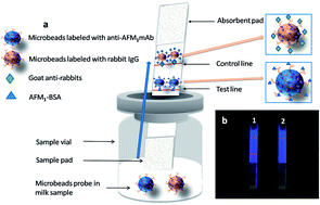
A highly-sensitive time-resolved fluorescent immunochromatographic assay (TRFICA) was developed to detect aflatoxin M1 (AFM1) in raw milk samples within 6 minutes without any sample pretreatments. This method could meet the requirement for rapid and sensitive milk monitoring in dairy farms and milk industries. Based on a competitive format and the home-made monoclonal antibody 2C9 against AFM1, this assay enhanced the sensitivity from 0.3 ng mL−1 (by using nanogold-strip method previously reported) to 0.03 ng mL−1 (by using this TRFICA method). The improved sensitivity could probably result from the increases in both the higher affinity of monoclonal antibody 2C9 against AFM1 and detection signals of immunoassay probes (with each europium microbead coupled with numbers of 2C9 antibodies). The TRFICA method showed a considerable dynamic range of 0.1–2.0 ng mL−1 and spiked recoveries of 80–110% for AFM1 quantification in raw milk. The results via TRFICA method was found to be high consistency (R2 = 0.988) with results via standard high-performance liquid chromatography (HPLC) method, when detecting AFM1 in 17 blind milk samples.


Introduction

Aflatoxins are highly toxic mycotoxins, naturally produced by Aspergillusflavus, Aspergillusparasiticus. Among the major aflatoxins B1, B2, G1, G2, M1, Aflatoxin M1 (AFM1), a major metabolic product of aflatoxin B1 (AFB1), is excreted from lactating animals that ingest feed contaminated with AFB1.1 The formation of AFM1 comes from the hydroxylated derivative of AFB1 in the liver via P450 cytochrome enzymes and is secreted into the milk through the mammal. It is found that AFM1 derivatives can be determined in milk within 12–24 hours after AFB1 intake.2 Because of its extremely high, chronic, acute toxicity, AFM1 has been classified as group 2B human carcinogens. AFM1 can be extensively found in milk and milk products in both developed and developing countries,3,4 threatening consumers' health. Therefore, most countries and districts, such as European Union (EU), China, United States (US), etc., have set the maximum residue levels (MRLs) for AFM1. The current MRLs of AFM1 in milk is 0.5, 0.5, 0.05 μg L−1 in milk set by US, China, and EU, respectively.5–8

Numbers of detecting methods for AFM1 have been developed, such as HPLC-FLD,9,10 HPLC-MS (-MS/MS),11 ELISA,12,13 colloid gold immunochromatographic assay,14,15 immunochip.16 Although their accuracy and sensitivity, the use of HPLC-FLD and HPLC-MS (-MS/MS) require specific high-cost instruments and skilled operators, and they are rather time/labor-consuming. For the developing countries, these methods could not be extensively employed in daily life to ensure the milk safety. Moreover, with the emerging trend of the in-field AFM1 detection, there is a request to simplify the sample preparation of raw milk. The current sample preparation of raw milk is rather complicated, suggesting that the instruments-based determination method could not be suitable for in-field AFM1 determination, especially for dairy farms and industries. In this regard, it is urgently required to develop a rapid, in-field, and sensitive quantification for AFM1 in milk. As an emerging advanced rapid assay method, immunochromatographic strip based on gold nanoparticle, quantum dots has been employed in AFM1 determination due to its sensitivity, rapidness, and reliability,15 allowing a limit of detection of 1.0 ng mL−1,15 0.3 ng mL−1,17 and 0.1 ng mL−1 (ref. 14) for AFM1 detection, respectively. However, these sensitivity and practicability could not still meet the request of high sensitive, stabile in-field detection of AFM1 in milk.

To enhance immunochromatographic sensitivity and quantification, we developed a time-resolved fluorescent immunochromatographic assay (TRFICA) for AFM1 in raw milk without sample pretreatments. Instead of nanogold particles, 190 nm-based europium microbeads was employed in prepare high-affinity antibody probes. This TRFICA method combined the advantages of immunochromatographic assay and time-resolved fluorescence for AFM1 detection. Based on a competitive format, this assay format has unique properties, compared with traditional time-resolved fluorescent detection.18 In the TRFICA method, the total internal reflection fluorescence time-resolved luminescence results in high specific signals with lower background noises, larger Stokes shifts, narrower emission bands and longer fluorescence lifetimes.19,20 It could suggest that this TRFICA method pose potential application of determination other food toxins.

Experimental

Instrument

AnXYZ3050Dispensing Platform, CM4000 Guillotine Cutter and LM4000 Batch Laminator (Bio Dot, Irvine, CA, USA) were used to prepare test strips. The vacuum freeze drier was obtained from Thermo Electron Corporation (Rockford, IL, USA). The ultraviolet spectrum was obtained using a Spectra Max M2e microplate reader (Molecular Devices Corp., Sunnyvale, CA, USA). The high-speed freezing centrifuge (CF16RX) was from Hitachi (Tokyo, Japan). Nitrocellulose membranes, sample pads, and absorbent pads were purchased from Millipore Corp. (Bedford, MA, USA). Sonicator 3000 was from Misonix (USA). A home-made portable fluorescence spectrophotometer was employed, including a Xe lamp with a clock-pulse generator, a side-window photomultiplier tube, an interference band-pass filters, a rapid preamplifier-discriminator and pulse counter, and a readout component (data not shown).

Reagents

1-Ethyl-3-(3-dimethylaminopropyl)carbodiimide (EDC), boric acid, rabbit immunoglobulin (IgG), goat anti-rabbit IgG and bovine serum albumin (BSA) were all purchased from Sigma-Aldrich and then directly used without processing. Anti-aflatoxin M1 monoclonal antibody (mAb) 2C9 was produced in our laboratory, and the mAb 2C9 exhibited high affinity for AFM1 of 1.74 × 10⁹ L mol−1, its competitive ELISA's IC50 (50% inhibition concentration of AFM1) was 0.067 ng mL−1, and its cross-reactivity to aflatoxin B1, B2, G1 and G2 was less than 0.1% (ref. 21 and 22) (the high specificity might result from that the antibody cavity was not fit for aflatoxin B1, B2, G1 or G2, but just for AFM1, and that the group of –OH played an important role in the interaction of AFM1-antibody). Microbeads were provided by You Ni Biotechnology Company. Deionized water was used in all experiments.

Microbead probe preparation

An 800 μL of boric acid buffer solution (pH 8.18) was mixed with 200 μL of microbeads. After treated by a sonicator for 3 s twice, 40 μL EDC solution of 15 mg mL−1 was added and mixed for 15 min. Then, the suspension was separated by centrifugation at 14[thin space (1/6-em)]000 rpm for 10 min, the upper aqueous layer was removed, the residue was resuspended in 1 mL boric acid buffer using a sonicator for 3 s. After 15, 25, 35, 40 and 50 μL monoclonal anti-AFM1 antibodies of 1 mg mL−1 were added, the mixture was shaken for 12 h before being separated by centrifugation at 14[thin space (1/6-em)]000 rpm for 10 min. The residue was resuspended in 1 mL boric acid buffer (0.5% BSA), and the reaction continued for another 2 h under shaking at 20 °C. Finally, 0.5 mL solution was placed into each tube and stored at 4 °C for later use. Microbeads labeled with rabbit immunoglobulin were also coupled under the same condition.

Microbeads labeled with anti-aflatoxin M1 mAb (anti-AFM1 mAb-microbeads) reacting with the AFM1–BSA on the test line were diluted properly in the protective reagent containing 2.0% (w/v) BSA, 0.5% (w/v) sucrose and 0.5% (v/v) Tween-20. Microbeads labeled with rabbit immunoglobulin (IgG-microbeads) reacting with the goat anti-rabbit IgG on the control line were diluted properly in the protective reagent. The protective reagents containing diluted anti-AFM1 mAb-microbeads and IgG-microbeads were separated loading in each sample vial, dried with the vacuum freeze drier, and stored at 4 °C.

Preparation of immunochromatographic test strips

An immunochromatographic test strip has a test line coated with the AFM1–BSA conjugate and a control line coated with the goat anti-rabbit IgG. Both the AFM1–BSA conjugate and the goat anti-rabbit IgG were spurted onto the nitrocellulose membrane at (HF07502S25, Millipore) the rate of 0.75 μg cm−1. The nitrocellulose membrane was dried for 2 hours at 37 °C and then pasted to a plastic scaleboard, on top of which an absorbent pad (glass fiber) was assembled. The absorbent pad was employed without treatment. The sample pad (glass fiber) was treated with blocking buffers (pH 8.0) containing 20 mmol L−1 sodium borate, 2.0% (w/v) sucrose, 2.0% (w/v) BSA and 0.1% (w/v) NaN3 and dried overnight at 37 °C, and then it overlapped the nitrocellulose membrane by 1 mm. Then, the assembly was cut into 4 mm × 60 mm strips with CM4000 Guillotine Cutter. Finally, the strips were stored at 4 °C in a plastic bag with desiccant.

TRFICA optimization

The lyophilized reagent was dissolved in milk sample in vial and mixed for 5 s, an IC strip was inserted into the vial, and the mixture was incubated at 37 °C. Then, the line intensity was measured by fluorescence with a portable scanner (with the excitation wavelength of 365 nm ± 5 nm and emission wavelength of 615 nm ± 5 nm). The density peaks obtained from the development of the test line and control line were transferred by the automated software function.
Optimum concentration of immunoreagents. The concentrations of immunoreagents were screened similar to a checkerboard titration in ELISA. The concentrations of AFM1–BSA and goat anti-rabbit IgG were prepared with serial dilutions from 0.8 to 0.1 ng mL−1 by a dilution factor of 2 in water. The anti-AFM1 mAb-microbeads and IgG-microbeads were diluted to 1[thin space (1/6-em)]:[thin space (1/6-em)]50, 1[thin space (1/6-em)]:[thin space (1/6-em)]100, and 1[thin space (1/6-em)]:[thin space (1/6-em)]200 with protective reagents. The optimum concentrations were defined with IC50. (concentration at which spiking of the AFM1 to the AFM1–BSA is inhibited by 50%).
Reaction volume. AFM1 standard (0.25 ng mL−1) was prepared using blank milk sample. Then, spiked milk samples with different volumes (60, 150, 300, 400, and 500 μL) were put into the sample vial. The microbead probe was completely dissolved in the milk sample and mixed for 5 s. After that, the IC strip was inserted into the sample vial incubated at 37 °C for several minutes and then inserted into the portable scanner for quantification to evaluate the volume needed for the antigen–antibody reaction to reach equilibrium.
Incubation time. The prepared 0.25 ng mL−1 milk sample was added into the sample vial and mixed for 5 s. The strip was inserted into the sample vial incubated for different lengths of time (2 min, 3 min, 6 min, 8 min and 10 min), and then the strip was inserted into the portable scanner for quantification to evaluate the time needed for the antigen–antibody reaction to reach stability.
Interference test. In view of the common chemical residues found in raw milk, interference test was conducted to confirm TRCFIA's specificity, reliability and validity by using spiked chemical residues in raw milk. Four antibiotics (penicillin sodium, erythrocin, oxytetracycline, and aureomycin with a concentration of 100 ng L−1, respectively), two hormone (estradiol, diethylstilbestrol with a concentration of 100 ng L−1, respectively), as well as stale milk sample were selected as distracters. Milk samples were spiked by AFM1 with concentrations of 0.3 ng mL−1, followed by determined via TRFICA.

TRFICA evaluation

Sensitivity and dynamic range. The AFM1 standard solution was mixed with blank milk sample at different concentrations (0.06, 0.12, 0.25, 0.50, 1.0 and 2.0 ng mL−1). Each milk sample concentration was determined by TRFICA five times, while the negative sample was determined by TRFICA 20 times.
Accuracy and precision. Recovery was used to evaluate the TRFICA accuracy. The AFM1 standard solution was spiked in blank milk sample to 0.1, 0.2, 0.3, 0.5, 1.0 and 1.8 ng mL−1. Six different concentrations of milk samples were determined 5 times by TRFICA. The TRFICA precision was assessed by analyzing the AFM1 replicates in the spiked milk samples. The intra-assays precision was obtained by 11 replicates in the same day, whereas the inter-assays precision was obtained by 11 replicates in 11 different days.

Application and comparison with the standard method

The liquid milk samples were gathered from different dairy farms and raw milk stations in China and directly analyzed by TRFICA without any pretreatment. Meanwhile, the milk samples were cleaned up by immunoaffinity chromatography and determined by HPLC (GB 5413.37-2010), the HPLC system equipped with a 250 × 4.6 mm, C8 column was used. The mobile phase consisted of acetonitrile and water at a volume ratio of 1[thin space (1/6-em)]:[thin space (1/6-em)]4, delivered to the column at a rate of 1 mL min−1. LOD and LQD of HPLC method is 0.008 μg L−1 and 0.02 μg L−1, respectively. To evaluate the method applied to real samples, 17 blind milk samples were determined by both HPLC and TRFICA for comparison.

Results and discussion

Preparation of microbead probe

The fluorescence spectrophotometer was used to confirm the reactivity of microbeads with anti-AFM1 mAb and rabbit IgG. Fig. 1 shows the fluorescence spectra of anti-AFM1 mAb-microbeads, rabbit IgG microbeads and microbeads. The emission wavelengths of them can be seen at 617 nm, which indicate that the optical properties of microbeads will not be changed after the microbeads are coupled with the antibody and IgG. The fluorescence of both microbeads labeled with rabbit IgG (Fig. 1A-b) and microbeads labeled with anti-AFM1 antibodies (Fig. 1A-a) is lower than that of microbeads (Fig. 1A-c), which further confirms that the anti-AFM1 antibody and rabbit IgG have been successfully formed on the microbeads. Fig. 2 shows the TEM images of the microbeads and the coupled microbeads. To evaluate the optimal concentration of the antibody coupled with microbeads, monoclonal anti-AFM1 antibodies elution and rabbit IgG were performed at 1 mg mL−1 using a discontinuous volume gradient, with steps at volume 15, 25, 35, 40 and 50 μL, respectively. The result showed the lowest IC50 value when we added 25 μL monoclonal anti-AFM1 antibodies, as well as the volume of rabbit IgG chose 40 μL when the results showed the best sensitivity. Sensitivity was determined by comparing the IC50 values (half maximal inhibitory concentration) of analytes. For microbeads of the 190 nm diameter, about 20 hundred millions of Eu3+ were bundled in each microbead and much more antibodies were coupled with the microbeads. Therefore, the microbead probe had strong fluorescence responses and good affinity (Fig. 1B).
image file: c4ra12097c-f1.tif
Fig. 1 Comparison of fluorescent emission spectra (A) between the microbeads (c) and the corresponding conjugates with the mAb 2C9 (a) or rabbit IgG (b). Microbeads labeled with anti-AFM1 mAb (B).

image file: c4ra12097c-f2.tif
Fig. 2 (A) TEM images of microbeads; (B) TEM images of the corresponding conjugates with mAb 2C9; (C) TEM images of the corresponding conjugates with rabbit IgG.

Principle of the lateral flow test strip

The milk sample was added into the sample vial after microbead probes were stored in advance, and the specific reaction of AFM1-antibody may occur after an intensive mixing. The TRFICA strip's sample pad was dipped into the mixture and the solution laterally flew up along the strip via capillary action. When the mixture flowed to the test line, AFM1 in the positive sample, if any, competed with AFM1–BSA for limited antibody binding sites (Fig. 3). The fluorescence on test line and control line was measured by a TRFICA Tester. The intensity of fluorescence on the test line was inversely proportional to AFM1 concentration. Additionally, the test line and the control line could be seen using an ultraviolet light. Thus, the detection result could be observed directly, according to appearance or absence of the test line, which was similar to gold particle-based immunochromatographic assay.
image file: c4ra12097c-f3.tif
Fig. 3 (A) Principle of the lateral flow time-resolved fluorescent immunochromatographic assay. The control line was coated with goat anti-rabbits, and the test line was coated with AFM1–BSA. As the milk sample was added into the sample vial, the antibody labeled on the microbeads reacted with the AFM1 first (for the positive sample), and then the compound would pass over the test line due to the capillary action, and the AFM1 in the positive sample competes with AFM1–BSA on the test line for the antibody binding sites. The rabbit IgG labeled on the microbeads moves to the control line and reacts with the goat anti-rabbit IgG. (1) Microbeads labeled with anti-AFM1 mAb; (2) microbeads labeled with rabbit IgG; (3) goat anti-rabbits; (4) AFM1–BSA; (5) sample vial; (6) sample pad; (7) microbeads probe in milk sample; (8) absorbent pad; (9) control line; (10) test line. (B) Photo of readout (a) negative; (b) positive.

TRFICA optimization

Reaction system of the test line and control line. The AFM1–BSA concentration and amount of anti-AFM1 mAb-microbeads directly affect the fluorescence response of the test line on the nitrocellulose membrane. The optimal coating AFM1–BSA concentration and amount of anti-AFM1 mAb-microbeads on the test line for the assay were tested by checkerboard. The strip has the lowest IC50 value and minimum reagent expense when using 0.20 ng mL−1 and 100 dilution factors of anti-AFM1 mAb-microbeads (Table 1). Based on these conditions, the optimal coating goat anti-rabbit concentration and amount of IgG-microbeads on the control line for the assay were studied with the same method. Finally, 0.40 ng mL−1 was selected for goat anti-rabbit IgG and a 1[thin space (1/6-em)]:[thin space (1/6-em)]200 dilution of the IgG-microbeads was used for the reaction, because the strip has the lowest IC50 (Table 2).
Table 1 Analysis results of the test line
Group Concentrationa (ng mL−1) Dilution factorb IC50 value (ng mL−1 ± SD)
a The concentration of AFM1–BSA coated on the test line.b The dilution factor of anti-AFM1 mAb-microbeads in the sample vial.
1 0.10 50 0.27 ± 0.06
2 0.20 50 0.27 ± 0.04
3 0.40 50 0.30 ± 0.07
4 0.80 50 0.32 ± 0.06
5 0.10 100 0.28 ± 0.05
6 0.20 100 0.25 ± 0.04
7 0.40 100 0.28 ± 0.05
8 0.80 100 0.31 ± 0.06
9 0.10 200 0.28 ± 0.06
10 0.20 200 0.29 ± 0.05
11 0.40 200 0.30 ± 0.06
12 0.80 200 0.29 ± 0.06


Table 2 Analysis results of the control line
Group Concentrationa (ng mL−1) Dilution factorb IC50 value (ng mL−1 ± SD)
a The concentration of goat anti-rabbit IgG coated on the control line.b The dilution factor of IgG-microbeads in the sample vial.
1 0.10 50 0.29 ± 0.07
2 0.20 50 0.29 ± 0.09
3 0.40 50 0.30 ± 0.08
4 0.80 50 0.32 ± 0.08
5 0.10 100 0.34 ± 0.04
6 0.20 100 0.26 ± 0.04
7 0.40 100 0.34 ± 0.01
8 0.80 100 0.38 ± 0.06
9 0.10 200 0.28 ± 0.04
10 0.20 200 0.25 ± 0.06
11 0.40 200 0.23 ± 0.06
12 0.80 200 0.29 ± 0.05


Reaction volume. The optimal reaction volume of the milk sample was obtained by studying different volumes of the samples reacting with the solution in the sample vial. The IC50 value decreased between 60 and 150 μL, after that it is stable (Fig. 4A). When the 60 μL milk sample was selected for dissolving the solution in the sample vial, it took 4 min for the compound solution to infiltrate the whole membrane, indicating that much more time will be consumed to complete the antigen–antibody reaction. Considering the TRFICA operability, we selected 300 μL sample as the optimal reaction volume under the same time.
image file: c4ra12097c-f4.tif
Fig. 4 Effects of (A) the volume of milk sample, (B) incubation time for the antigen–antibody reaction.
Incubation time. Incubation-time-dependent development of IC50 was studied. The IC50 value decreased between 2 and 3 min, and then remains stable (Fig. 4B). In consideration of rapid assay, a period of six minutes was selected as a viable incubation time for the antigen–antibody reaction.
Interference resistance to the other components in sample. Some chemical residues can be found in real milk samples, including antibiotics, hormone. The interference resistance of TRFICA to those chemical residues was tested by using spiked 0 and 0.3 ng mL−1 of AFM1 (Table 3). It was found the as-developed TRFICA could be hardly affected by the antibiotic, hormone. Stale milk could interfere to TRFICA results, probably because that the decreased pH value in stale milk prevented sufficient immunoreaction between antigen and antibody on the TRFICA strip, and that the agglomerated milk protein in stale milk caused ineffective dissolvent of antibody in sample vial. The anti-interference performance suggested this method could be extensively employed in various environment and different milks.

TRFICA evaluation

Sensitivity. In order to assess TRFICA sensitivity, a blank milk sample was analyzed 20 times. The limit of detection (LOD) of the portable scanner was 0.03 ng mL−1, defined as the negative milk sample given three times the SD of the T/C are obtained, and the limit of quantification (LOQ) was 0.10 ng mL−1, defined as the negative milk sample given 10 times the SD of the T/C are obtained. The IC50 value was 0.25 ng mL−1. According to previous reports, a lateral flow of nanogold strip assay had a visual detection limit (VDL) for AFM1 of 0.3 ng mL−1 (using antibody 2C9),23 the limit of detection was enhanced from 0.3 to 0.03 ng mL−1. Another report showed the detection limit of 1.0 ng mL−1 for AFM1 in milk sample.14
Dynamic range. Aflatoxin M1 at a series of concentrations was spiked to blank milk samples for calibration. The goat anti-rabbit antibody on the control line only captured IgG-microbeads, producing a control line as a confirmation of the particle flow. To obtain steady signals of the control line, the microbeads labeled with rabbit IgG should be superfluous compared with the goat anti-rabbit immunoglobulin. The control line should be used as an appropriate normalizing factor in the curve to minimize the variability from strip to strip. As a result, the calibration curve established here was obtained by plotting the measured intensity ratio of the detection line to the control line (T/C). This may impose restrictions on the linear range of the calibration curve and on the potential applications of the assay. Considering the accuracy of the results, a dynamic range of 0.1–2.0 ng mL−1 AFM1 was obtained for the spiked samples. The results show that the method is sensitive and are able to detect AFM1 at a level lower than 0.5 ng mL−1, meeting the requirement of current legislative of China.
Table 3 Results of resistance matrix impact text
Distractors (ng mL−1) Spiked concentration of AFM1 (ng mL−1) Results (ng mL−1) mean ± SD
Milk 0 0.00 ± 0.00
Mixed antibiotic 0 0.00 ± 0.00
Mixed hormone 0 0.03 ± 0.00
Stale milk 0 0.06 ± 0.005
Milk 0.3 0.33 ± 0.04
Mixed antibiotic 0.3 0.37 ± 0.02
Mixed hormone 0.3 0.34 ± 0.03
Stale milk 0.3 0.11 ± 0.05


Accuracy and precision. To evaluate the accuracy and precision of the TRFICA, six standard samples containing various concentrations of AFM1 (0.1, 0.2, 0.3, 0.5, 1.0 and 1.8 ng mL−1) were prepared by spiking standard AFM1 into the blank milk sample, which had determined by HPLC. Each sample was assayed 5 times based on the fluorescence signals from the samples and the dynamic range. All the parameters, including mean value, recovery, RSD at each concentration were concluded in Table 4. When the spiked level ranged from 0.1 to 0.3 ng mL−1, the recovery was 80.0–110.0%. When the spiked level was higher than 0.3 ng mL−1, the recovery was 90.0–98.0%, showing that the TRFICA has good accuracy for milk. Moreover, 11 replicates intra-assays and inter-assays showed good reproducibility.
Table 4 Analysis results of the spiked milk samples
  Spiked concentration (ng mL−1) Mean ± SD (ng mL−1) Recovery (%) RSD (%)
a The assays are carried out in eleven replicates in the same day.b The assays are carried out in eleven different days.
Intradaya 0.10 0.08 ± 0.03 80.0 9.46
0.20 0.17 ± 0.07 85.0 6.24
0.30 0.32 ± 0.09 106.6 9.37
0.50 0.47 ± 0.06 94.0 5.26
1.00 0.98 ± 0.10 98.0 9.85
1.80 1.66 ± 0.08 92.7 9.84
Interdayb (n = 11) 0.10 0.08 ± 0.06 80.0 10.80
0.20 0.17 ± 0.09 85.0 7.50
0.30 0.33 ± 0.12 110.0 12.77
0.50 0.45 ± 0.10 90.0 9.15
1.00 0.96 ± 0.06 96.0 5.26
1.80 1.71 ± 0.10 95.0 9.86


Application and validation

Further examination was carried out to assess the TRFICA performance in real blind samples with 17 milk samples gathered from milk stations, and the results were validated by HPLC. The results obtained from TRFICA and HPLC for AFM1 detection in milk samples are summarized in Table 5. Four samples were found to be negative samples. When AFM1 content was lower than 0.3 ng mL−1 in milk sample, the TRFICA results were observed relatively lower, in comparison with those via HPLC method. On the contrary, when AFM1 content was over 0.3 ng mL−1, results showed in good agreement with those via HPLC method. In general, the proposed TRFICA method could be applied in real milk assay.
Table 5 Detection results of the TRFICA and HPLC for contaminated milk samples
Sample HPLC (n = 5) TRFICA (n = 5)
Mean (ng mL−1) Mean (ng mL−1)
a ND: not detected.
1 0.22 0.17
2 NDa ND
3 0.15 0.12
4 0.36 0.32
5 0.10 0.07
6 0.25 0.20
7 0.11 0.08
8 0.52 0.51
9 0.42 0.39
10 0.33 0.30
11 0.34 0.30
12 0.16 0.12
13 ND ND
14 0.01 ND
15 0.46 0.50
16 ND ND
17 0.23 0.18


Conclusions

Herein, a highly-sensitive lateral flow time-resolved fluorescent immunochromatographic assay for AFM1 in raw milk was developed to meet rapid monitoring requirement. The assay was based on a competitive format and relied on antibody–antigen interaction. Microbeads coated with anti-AFM1 monoclonal antibodies improved the sensitivity. Results showed high sensitivity. Seventeen samples were analyzed, and the concordant results were obtained when the data were compared with the HPLC method. The shortcoming of the TRFICA is narrow range for quantitative detection of AFM1 in milk, which means the assay will be not accurate when the concentration beyond the dynamic range. Although its dynamic range could be improved in the future, the TRFICA could be used for rapid detection of aflatoxin M1 in milk samples.

Conflict of interest

The authors declare no competing financial interest.

Acknowledgements

This work was supported by the Project of National Science & Technology Pillar Plan (2012BAB19B09), the Special Fund for Agro-scientific Research in the Public Interest (201203094), and the National Natural Science Foundation of China (31101299, 21205133).

References

  1. A. Prandini, G. Tansini, S. Sigolo, L. Filippi, M. Laporta and G. Piva, Food Chem. Toxicol., 2009, 47, 984–991 CrossRef CAS PubMed.
  2. A. E. van Herwaarden and A. H. Schinkel, Trends Pharmacol. Sci., 2006, 27, 10–16 CrossRef CAS PubMed.
  3. M. Mulunda and D. Mike, Food Control, 2014, 39, 92–96 CrossRef CAS PubMed.
  4. O. Golge, Food Control, 2014, 45, 150–155 CrossRef CAS PubMed.
  5. P. W. Li, Z. W. Zhang, Q. Zhang, N. Zhang, W. Zhang, X. X. Ding and R. Li, Electrophoresis, 2012, 33, 2253–2265 CrossRef CAS PubMed.
  6. Z. W. Zhang, P. W. Li, X. F. Hu, Q. Zhang, X. X. Ding and W. Zhang, Sensors, 2012, 12, 9234–9252 CrossRef CAS PubMed.
  7. P. W. Li, Z. W. Zhang, X. F. Hu and Q. Zhang, Mass Spectrom. Rev., 2013, 32, 420–452 CAS.
  8. Z. W. Zhang, L. Yu, L. Xu, X. F. Hu, P. W. Li, Q. Zhang, X. X. Ding and X. J. Feng, Electrophoresis, 2014, 35, 1547–1559 CrossRef CAS PubMed.
  9. M. H. Iha, C. B. Barbosa, R. M. D. Favaro and M. W. Trucksess, J. AOAC Int., 2011, 94, 1513–1518 CrossRef CAS PubMed.
  10. Y. T. Wang, X. B. Liu, C. X. Xiao, Z. L. Wang, J. G. Wang, H. F. Xiao, L. Cui, Q. S. Xiang and T. L. Yue, Food Control, 2012, 28, 131–134 CrossRef CAS PubMed.
  11. H. Wang, X. J. Zhou, Y. Q. Liu, H. M. Yang and Q. L. Guo, J. Agric. Food Chem., 2011, 59, 3532–3538 CrossRef CAS PubMed.
  12. S. C. Pei, Y. Y. Zhang, S. A. Eremin and W. J. Lee, Food Control, 2009, 20, 1080–1085 CrossRef CAS PubMed.
  13. K. Kav, R. Col and K. K. Tekinsen, Food Control, 2011, 22, 1883–1886 CrossRef CAS PubMed.
  14. Y. M. Huang, D. F. Liu, W. H. Lai, Y. H. Xiong, W. C. Yang, K. Liu and S. Y. Wang, Chin. J. Anal. Chem., 2014, 42, 654–658 CAS.
  15. J. J. Wang, B. H. Liu, Y. T. Hsu and F. Y. Yu, Food Control, 2011, 22, 964–969 CrossRef CAS PubMed.
  16. C. O. Parker, Y. H. Lanyon, M. Manning, D. W. M. Arrigan and I. E. Tothill, Anal. Chem., 2009, 81, 5291–5298 CrossRef CAS PubMed.
  17. D. H. Zhang, P. W. Li, Q. Zhang, Y. Yang, W. Zhang, D. Guan and X. X. Ding, Anal. Methods, 2012, 4, 3307–3313 RSC.
  18. B. Huang, W. Y. Tao, J. Shi, L. H. Tang and J. Jin, Arch. Toxicol., 2006, 80, 481–485 CrossRef CAS PubMed.
  19. G. I. Groma, Z. Heiner, A. Makai and F. Sarlos, RSC Adv., 2012, 2, 11481–11490 RSC.
  20. A. Das, R. Thakur and A. Chakraborty, RSC Adv., 2013, 3, 19572–19581 RSC.
  21. D. Guan, P. W. Li, Q. Zhang, W. Zhang, D. H. Zhang and J. Jiang, Food Chem., 2011, 125, 1359–1364 CrossRef CAS PubMed.
  22. P. W. Li, Z. W. Zhang, Q. Zhang, X. X. Ding and W. Zhang, Chinese patent, ZL 201310115849.201310115849, 2014.
  23. D. H. Zhang, P. W. Li, Q. Zhang and W. Zhang, Biosens. Bioelectron., 2011, 26, 2877–2882 CrossRef CAS PubMed.

Footnote

These authors contributed equally.

This journal is © The Royal Society of Chemistry 2015