Growth mechanism of bioglass nanoparticles in polyacrylonitrile-based carbon nanofibers

Xiaolong Jiaab, Tianhong Tanga, Dan Chenga, Lijuan Guoa, Cuihua Zhanga, Qing Cai*a and Xiaoping Yangab
aState Key Laboratory of Organic-Inorganic Composites, College of Materials Science and Engineering, Beijing University of Chemical Technology, Beijing 100029, P. R. China. E-mail: caiqing@mail.buct.edu.cn; Fax: +86 10 64412884
bChangzhou Institute of Advanced Materials, Beijing University of Chemical Technology, Jiangsu 213164, P. R. China

Received 11th October 2014 , Accepted 19th November 2014

First published on 19th November 2014


Abstract

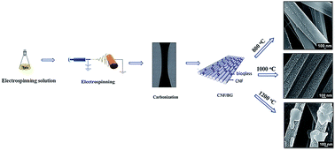
Polyacrylonitrile (PAN) electrospinning in combination with sol–gel method has been a common technique to produce inorganic nanoparticles containing composite carbon nanofibers (CNFs) for diverse applications. To investigate the morphology evolution and crystal transformation of inorganic components along with CNF formation, bioactive glass (BG) containing CNFs (CNF/BG) were prepared by sintering as-spun PAN/precursor composite nanofibers in a nitrogen atmosphere at temperatures of 800, 1000 and 1200 °C. Comprehensive characterizations were performed with TEM, SEM-EDXA and XRD. For samples sintered at 800 °C, numerous BG nanoparticles were observed inside the CNFs and mainly in an amorphous state. With the sintering temperature raised to 1000 °C, a number of spherical BG nanoparticles were detected on the surface of the resulting CNFs, with a crystal structure of wollastonite (β-CaSiO3) polycrystals. When the samples were sintered at 1200 °C, the BG nanoparticles on the surface of CNFs merged into forms with cuboid-like geometry, mainly consisting of pseudowollastonite (Ca3(Si3O9)) single crystals. Based on the geometry evolution and dynamic size distribution function analyses (Ostwald ripening and Smoluchowski equations), it was concluded that the growth of BG nanoparticles conformed to the ripening mechanism at 800 °C and migration–coalescence mechanism at 1200 °C, while the process involved both ripening and migration–coalescence mechanisms at 1000 °C.


1. Introduction

With similar advantages in terms of mechanical strength, thermal stability, electrical conductivity and specific surface area to carbon nanotubes (CNTs), carbon nanofibers (CNFs) have been considered as an excellent carrier of metal/metal oxide nanoparticles (M/MOx).1–3 Traditionally, composite CNFs containing inorganic nanoparticles (CNF/M or CNF/MOx) have been fabricated by combining the processes of chemical vapor deposition (CVD) and surface chemical modification. The flexibility in controlling the chemical compositions of metal/metal oxide nanoparticles have resulted in CNF/M or CNF/MOx composites as promising substrates for various applications as catalysts, batteries, super-sensitive chemical/biological sensors and for tissue repair.3–5 However, the purification of CVD-produced CNFs is a complicated and time-consuming procedure because the residual impurities like Co/Ni catalysts and graphite particles are hard to remove completely. The structural integrity of CNFs was usually deteriorated by covalently/non-covalently decorated inorganic nanoparticles.6,7 Polyacrylonitrile (PAN)-based CNFs produced from solution electrospinning, pre-oxidation and carbonization have provided a feasible way to modify CNFs and improve the performances of currently available CNFs.8–11 The process showed advantages of catalyst-free preparation and strong flexibility in tailoring the strength and elastic modulus of CNFs by regulating the carbonization temperatures and alignment degrees.12 By dissolving organic precursors of metal/metal oxide into the electrospinning PAN solution, CNF/M or CNF/MOx composites could be readily prepared.13–16 In comparison with cases of introducing M or MOx nanoparticles into electrospinning PAN solution directly, the electrospinning/sol–gel technique produced CNF/M or CNF/MOx composites conquered the disadvantage of serious aggregation of nanoparticles in the solution and in resulting nanofibers. In our previous studies, different functionalized CNF/M or CNF/MOx composites were obtained by electrospinning/sol–gel technique and carbonization.13–16 Materials including CNF/β-TCP,13,14 CNF/Sn,15 CNF/TiO2,16–18 CNF/SnOx,19 CNF/Fe3O4,20 CNF/MnOx,8,21 CNF/Ni22 and CNF/Cu,22,23 had been intensively studied as photocatalyst, Li-ion battery anode, energy storage container and bone tissue regeneration substrate, etc.

Noticeably, physicochemical properties and surface morphology of CNF/M or CNF/MOx composites were found significantly depending on preparation parameters such as pre-oxidation conditions and sintering conditions, etc.13–15,20,24 Wang et al. reported that the size of Fe3O4 nanoparticles in CNF/Fe3O4 composite increased remarkably along with higher sintering temperature. They found the electrical properties of CNF/Fe3O4 films were optimized for samples sintered at 600 °C.20 Our previous works revealed that Sn nanoparticles had strong tendency to migrate to the surface of CNFs when the sintering temperature in producing CNF/Sn composites was raised from 550 to 1000 °C. At the same time, the initial amorphous Sn transformed into crystalline Sn. These transitions led to CNF/Sn composites sintered at 850 °C showing the best electrical performance.24 The variances in publications distinctly demonstrated the complexity in preparing such CNF composites and in relating the specific features of M (or MOx) nanoparticles to the final performance. There was a large uncertainty of understanding the morphology evolution and formation mechanism of M (or MOx) nanoparticles during the preoxidation and carbonization of PAN/precursor nanofibers at different sintering temperatures and times. To design and fabricate high performance CNF/M or CNF/MOx composites, thus, it was necessary to investigate the effective approach to control the structure, morphology and size of M (or MOx) nanoparticles in CNFs, which would help to provide theoretical foundation.

Several mechanisms (e.g. Ostwald ripening (OR) and particle coalescence) have been discussed in the context of particle growth dependent on the dominating process of material transport.25–27 It was stated that mass transport was the dominant process to control the particle growth process, although the driving force was associated with the decrease in chemical potential due to the reduction of interfacial area. The difference between Ostwald ripening and particle coalescence mechanism was in their mass transport process. In particular, Ostwald ripening was caused by the diffusion of atoms through the matrix leading to the growth of larger particles at the expense of smaller ones, and atomic diffusion could occur in solid embedding media. While coalescence mechanism involved the collision of migrating particles and subsequent recrystallization and reshaping of aggregates, in which, particle migration required a mobile environment or self-mobile ability. Thus, diffusion-driven aggregation and coalescence were expected only at temperatures above the softening temperature of the embedding medium or the melting temperature of the particles. These mechanisms provided feasible entry paths to look into the evolution of M (or MOx) nanoparticles in preparing CNF/M or CNF/MOx composites.

In this study, bioactive glass (BG) containing CNFs (CNF/BG) was chosen as the model system for the purpose to investigate the morphology evolution and crystal transformation of inorganic components along with CNF formation. As bioceramic material, BGs have been long investigated for applications in bone regeneration.28–30 They were bioactive and able to induce apatite formation by releasing sodium, calcium and silicium ions etc., when they were in contact with body fluid.28,29 In addition to their excellent bone bonding properties, soft connective tissues were also reported able to form a bond with BGs.30 In our previous work,31 CNF/BG composites containing BG nanoparticles of different compositions have demonstrated strong stimulations in biomineralization and osteocompatibility in comparison to pure CNFs. It interested us to investigate the morphology evolution, crystallization behavior and growth kinetics of CNF/BG composites in detail, which was essential to develop new biomaterials for orthopedic applications. Additionally, the study could also provide understanding the growth mechanism of M/MOx nanoparticles in CNF composites prepared in similar procedures. Herein, CNF/BG composites were prepared by sintering as-spun PAN/precursor composite nanofibers in nitrogen atmosphere at temperatures of 800, 1000 or 1200 °C for different times. The resulting CNF/BG composites were comprehensively and quantitatively analyzed with characterizations including TEM, SEM-EDXA and XRD etc., to expose the dependence of growth mechanism of BG nanoparticles on sintering temperature and time.

2. Experimental section

2.1 Materials and sample preparation

The illustration of experiment process is shown in Fig. 1. Firstly, 10 ml of triethyl phosphate (TEP, Aldrich, USA) was dissolved in a mixed solvent of absolute ethyl alcohol (10.2 ml), distilled water (3.2 ml) and ammonia water (0.12 ml), and stirred thoroughly at 80 °C for 24 h to obtain a hydrolyzed TEP solution. Then, 0.16 ml of the hydrolyzed TEP solution, 0.42 g of calcium nitrate tetrahydrate (CN, Aldrich, USA) and 0.65 ml of tetraethoxysilane (TEOS, Aldrich, USA) were added in turn into 20 ml of N,N-dimethyl formamide (DMF, Tianjin Fine Chemical Co., China) containing 10 wt% of PAN (Mw = 100[thin space (1/6-em)]000 g mol−1, composed of 93.0 wt% acrylonitrile, 5.3 wt% methylacrylate and 1.7 wt% itaconic acid, Courtaulds Co., UK). The system was stirred at room temperature for 48 h to obtain a homogeneous solution for electrospinning. The electrospinning process was carried out by following our previous works.13–16,31 The as-spun nanofibers were pre-oxidized at 270 °C along with hot-stretching in air for 0.5 h, and then sintered at 800, 1000 or 1200 °C, for various retention time periods in N2 atmosphere to obtain the BG-containing CNFs. The BG was named 58S, with composition of SiO2 (58 mol%)–CaO (33 mol%)–P2O5 (9 mol%). CNF/BG composites sintered at 800, 1000 and 1200 °C were named in terms of CNF/BG-800, CNF/BG-1000 and CNF/BG-1200, respectively. Similarly, pure BGs sintered at 800, 1000 and 1200 °C were named in terms of BG-800, BG-1000 and BG-1200.
image file: c4ra12177e-f1.tif
Fig. 1 Schematic illustration for experimental preparation of CNF/BG composites.

2.2 Characterizations

Morphologies of CNF/BG composites were observed by scanning electron microscope (SEM, S-250, UK) at accelerating voltages of 15–20 kV and transmission electron microscopy (TEM, JEOL 2000 EX, Japan) at an accelerating voltage of 200 kV. Before SEM observation, the sample surface was coated with a thin layer of a gold alloy. TEM imaging was done by amplitude and phase contrast, and images were acquired using a Gatan Orius SC600 high resolution camera. Crystalline microstructures of CNFs and BG nanoparticles were investigated by high resolution transmission electron microscope (HR-TEM, Hitachi H-800, Japan) at an operating voltage of 200 kV and an X-ray diffractometer (XRD, Rigaku D/max 2500 VB2+/PC, Japan) operating at 40 kV and 200 mA. Average fiber diameters and particle sizes were calculated with SEM images by using ImageJ software (National Institutes of Health, USA) to measure at least 500 nanofibers or nanoparticles.

3. Results and discussion

3.1 Morphology evolution of PAN/BG precursor composite nanofibers

Fig. 2 shows the morphologies of as-spun and pre-oxidized PAN/BG precursor composite nanofibers. The as-spun composite nanofibers were bead-free with smooth surface and average fiber diameter of 550 nm (Fig. 2(a)). The inserted TEM image showed the as-spun composite nanofibers were uniform, indicating the uniform dispersion of BG precursor in PAN matrix. The partial orientation was attributed to the fiber collection by using a rolling cylinder. The orientation of the nanofibers was beneficial to increasing the mechanical properties of CNFs as found in our previous work.32 After pre-oxidation, the surface of nanofibers remained smooth, and the nanofibers were more parallel due to the hot-stretching treatment (Fig. 2(b)). During the pre-oxidation progress, loss of the methylacrylate comonomer and other substitutional groups of PAN would occur as results of the cyclization, decyanation and denitrogenation processes.33,34 Accordingly, the average fiber diameter of pre-oxidized PAN/BG precursor composite nanofibers decreased to 500 nm. As shown by the inserted TEM image, no aggregation formed during the pre-oxidization process, showing no phase separation between PAN and BG precursor taking place at this stage.
image file: c4ra12177e-f2.tif
Fig. 2 Morphologies of (a) the as-spun and (b) pre-oxidized PAN/BG precursor composite nanofibers observed by SEM and TEM (the inserts). The insets are the diameter distribution of nanofibers.

Fig. 3 shows morphology evolution of CNF/BG-800 with various sintering time periods at 800 °C. Upon high-temperature sintering, PAN was carbonized into carbon and BG precursors would transform into BG. In this transition, the fiber diameter would decrease gradually with the loss of non-carbon elements. As shown in Fig. 3(a1), (b1) and (c1), the average diameters of CNF/BG-800 composites decreased with longer retention time at 800 °C, and were smaller than that of the pre-oxidized nanofibers. Some nano-scaled grains began to appear on the surfaces of CNFs, and the particle number increased slowly with sintering time prolonging. After 1 h sintering at 800 °C, numerous grains with average size of 28 nm were observed inside CNFs (Fig. 3(a2)), although the fiber surface remained smooth. The grains were identified BG nanoparticles as found in our previous work.31 When the sintering time reached 2 h, the emerged grains increased the surface roughness of CNF/BG-800 (Fig. 3(b1)), and the grains gradually grew into bigger size at the third hour of sintering (Fig. 3(c1)). Along with BG grains showing the tendency of migration towards outer surfaces of fibers, the TEM images revealed that those grains inside CNFs also grew bigger with broadened size distribution and declined distribution density. This change apparently met the quality conservation laws. It could be summarized from the results that 3 h sintering at 800 °C was not effective enough in propelling BG nanoparticles to the surface of CNFs.


image file: c4ra12177e-f3.tif
Fig. 3 SEM (left columns) and TEM (right columns) images of CNF/BG-800 with various sintering time periods of (a1 and a2) 1 h, (b1 and b2) 2 h and (c1 and c2) 3 h. The insets in SEM and TEM images are the diameter distribution of CNFs and the size distribution of BG nanoparticles, respectively.

When the sintering temperature was raised to 1000 °C, completely different morphologies were detected in resulting CNF/BG-1000 composites. As shown in Fig. 4, a mass of BG nanoparticles sprung up on surfaces of CNFs, with average sizes increasing from 45 to 75 nm as sintering time proceeding from 1 h to 3 h. The BG nanoparticles were spherical initially, and then evolved to irregular shaped aggregates of bigger sizes by fusion of individual nanoparticles. The particle sizes and distributions of BG nanoparticles inside CNFs changed accordingly. Bigger and denser BG grains were found on fiber surfaces, at the same time, bigger but less denser BG grains were found inside the fibers. These implied the nanoparticles got more thermodynamical driving force by raising sintering temperature to 1000 °C, which promoted them to migrate from the inside to the outside of the nanofibers. The driving force was associated with the decrease in chemical potential due to the reduction of interfacial area between BG nanoparticles and CNF matrix. After migration to the surface of CNFs, BG nanoparticles existed in a more stable thermodynamical state. With sintering time prolonging, the morphology of BG nanoparticles evolved from spherical to irregular shape due to the random and isotropic diffusion, migration, collision and mergence of small nanoparticles. It could be seen clearly in Fig. 4(b1), (b2), (c1) and (c2), some nanoparticles have merged together, but the contours of the original nanoparticles were still apparent as marked with blue rectangles. Some nanoparticles just collided together as marked with red ellipse, tending to form contact angle which built up the prerequisite for further mergence. The differences in morphology evolution of CNF/BG-1000 and CNF/BG-800 indicated that the formation mechanism of BG nanoparticles should be different. In addition, the degree of carbonization of PAN at 800 °C and 1000 °C was also different. In comparison with CNFs carbonized at 800 °C, the CNFs carbonized at 1000 °C were apparently thinner, indicating the loss of non-carbon elements to higher degrees.


image file: c4ra12177e-f4.tif
Fig. 4 SEM (left columns) and TEM (right columns) of CNF/BG-1000 with various sintering time periods of (a1 and a2) 1 h, (b1 and b2) 2 h and (c1 and c2) 3 h. The insets in SEM and TEM images are the diameter distribution of CNFs and the size distribution of BG nanoparticles, respectively. The collision of nanoparticles which tended to further mergence was marked with red ellipse. And the contours of the original nanoparticles which have merged were marked with blue rectangles.

By further increasing the sintering temperature to 1200 °C, the morphology of CNF/BG-1200 was found definitely different from the two previous cases. As seen from TEM images in Fig. 5, the majority of BG nanoparticles had migrated to the outside of nanofibers and only a few of particles stayed inside the nanofibers. The growth and migration of nanoparticles were significantly accelerated because the thermodynamical driving force went stronger at higher temperature. The BG nanoparticles of CNF/BG-1200 presented cuboid-like geometry at sintering time points of 1 h and 2 h (Fig. 5(a1) and (b1)), which were different from those spherical BG nanoparticles in cases of CNF/BG-800 and CNF/BG-1000, implying the different formation. mechanisms of BG nanoparticles at different sintering temperatures. Along with longer sintering time, significant fusion between nanoparticles took place to result in large size (107 nm), irregular aggregates (Fig. 5(c1)). Noticeably, the diameters of CNFs resulting from carbonization at 1200 °C were further decreased in comparison with those carbonized at 800 and 1000 °C. In addition to the increased thermodynamical driving force that accompanying with the increasing temperature, the continuously decreasing fiber diameter was believed another reason to cause BG nanoparticles migrating to fiber surface. During carbonization, the microstructure of PAN-based carbon fiber transformed from two-dimensional disordered graphite structure to three-dimensional laminar structure with loss of non-carbon elements, leading to structure densification at the same time.35,36 The densification was determined by both the rate of denitrogenation and the rearrangement of carbon atoms, which were proportional to sintering temperature. The denser carbon network might create stronger expelling force to “squeeze out” BG nanoparticles due to their chemical incompatibility.


image file: c4ra12177e-f5.tif
Fig. 5 SEM (left columns) and TEM (right columns) of CNF/BG-1200 with various sintering time periods of (a1 and a2) 1 h, (b1 and b2) 2 h and (c1 and c2) 3 h. The insets in SEM and TEM images are the diameter distribution of CNFs and the size distribution of BG nanoparticles, respectively. The collision of nanoparticles which tended to further mergence was marked with red ellipse. And the contours of the original nanoparticles which have merged were marked with blue rectangles.

3.2 Crystallization behavior of BG nanoparticles in CNFs

Fig. 6, 7 and 8 show high resolution TEM (HR-TEM) images and fast Fourier transform (FFT) patterns of BG nanoparticles in CNF/BG-800, CNF/BG-1000 and CNF/BG-1200 with various sintering time periods, respectively. As seen in Fig. 6(a1), (b1) and (c1), all HR-TEM images showed the isotropic amorphous structure of BG nanoparticles, which was consistent with the results of the obscure diffuse rings observed from FFT patterns (Fig. 6(a2), (b2) and (c2)). This demonstrated that BG component did not form crystalline structure at 800 °C within 3 h, indicating the temperature being lower than that required for the crystallization of BG37,38 From Fig. 7 and 8, all HR-TEM images showed clear lattice structure with high degree crystallization for nanoparticles in CNFs. The corresponding FFT patterns emerged with the specific diffraction mode, which confirmed the BG precursor had transformed into crystalline forms at both 1000 and 1200 °C within 3 h. In both cases, the perfectness of crystallized lattice was enhanced with the prolonging of sintering time. These observations were highly consistent with the XRD patterns of CNF/BG composites. As shown in Fig. 9(a), no characteristic sharp peaks were found in the XRD pattern of CNF/BG-800, except the broad and weak diffraction peaks at 2θ = 24.7° and 44.5°, which were indexed as the (0 0 2) and (1 0 1) reflections of graphitic carbon,39 indicating the formation of CNFs. To make the situation more clearly, pure BG sintered at the same condition (BG-800) was also analyzed with XRD. Similarly, no characteristic sharp peaks were found (Fig. 9(b)). These data revealed that the BG obtained at 800 °C was amorphous within the experimental time. In contrast, a characteristic diffraction peak at 2θ = 32.8° was detected for CNF/BG-1000, and a sharp peak at 2θ = 31° was detected for CNF/BG-1200. Corresponding peaks were also identified in pure BG-1000 and BG-1200, respectively. Referring to the matching analysis of JADE software and literatures,40–42 the peak at 2θ = 32.8° was assigned as the (1 0 0) reflection of wollastonite (β-CaSiO3) (JCPDS 42-0547), and the peak at 2θ = 31° was assigned as the (1 0 1) reflection of pseudowollastonite (Ca3(Si3O9)) (JCPDS 74-0874).
image file: c4ra12177e-f6.tif
Fig. 6 HR-TEM images (left columns) and FFT patterns (right columns) of BG nanoparticles in CNF/BG-800 with various sintering time periods of (a1 and a2) 1 h, (b1 and b2) 2 h and (c1 and c2) 3 h.

image file: c4ra12177e-f7.tif
Fig. 7 HR-TEM images (left columns) and FFT patterns (right columns) of BG nanoparticles in CNF/BG-1000 with various sintering time periods of (a1 and a2) 1 h, (b1 and b2) 2 h and (c1 and c2) 3 h.

image file: c4ra12177e-f8.tif
Fig. 8 HR-TEM images (left columns) and FFT patterns (right columns) of BG nanoparticles in CNF/BG-1200 with various sintering time periods of (a1 and a2) 1 h, (b1 and b2) 2 h and (c1 and c2) 3 h.

image file: c4ra12177e-f9.tif
Fig. 9 XRD patterns of (a) CNF/BG composites and (b) pure BGs sintered at different temperatures for 3 h.

Accordingly, the well-defined lattice fringes with an average d-spacing of 0.71 nm found in the HR-TEM images of CNF/BG-1000 (Fig. 7), could confirm the formation of the (1 0 0) lattice plane of wollastonite structure.43,44 Similarly, the well-defined lattice fringes with an average d-spacing of 1.41 nm found in the HR-TEM images of CNF/BG-1200 (Fig. 8) could also confirm the formation of the (1 0 1) lattice plane of pseudowollastonite structure.45,46 Wollastonite and pseudowollastonite had different crystal structures. The former possessed chain structure while the latter possessed ring structure in the space of crystals, which resulted in the differences in the formation temperatures and the relating thermal stability. In addition, all these findings were consistent with corresponding FFT patterns. In Fig. 7, the crystallized lattices were observed disordered and the FFT patterns showed concentric diffraction rings associated with some diffuse diffraction spots, indicating wollastonite-type BG nanoparticles in CNF/BG-1000 mainly being in the polycrystalline form. From Fig. 8, highly ordered lattices with uniform interplanar spacing were observed and the corresponding FFT patterns displayed specific discrete diffraction spots, suggesting pseudowollastonite-type BG nanoparticles in CNF/BG-1200 being in the single crystalline form. In literatures, evidences showed that pseudowollastonite phase was a stable phase at high temperature, and the transformation from wollastonite to pseudowollastonite phase took place at temperatures over 1100 °C.47,48 Therefore, in a word, BG nanoparticles were in the form of polycrystalline wollastonite when the sintering temperature was 1000 °C, and transformed into more stable pseudowollastonite form at higher sintering temperature of 1200 °C.

3.3 Growth mechanism of BG nanoparticles in CNFs

As shown in Fig. 3, 4 and 5, morphology evolution of BG nanoparticles in CNF/BG composites were much different when different sintering temperatures were applied. As the sintering time was fixed at 1 h, the BG nanoparticles in both CNF/BG-800 and CNF/BG-1000 composites were in spherical geometry (Fig. 3(a1) and 4(a1)), while the BG nanoparticles in CNF/BG-1200 composites exhibited the cuboid-like geometry (Fig. 5(a1)). In the case of CNF/BG-800, most of the BG nanoparticles were still inside the CNFs at this stage. As the sintering time prolonged, more BG nanoparticles emerged on the surface of CNFs in all cases. However, the average size of the initial spherical nanoparticles in CNF/BG-1000 increased continuously with longer sintering time, at the same time, the particles gradually collided to form larger and more irregular geometry (Fig. 4(b1) and (b2)). As for CNF/BG-1200, the BG nanoparticles could be seen merged together with longer sintering time, which led to large-sized and irregular BG particles that showed a tendency to drop off from the CNFs (Fig. 5(b1) and (b2)). These distinct differences found in preparing CNF/BG composites implied that the growth mechanism and coarsening kinetics of BG nanoparticles were tightly controlled by the sintering temperature.

In Ostwald ripening mechanism, a characteristic stated is the formation of spherical-type particles, for the material diffusion in the atomistic level was isotropic as mentioned in our previous study27 and literature.26,49,50 In coalescence mechanism, whereas, the feature is the formation of irregular-shaped particles as a result of anisotropic and random diffusion–collision of sub-particles.25,51,52 Therefore, the growth of BG nanoparticles with well-distributed spherical shape along CNFs was dominated by Ostwald ripening mechanism at the sintering temperature of 800 °C and the initial stage of the sintering temperature of 1000 °C. As the sintering going on, the spherical BG nanoparticles in CNF/BG-1000 began to merge into irregular-shaped particles, which meant coalescence had taken part in the morphology evolution. While in the case of CNF/BG-1200, the initial cuboid-like BG nanoparticles was apparently controlled by coalescence mechanism from the beginning at the sintering temperature of 1200 °C, which led to irregular-shaped particles in large size liable to fall off.

Theory analysis showed that each mechanism would result in obvious scale invariant distribution functions for the particle size, F(d/〈d〉) vs. d/〈d〉, in the asymptotic limit. In addition to using geometry evolution of nanoparticles, therefore, the size distributions of BG nanoparticles obtained under different conditions could be supplemental evidences to deduce the mechanism that engendered coarsening. Accordingly, the size distribution function of BG nanoparticles in CNFs was analyzed by normalizing the cluster size frequency distribution at each time point as shown in Fig. 10. It was found that all the data did not collapse onto a single curve, as expected for a self-similar coarsening process. As shown in Fig. 10(a) and (b), the experimental particle distribution of BG nanoparticles obtained at 800 °C and at earlier time period (≤2 h) of 1000 °C showed the best agreement with the diffusion-controlled Ostwald ripening equation (eqn (1)):53,54

 
image file: c4ra12177e-t1.tif(1)
however, the features of the experimental particle distribution of BG nanoparticles obtained at 1200 °C and at later time period (3 h) of 1000 °C were better captured by the analytical solution to the coalescence-controlled Smoluchowski equation (eqn (2)):55,56
 
image file: c4ra12177e-t2.tif(2)
where W = Γ(−α + 1)/Γ(−α + 2/3), Γ is gamma function and α is a scaling exponent that quantifies the dependence of the cluster diffusion coefficient with its mass. This long-time analytical solution accorded with the expected power-law scaling behavior of the average particle size, 〈d〉 ∼ tβ, with β = 1/(2 − 3α).


image file: c4ra12177e-f10.tif
Fig. 10 Comparison of the theoretical and experimental normalized distribution functions of BG nanoparticles in (a) CNF/BG-800, (b) CNF/BG-1000 and (c) CNF/BG-1200. The experimental distributions were plotted with unfilled markers. The solid black line represented the distribution characteristic of diffusion-limited Ostwald ripening, and the dotted lines denoted the Smoluchowski distributions that best fit the data at each stage: the long dotted curve corresponded to α = −1.6, and the short dotted curve corresponded to α = −0.1.

The relative contributions of both Ostwald ripening and coalescence process could also be evaluated from the first and third moments of the nanoparticle size distribution, μ1 = d3/dh and μ3 = 〈d〉/d3, where 〈d〉 = ∑di/n is the arithmetic mean radius, d3 = (∑di3/n)1/3 is the cube-mean radius, Rh = n/(∑(1/Ri)) is the harmonic mean radius. Both moments were unity for monodisperse particles resulted from condensation, and the growth of the particles was only predominated by coalescence mechanism when μ1 > 1.25 and μ3 < 0.905.57,58 If 1 < μ1 < 1.25 and 0.905 < μ3 < 1, the growth of nanoparticles was controlled by both mechanisms. Fig. 11 shows the first (μ1) and third (μ3) moments of the size distribution function of BG nanoparticles in CNFs. For example, the moments of the particle size distribution at t = 3 h at 1000 °C were μ1 = 1.64 and μ3 = 0.81 (Fig. 11(b)), implying that the coalescence should play a dominating role in the growth of BG nanoparticles at this stage. Therefore, the calculations confirmed that the growth of BG nanoparticles at 800 and 1200 °C was predominately controlled by Ostwald ripening and coalescence mechanism, respectively, while the growth of BG nanoparticles at 1000 °C was firstly controlled by Ostwald ripening mechanism followed by coalescence mechanism with longer sintering time.


image file: c4ra12177e-f11.tif
Fig. 11 First (μ1) and third (μ3) moments of the size distribution function of BG nanoparticles in (a) CNF/BG-800, (b) CNF/BG-1000 and (c) CNF/BG-1200. The μ1 and μ3 were, respectively, marked with black rectangle and red circle, which gradually diverged from unity as the polydispersity of particles in CNFs grew.

Based on all the above results, evolution mechanism of BG nanoparticles in CNFs sintered at various conditions was proposed as illustrated in Fig. 12. It was found that the sintering temperature and sintering time were the determinant factors in controlling the morphology and structure of BG nanoparticles in CNFs. The quantity of BG nanoparticles migrating onto the surfaces of CNFs was proportional to the sintering temperature and sintering time, which obviously dominated the size and the size distribution of BG nanoparticles onto CNFs. This was due to the enhancement of the thermodynamical driving force for particle migration by increasing sintering temperature or prolonging sintering time. At low sintering temperature (e.g. 800 °C), BG nanoparticles possessed small size and most of them remained inside CNFs. At high sintering temperature (e.g. 1200 °C), BG nanoparticles with big size migrated onto the surfaces of CNFs. Rationally, BG nanoparticles grew into bigger size with the increasing of sintering time. However, sintering temperature was identified the primary factor in controlling the crystallization behavior and growth mechanism of BG nanoparticles. At low sintering temperature (e.g. 800 °C), only numerous BG nanoparticles with amorphous structure were detected, and the growth of BG nanoparticles could be well explained by the ripening mechanism. At high sintering temperature (e.g. 1200 °C), BG nanoparticles emerged in the cuboid-like geometry, mainly composed of pseudowollastonite (Ca3(Si3O9)) single crystals, which were dominated by migration–coalescence mechanism. However, the growth of BG nanoparticles was controlled by both ripening and migration–coalescence mechanisms at the sintering temperature of 1000 °C. The resulting BG nanoparticles were composed of wollastonite (β-CaSiO3) polycrystals. In the process of BG nanoparticles formation and migration, the structure transformation and densification of CNF matrix along with carbonization were another possible factor playing important role in the morphology and structure evolution of BG particles. When more three-dimensional laminar structure in CNFs were formed along with the significant structure densification, more BG-related materials should be expelled under thermodynamical driving force generated at higher sintering temperature or longer sintering time.


image file: c4ra12177e-f12.tif
Fig. 12 Evolution mechanism of BG nanoparticles in CNFs sintered at various conditions.

4. Conclusions

The results of the morphology and structure evolution of BG nanoparticles in CNF/BG composites in this study clearly revealed that sintering temperature and time were critical factors in controlling the features of resulting CNF/M (or MOx) composites that were prepared by PAN/sol–gel electrospinning and sintering technique. From low to high sintering temperature, the BG nanoparticles demonstrated changes from amorphous state to wollastonite polycrystals and further to pseudowollastonite single crystals. At lower or higher sintering temperatures, the transformation of BG nanoparticles from the precursors undergone ripening or coalescence mechanism to result in isotropic spherical or irregular-shaped particles. The BG particles were expelled from the CNFs gradually by both the increased thermodynamical driving force generated at higher sintering temperature or longer sintering time, and the structure transformation and densification of CNF matrix along with carbonization. When the sintering temperature and time reached critical points, mass of BG nanoparticles migrated to the surface of CNFs, merged together and fell off.

Acknowledgements

The authors are very pleased to acknowledge financial support from National Natural Science Foundation of China (no. 51102008 and 51373016), the Research Fund for the Doctoral Program of Higher Education (no. 20110010120014), the Fundamental Research Funds for the Central Universities (no. ZY1106) and Program for New Century Excellent Talents in University (NCET-11-0556).

Notes and references

  1. H. Q. Li, C. P. Li, J. Bai, C. L. Zhang and W. Y. Sun, RSC Adv., 2014, 4, 48362 RSC.
  2. B. Zhang, L. X. Zhang, L. Xu and K. M. Jang, RSC Adv., 2014, 4, 12298 RSC.
  3. G. Che, B. B. Lakshmi, C. R. Martin and E. R. Fisher, Chem. Mater., 1998, 10, 260 CrossRef CAS.
  4. R. S. Chen, Y. Li, K. F. Huo and P. K. Chu, RSC Adv., 2013, 3, 18698 RSC.
  5. B. S. Harrison and A. Atala, Biomaterials, 2007, 28, 344 CrossRef CAS.
  6. Z. M. Huang, Y. Z. Zhang, M. Kotaki and S. Ramakrishna, Compos. Sci. Technol., 2003, 63, 2223 CrossRef CAS.
  7. M. Inagaki, Y. Yang and F. Y. Kang, Adv. Mater., 2012, 24, 2547 CrossRef CAS.
  8. Y. X. Du, X. Zhao, Z. L. Huang, Y. Z. Li and Q. H. Zhang, RSC Adv., 2014, 4, 39087 RSC.
  9. L. W. Ji, Z. Lin, A. J. Medford and X. W. Zhang, Carbon, 2009, 47, 3346 CrossRef CAS.
  10. H. J. Dai, Acc. Chem. Res., 2002, 35, 1035 CrossRef CAS.
  11. N. M. Rodriguez, J. Mater. Res., 1993, 8, 3233 CrossRef CAS.
  12. S. K. Nataraj, K. S. Yang and T. M. Aminabhavi, Prog. Polym. Sci., 2012, 37, 487 CrossRef CAS.
  13. H. Y. Liu, Q. Cai, P. F. Lian, Z. Fang, S. Duan, X. P. Yang, X. L. Deng and S. Ryu, Mater. Lett., 2010, 64, 725 CrossRef CAS.
  14. H. Y. Liu, Q. Cai, P. F. Lian, Z. Fang, S. Duan, S. Ryu, X. P. Yang and X. L. Deng, Carbon, 2010, 48, 2266 CrossRef CAS.
  15. Y. Yu, Q. Yang, D. Teng, X. Yang and S. Ryu, Electrochem. Commun., 2010, 12, 1187 CrossRef CAS.
  16. D. Teng, Y. Yu, H. Liu, X. Yang, S. Ryu and Y. Lin, Catal. Commun., 2009, 10, 442 CrossRef CAS.
  17. S. Kim and S. K. Lim, Appl. Cat. B., 2008, 84, 16 CrossRef CAS.
  18. J. S. Im, M. I. Kim and Y. S. Lee, Mater. Lett., 2008, 62, 3652 CrossRef CAS.
  19. L. Zou, L. Gan, F. Y. Kang, M. X. Wang, W. C. Shen and Z. H. Huang, J. Power Sources, 2010, 195, 1216 CrossRef CAS.
  20. L. Wang, Y. Yu, P. C. Chen, D. W. Zhang and C. H. Chen, J. Power Sources, 2008, 183, 717 CrossRef CAS.
  21. L. W. Ji, A. J. Medford and X. W. Zhang, J. Mater. Chem., 2009, 195, 593 Search PubMed.
  22. L. Ji, Z. Lin, A. J. Medford and X. Zhang, Chem.–Eur. J., 2009, 15, 10718 CrossRef CAS.
  23. L. Ji, Z. Lin, R. Zhou, Q. Shi, O. Toprakci, A. J. Medford, C. R. Millns and X. Zhang, Electrochim. Acta, 2010, 55, 1605 CrossRef CAS.
  24. M. A. Atwater, J. Phillips and Z. C. Leseman, Carbon, 2010, 48, 1932 CrossRef CAS.
  25. W. W. Pai, A. K. Swan, Z. Zhang and J. F. Wendelken, Phys. Rev. Lett., 1997, 79, 3210 CrossRef CAS.
  26. F. Huang, H. Z. Zhang and J. F. Banfield, Nano Lett., 2003, 3, 373 CrossRef CAS.
  27. X. L. Jia, J. Listak, W. Velencia, E. E. Kalu, X. P. Yang and M. Bockstaller, Langmuir, 2010, 26, 12190 CrossRef CAS.
  28. S. Labbaf, O. Tsigkou, K. H. Muller, N. M. Steven, A. E. Porter and J. R. Jones, Biomaterials, 2011, 32, 1010 CrossRef CAS.
  29. A. Hoppe, N. S. Guldal and A. R. Boccaccini, Biomaterials, 2011, 32, 2757 CrossRef CAS.
  30. Q. Hu, Y. L. Li, G. H. Miao, N. R. Zhao and X. F. Chen, RSC Adv., 2014, 4, 22678 RSC.
  31. Q. Yang, G. Sui, Y. Z. Shi, S. Duan, J. Q. Bao, Q. Cai and X. P. Yang, Carbon, 2013, 56, 288 CrossRef CAS.
  32. J. Y. Ji, G. Sui, Y. H. Yu, Y. X. Liu and Z. J. Du, J. Phys. Chem. C, 2009, 113, 4779 CAS.
  33. M. G. Dunham and D. D. Edie, Carbon, 1992, 30, 435 CrossRef CAS.
  34. E. Fitzer and D. J. Muller, Carbon, 1975, 13, 63 CrossRef CAS.
  35. A. J. Gao, C. J. Su, S. Luo, Y. J. Tong and L. H. Xu, J. Phys. Chem. Solids, 2011, 72, 1159 CrossRef CAS.
  36. B. Wang, C. Zhao, S. J. Xiao, J. Zhang and L. H. Xu, J. Appl. Polym. Sci., 2012, 125, 3545 CrossRef CAS.
  37. J. L. Liu and X. G. Miao, Ceram. Int., 2004, 30, 1781 CrossRef CAS.
  38. S. Padilla, J. Román, A. Carenas and M. Vallet-Regí, Biomaterials, 2005, 26, 475 CrossRef CAS.
  39. C. J. Lu, Z. P. Xia, Y. Zhou and Z. Luo, Carbon, 2007, 45, 1686 CrossRef.
  40. P. N. De Aza, F. Guitian, S. De Aza and F. J. Valle, Analyst, 1998, 123, 681 RSC.
  41. A. M. Minarelli-Gaspar, S. Saskal, L. R. da Cunha, P. D. A. Bolini, R. G. Carrodequas, A. H. De Aza, P. Pena, P. N. De Aza and S. De Aza, Key Eng. Mater., 2008, 1083, 361 Search PubMed.
  42. H. X. Yang and C. T. Prewitt, Am. Mineral., 1999, 84, 929 CAS.
  43. J. L. Hutchison and A. C. McLaren, Contrib. Mineral. Petrol., 1976, 55, 303 CrossRef CAS.
  44. Y. Ohashi, Phys. Chem. Miner., 1984, 10, 217 CrossRef CAS.
  45. M. A. Bouhifd, G. Gruener, B. O. Mysen and P. Richet, Phys. Chem. Miner., 2002, 29, 655 CrossRef CAS.
  46. P. N. De Aza, Z. B. Luklinska, M. R. Anseau, F. Guitian and S. De Aza, Acta Microsc., 2001, 201, 33 CrossRef CAS.
  47. O. M. Goudouri, X. Chatzistavroul, E. Kontonasaki, N. Kantiranis, L. Papadopoulou, K. Chirissafis and K. M. Paraskevopoulos, Key Eng. Mater., 2009, 131, 396 Search PubMed.
  48. P. Richet and R. A. Robie, Eur. J. Mineral., 1991, 3, 475 CrossRef CAS.
  49. B. F. Zou, Y. F. Liu and Y. Q. Wang, RSC Adv., 2013, 3, 23327 RSC.
  50. J. D. Ng, B. Lorber, J. Witz, A. Theobald-Dietrich, D. Kern and R. Giege, J. Cryst. Growth, 1996, 168, 50 CrossRef CAS.
  51. S. Arditty, C. P. Whitby, B. P. Binks, V. Schmitt and F. Leal-Calderon, Eur. Phys. J. E, 2003, 11, 273 CrossRef CAS.
  52. J. P. Hare, H. W. Kroto and D. R. M. Walton, Chem. Mater., 1999, 11, 2709 CrossRef.
  53. P. W. Voorhees, J. Stat. Phys., 1985, 38, 231 CrossRef.
  54. A. D. Brailsford and P. Wynblatt, Acta. Metall. Sin., 1979, 27, 489 CrossRef CAS.
  55. K. M. Hong and J. Noolandi, J. Chem. Phys., 1978, 68, 5163 CrossRef CAS.
  56. G. Wilemski, J. Stat. Phys., 1976, 14, 153 CrossRef.
  57. G. G. Naumis, J. Phys.: Condens. Matter, 1999, 37, 7143 CrossRef.
  58. E. Appelbaum and A. Ullah, Rev. Econ. Stat., 1997, 79, 631 CrossRef.

This journal is © The Royal Society of Chemistry 2014