Sunil R. Kadam,
Vivek R. Mate,
Rajendra P. Panmand,
Latesh K. Nikam,
Milind V. Kulkarni,
Ravindra S. Sonawane and
Bharat B. Kale*
Centre for Materials for Electronics Technology, Department of Electronics and Telecommunication (DeitY), Govt. of India, Panchawati, Off Pashan Road, Pune-411008, India. E-mail: bbkale@cmet.gov.in
First published on 23rd October 2014
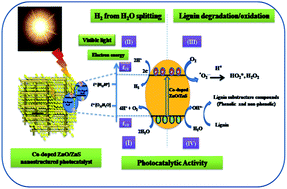
Herein, we have reported the simultaneous water splitting and lignin (biomass) degradation using C, N and S-doped ZnO nanostructured materials. The synthesis of C, N and S-doped ZnO was achieved via calcination of bis-thiourea zinc acetate (BTZA) complex. Calcination of the complex at 500 °C results in the formation of C, N, and S doping in a mixed phase of ZnO/ZnS, whereas calcination at 600 °C gives a single phase of ZnO with N and S-doping, which is confirmed by XRD, XPS and Raman spectroscopy. The band gap of the calcined samples was observed to be in the range of 2.83–3.08 eV. Simultaneous lignin (waste of paper and pulp mills) degradation and hydrogen (H2) production via water splitting under solar light has been investigated, which is hitherto unattempted. The highest degradation of lignin was observed with the sample calcined at 500 °C, i.e., C, N, S-doped ZnO/ZnS when compared to the sample calcined at 600 °C, i.e., N and S doped ZnO. The degradation of lignin confers the formation of a useful fine chemical as a by-product, i.e., 1-phenyl-3-buten-1-ol. However, excellent H2 production, i.e., 580, 584 and 643 μmol h−1 per 0.1 g, was obtained for the sample calcined at 500, 550 and 600 °C, respectively. The photocatalytic activity obtained is considerably higher as compared to earlier reported visible light active oxide and sulfide photocatalysts. The reusability study shows a good stability of the photocatalyst. The prima facie observations show that lignin degradation and water splitting is possible with the same multifunctional photocatalyst without any scarifying agent.
On the other hand, attention on non-conventional energy for domestic purposes has increased due to increased fuel prices, which arise either by the depletion of oil resources or its dependency on its supply from a producer. Amongst the renewable energy resources, solar energy is the largest exploitable resource, and it is estimated that ∼0.014% of the solar energy reaching the earth is enough to sustain domestic requirements.14 The efficient and direct conversion of solar energy into chemical energy, preferably solar light driven photocatalytic water splitting and degradation of lignin, has an immense importance.15,16 The number of photocatalysts, such as TiO2, TiO2–graphene, tungsten carbide (WC), zinc oxide (ZnO), zinc sulfide (ZnS) and tin oxide SnO2 that have been reported are considered to be the best with respect to H2 production and lignin degradation in UV light.6,10,13,17–28 However, widespread use of both TiO2 and ultraviolet (UV) light are not economical for large-scale water treatment, thereby interest has been focused towards visible light driven photocatalysis.
To the best of our knowledge there is no report on the simultaneous water splitting and degradation of lignin via visible light driven photocatalysis. Therefore, in this manuscript an attempt has been made to examine both the photocatalytic water spitting and degradation of lignin by a multifunctional visible light active photocatalyst. The complete degradation of lignin (sodium salt of lignosulfonate) obtained from an industrial site (Vikarabad pulp & paper mills Pvt. Ltd., India) and Aldrich chemicals as well as simultaneous H2 production via water spitting are successfully performed using carbon, nitrogen- and sulfur-doped ZnO/ZnS (C, N, S-doped ZnO/ZnS) as a solar light active photocatalyst. Research on the photocatalytic degradation of lignin using solar light is an effective solution to trim down the pollution caused due to paper & pulp mills. Therefore, the degradation of lignin has also been explored under solar light irradiation. The degradation of lignin conferred a useful fine chemical as a by-product, i.e., 1-phenyl-3-buten-1-ol. From this, we can achieve maximum clean energy (H2 production) and at the same time environmental problems caused by lignin contamination in water can be minimised.
![]() | (1) |
:
ZnS was found to be 1
:
1.5, which was calculated from area under the deconvoluted peaks of ZnO at 2θ = 31.8° (100), 34.4° (002), 36.3° (101), 68.0° (112) and ZnS peak at 2θ = 27.5° (100) (ESI S-1 Table SI-1 and Fig. SI-2†). We thought that heating the sample at an intermediate temperature of 550 °C will produce a mixed phase composition 1
:
1, but it shows a significant ZnO phase (JCPDS = 36-1451) (Fig. 1). The XRD of the sample calcined at 600 °C (S3) and 700 °C (S4) show the existence of a hexagonal wurtzite ZnO structure and the as-prepared complex shows characteristics peaks of the BTZA complex (ESI S-1 Fig. SI-1†). The crystallite size, as determined by the Scherrer equation, was found to be 18, 20, 22 and 32 nm, for the S1, S2, S3 and S4 samples, respectively. It is quite evident that the average crystallite size increases with temperature as per the crystal growth phenomenon, i.e., Ostwald ripening.
The extent of C, N and S doping into the interstitial positions of sample S1 and S3 were determined by XPS (Fig. 2(A)–(F)). During the thermal treatment, the complex decomposes into ZnS and ZnO at 500 °C (S1), but the sample calcined at a higher temperature of 600 °C (S3) shows a single phase of ZnO as observed in XRD and the Raman spectra. The XPS survey spectrum of sample S1 and S3 shows the presence of peaks assigned to Zn, O, C, N and S (Fig. 2(A)). High resolution XPS spectra of the Zn 2p and O 1s lines are shown in Fig. 2(B). The shifting in core lines at Zn 2p3/2 and 2p1/2 was from 1022.1 to 1021.6 eV and from 1045.2 to 1044.8 eV for sample S1 and S3, respectively. This shifting of Zn 2p3/2 and 2p1/2 core lines was by ∼0.5 and 0.4 eV, respectively, which is due to doping of C, N and S into the interstitial positions of S1.29–31 However, calcination at higher temperature (600 °C) caused a decrease in the extent of doping in sample S3 (Fig. 3(B)).29–31
![]() | ||
| Fig. 2 XPS spectra of the S1 and S3 samples. (A) XPS survey spectrum, (B) zinc, (C) oxygen, (D) carbon, (E) nitrogen and (F) sulfur elements. | ||
The XPS spectrum of O 1s is asymmetric in nature, which indicates some doping over the surface, i.e., the possibility of multi-component oxygen species on the surface region of the samples (Fig. 2(C)). Therefore, spectra were fitted by three Gaussian–Lorentzian located at ∼530, 531.5, 531.6 and 533.5 eV (ESI S-2 Fig. SI-1†). The peak at 531.6 eV corresponds to O2 adsorbed on the sample surface probably as –OH sites.32 The high energy peaks centred at about 530 and 531.5 eV are (O 1s) due to the chemisorbed O2 atoms coordinated with ZnO/ZnS (S1) and surface –OH groups, respectively. The peak at 533.5 eV is probably due to R–C
S group associated with either Zn or O.32 This indicates that the doping of C, N and S into the interstitial positions of the ZnO surface was partially via coordinative bond with oxygen. However, the intensity of O 1s peak was slightly increased in case of sample S3 when compared to sample S1 calcined in air (Fig. 2(C)). This clearly indicates a decrease in oxygen deficiency due to calcination, and the oxidation of C, N and S. This decrease in doping (N and S) was also observed in UV, Raman and XPS (ESI S-2 Table SI-1†). The area under the main peaks (530 and 531.5 eV) was found to be higher in S3 than sample S1, which shows a decrease in the oxygen deficiency (ESI S-2 Table SI-1†).
The XPS spectrum for C 1s is shown in the Fig. 2(D). Fitting the spectrum with Gaussian–Lorentzian functions, it can be extracted into two components centred at binding energies of 279.9 and 284.6 eV in case of sample S1. The component (279.9 eV) can be assigned to the carbon contributions from the R–C
S associated with Zn or trapped interstitial positions in the ZnO/ZnS.33 The lower binding energy suggests that carbon may be incorporated into the interstitial positions of the ZnO lattice.34 These two components (279.9 and 284.6 eV) were only observed in the case of sample S1, whereas in the case of sample S3 a single component (284.6 eV) was observed, which was assigned to a standard XPS carbon component. This strongly suggests that carbon can be indeed incorporated into the interstitial positions of the ZnO/ZnS lattice in the case of sample S1, which is not observed in the case of sample S3.
Fig. 2(E) depicts that the N 1s peaks appearing at 398.4 eV were assigned to nitrogen incorporation in ZnO.29,35 The peaks appeared at 395.0, 396.7, 398.8 and 399.9 eV, which indicates nitrogen incorporation to form N–Zn, N–C, N–C
S and N–H bonds, respectively.29,36,37 However, with an increase in calcination temperature to 600 °C, the extent of nitrogen doping was lower in sample S3 when compared to that in sample S1 (ESI S-2 Table SI-1†). The XPS results are consistent with the interpretation of the Raman spectroscopy data, which demonstrates successful N-doping into the interstitial positions of both the samples.
The S 1s XPS peaks appearing at ∼161.3 and 168.6 eV were assigned to ZnS and R–C
S, respectively. They indicate the formation of ZnS along with C, N, S-doped ZnO in sample S1, which is in good agreement with the XRD and Raman data (Fig. 3(F)).38,39 The peak at 161.3 eV (ZnS) disappeared due to calcination at a higher temperature (600 °C), whereas a peak at 168.6 eV with lower intensity was observed in the case of sample S3, when compared to sample S1. This indicates that in both the samples, sulfur is incorporated into the interstitial positions of ZnO.
The percentage elemental composition of C, N, S doping present in the interstitial positions of ZnS/ZnO was found to be 34
:
17
:
49 and 0
:
28
:
72 for the S1 and S3 samples, respectively (ESI S-2 Table SI-1†). This clearly indicates that the percentage doping of the C, N and S elements is temperature dependent and one can easily tune this ratio by applying a suitable temperature. All the above observations confirm that sample S1 is a C, N, S-doped ZnO/ZnS mixed phase material, whereas sample S3 contains nitrogen and sulfur-doping with a single phase of ZnO.
Raman scattering was performed for the C, N, S-doped ZnO/ZnS and commercial ZnS samples, as shown in Fig. 3. In all the spectra, a common Raman peak was located at 437 cm−1 except in the commercial ZnS sample (428 cm−1). This peak corresponds to the E2 (high) vibrational mode, which implies the existence of a wurtzite structure. The peak located at 333 cm−1 corresponds to the second order (E2 (high)–E2 (low)).40 The other modes are at A1 (278 cm−1), A3 (509 cm−1) and A1 (LO) (580 cm−1) and can be assigned to the local vibration modes (LVMs) of nitrogen in ZnO.41 The Raman modes at 278 cm−1 were attributed to the localized vibration of Zn atoms, where parts of their first nearest oxygen atoms are partially replaced by nitrogen atoms in the ZnO lattice.42 Moreover, the Raman peaks observed at 509 and 580 cm−1 are related to the presence of nitrogen.43,44 It is observed that the intensity of the nitrogen related peaks gradually decrease due to calcination at higher temperature (S3 and S4), which is consistent with the XPS results. This clearly indicates the nitrogen escapes at high temperature.
Xue et al. reported an S-doped ZnO Raman band located at 1003 cm−1 associated with C–S group; however, this is observed at 979 cm−1 in our case and is in good agreement with the previously reported method.45 This might be due to sulfur doping via –C
S groups effectively shifting the Raman band.46,47 The 1385 cm−1 band was attributed to –CO vibrations.40 The 702 and 1100 cm−1 bands were attributed to C–S and C–N vibrations, respectively, which is matching with the commercial ZnS sample (Fig. 4).47 When the sample was heated at higher temperature (600 to 700 °C), a gradual decrease in the intensity of the nitrogen related Raman signal intensities were observed, as shown in the XPS analysis.
The morphology of the prepared compounds was determined by FESEM (Fig. 4(A)–(C)). Sample S1, i.e., a mixed phase of ZnO/ZnS, exhibits a three dimensional (3D) nanoplate-like structure. These nanoplates are arranged in such a way that they look like a honeycomb structure. Interestingly, the spherical nanoparticles are located in between the crossed nanoplates. FESEM also shows the formation of spherical nanoparticles on the surface of nanoplates. It seems that these nanoparticles originate from the secondary growth of nanoplates due to calcination. The thickness of the nanoplates was observed to be 30–50 nm and the nanoparticles between the crossed nanoplates were in the range of 30–40 nm. Further, the TEM investigations also show plate-like structures with nanoparticles on the surface. The careful observation conveys that there is porous honeycomb-like structure, which might be due to various processes occurring during open air calcination such as (1) superficial oxidation of ZnS sheets converted into spherical ZnO nanoparticles (30–40 nm), (2) the formation of a porous morphology due to evolution of gases such as CO2, NO2, and NH3 and (3) the formation of cross-linked plates as a result of –C
S, –SO2, etc., being associated with ZnO/ZnS, which requires high temperature to decompose. The formation of a ZnO/ZnS mixed phase material is also revealed from the XRD pattern. The FESEM of sample S3 shows slightly wavy nanoplates (Fig. 4(B1)). The higher magnified image shows highly crystalline nanoparticles (40–50 nm) on the surface of these wavy nanoplates. An almost similar morphology is observed in the case of the sample calcined at 700 °C (S4). This change from honeycomb-like porous morphology to spherical nanoparticles might be due to calcination at higher temperature (600 and 700 °C), which might be due to partial or complete oxidation of dopants such as C, N and S.
The crystalline nature and morphology of S1 and S3 were confirmed using TEM (Fig. 5). Sample S1 clearly shows the presence of nanoparticles on the surface of nanoplates (Fig. 5(a)). The size of the plates and nanoparticles was found to be ∼500 and 30 nm. At higher magnification, it is observed that the layer of nanoparticles is adhered to the nanoplates (Fig. 5(b2)). It suggests that, oxidation starts at the surface of sheets of ZnS, which is later converted into ZnO. The XRD and Raman studies also confirm the formation of a mixed phase ZnS and ZnO structure. Furthermore, the HRTEM image taken at the surface of ZnO the nanoparticles shows an interplanar distance of ∼0.28 nm corresponding to the (100) plane of wurtzite ZnO (Fig. 5(b) and (b1)). The electron diffraction pattern demonstrates the blur rings due to the polycrystalline nature of sample S1 (ESI S-9 Fig. SI-1(a)†), whereas, the TEM image of sample S3 reveals a porous sheet-like morphology, which is composed of ZnO nanoparticles. This might be a result of the complete oxidation of ZnS nanoplates. The size of the nanoparticles was observed to be ∼40–50 nm, which is consistent with the FESEM results. The HRTEM image was taken at the edge of the ZnO nanoparticles and shows their highly single crystalline nature with lattice fringes ‘d’ spacing 0.283 nm, which correspond to the (100) plane of wurtzite ZnO (Fig. 5(d)). The formation of ZnO nanoparticles takes place accompanied with lattice contraction (from 6.188 to 5.206 Å), and results in the spherical nanoparticles morphology.48–50 When the hexagonal ZnS is completely transformed into wurtzite ZnO at a calcination temperature of 600 °C, large numbers of nanopores were formed between the spherical nanoparticles as a consequence of lattice volume contraction during calcination and in this way the matrix released the tensile stress accumulated within it.49 It might be due to the doping of S and N, which is consistent with the Raman and XPS interpretations. Moreover, the FFT pattern (Fig. 5(d1)) exhibits the hexagonal crystal structure of ZnO. The electron diffraction pattern demonstrates the blur ring pattern due to the different orientation of nanoparticles (ESI S-9 Fig. SI-1(b)†).
The UV-visible absorption spectra of the doped ZnO samples annealed at different temperatures are presented in Fig. 6(a). The reported absorption cut off edge for the commercial ZnO and ZnS samples are ∼375 and 324 nm, respectively.19,35 The samples S1 to S4 show an absorption edge cut off at ∼438, 427, 403 and 396 nm, respectively. The corresponding band gap were found to be 2.8, 2.9, 3.1 and 3.13 eV for the as-prepared samples, which is slightly blue shifted due to the removal of C, N, S doping as shown in S1 to S4, respectively (ESI S-4 Fig. SI-1†). The energy difference between the aforementioned two features demonstrates the creation of a midgap (deep level acceptor) state in the band gap. However, in the case of sample S4, due to the higher calcination temperature (700 °C) there is an escape of dopants, and hence pure zinc oxide is formed. This suggests that the visible light absorption is due to the introduction of C, N and S into the ZnO/ZnS lattice.
![]() | ||
| Fig. 6 (a) UV-DRS spectra of S1, S2, S3 and S4 samples. (b) PL spectra of S1, S2, S3 and S4 samples. | ||
The photoluminescence (PL) is an effective method to investigate the optical characteristics of semiconductor nanomaterials because information, such as surface oxygen defects as well as separation and recombination of photoinduced charge carriers, can be obtained. Room temperature PL spectra of the as-prepared samples were obtained with an excitation wavelength of 350 nm and are shown in Fig. 6(b). The broad green emission band centred at 510 nm was due to the defects created by the deficiency of oxygen observed in sample S1 and S2, with an increase in calcination temperature (from 500 to 600 °C) the broad emission peak centred at 510 nm is normalized.51,52 Arbuj et al. reported that the visible emission generally designated as a deep level emission was probably related to the variation of the intrinsic defects in ZnO such as the vacancies created on the surface and at the interstitial positions due to zinc and oxygen. In other words, the origin of the visible emission in ZnO is highly controversial.20,53–56 It is clearly demonstrated that with an increase in the calcination temperature (from 500 to 700 °C), there was a steady decrease in C, N and S-doping due to oxidation of these elements. As a result, oxygen deficiency was minimized due to decrease in dopant concentration.
After careful evaluation of all the abovementioned results, the overall formation mechanism of C, N and S-doping either in mixed ZnO/ZnS or in a ZnO phase by the thiourea route can be observed by Scheme 1. eqn (1) and When zinc acetate and thiourea are mixed in an alcoholic medium (methanol) at room temperature, the BTZA complex is formed.27 We have carried out the reaction by maintaining the ratio of zinc acetate to thiourea at 1
:
2. Further, the complex sample was annealed at 500, 550, 600 and 700 °C on the basis of the precise thermal study using TG-DTA. The thermal study shows multistage weight losses to obtain ZnO/ZnS. Our foremost endeavour is to dope C, N and S using thiourea, which we have achieved at 500 and 600 °C (samples S1 and S3). On the basis of the critical assignment of weight loss and corresponding endothermic changes in the DTA observed during the thermal decomposition of the BTZA complex. We have proposed the reaction mechanism in Scheme 1. According to the TG-DTA (ESI: S-0, Fig. S-1†), further calcination at 185 °C shows a gradual decomposition of the BTZA complex and weight loss in the form of CO2, NO2, NH3, –CH3, etc. gases as explained in TG-DTA section. The thiourea and its decomposed products, such as a NH3, NO, –C
S, CO, and SO are in intimate contact with the complex, and hence the species containing C, N and S in these decomposed products can act as a source for doping in ZnO/ZnS, which is being formed at high temperature in atmosphere.57 The formation of a mixed phase of ZnO1−x(C, N and S)x and ZnS in the case at 500 °C (S1) is clearly seen in HRTEM. There is a formation of a ZnO grain domain on the ZnS plate as presented in Scheme 1 (S1). These ZnO grain domains are secondary growth of ZnO nanoparticles on the ZnS plates. At 600 °C (S3) a single phase of ZnO1−x(NS)x was already confirmed by XRD, XPS and Raman spectroscopy. In this case the plate-like structure is completely transformed into spherical nanoparticles as presented in Scheme 1. At 700 °C, pure phase ZnO (S4) was formed, which is also confirmed by XRD and Raman spectroscopy. This morphological transformation of ZnS to ZnO with C, N, S doping at different temperatures is presented in Scheme 1.
Considering the band gap of doped ZnO in the visible region, the photocatalytic activity of lignin degradation and H2 production by water splitting was performed under visible light. The levels of H2 production with and without lignin (WWL) over the prepared samples (S1 to S4), and commercial catalysts is shown in Table 1 and Fig. 7. The sample S1 showed 580 and 558 μmol h−1(0.1 g)−1 (5800 and 5580 μmol h−1 g−1) of H2 generation WWL, which is slightly lower than that of sample S2 (5840 and 5700 μmol h−1 g−1) and sample S3 (6430 and 6020 μmol h−1 g−1 WWL) photocatalysts (Table 1 and Fig. 7(a) and (b)). Surprisingly, sample S1 shows complete degradation of lignin as well as H2 production by water splitting under similar conditions (Table 1, ESI S-5 Fig. SI-1†). This was due to the band gap energy of 2.83 eV, which is ideally in the visible region. Hence, a significant amount of electron and holes can be generated by ZnO/ZnS under visible light illumination. The H2 generated by photocatalytic water splitting was analysed by GC (ESI S-6 Fig. SI-1†). In case of sample S1, the oxygen defects are more therefore, out of the total electrons generated, some of the electrons are trapped in the mid gap state created due to the ZnO/ZnS interface. Hence, H2 production is slightly lower when compared to that of sample S3, whereas the photogenerated holes are completely utilized for lignin degradation in the case of sample S1, which is not observed in sample S3. Sample S4 showed two times (276 and 223 μmol h−1 (0.1 g)−1 WWL) lower H2 production than sample S1, S2 and S3 (Table 1). Thus, the decrease in the activity of sample S4 can be clearly understood due to its higher band gap, i.e., 3.13 eV, which is in the UV region. Because there is no considerable difference in particle size, it is not wise to use this parameter to account for the enhancement, though it will contribute to some extent in all samples.
| Catalysts | Surface area (m2 g−1) | Average H2 generation (μmol h−1) | Apparent quantum yield | ||
|---|---|---|---|---|---|
| With Lignin | Without Lignin | With Lignin | Without Lignin | ||
| a Reaction conditions: catalyst, 0.1 g; water, 200 ml; lignin, 100 ppm; Xe lamp, 300 W (Oriel).b Standard deviation is 0.957 and 0.019, in the case of volume of H2 generated and apparent quantum yield, respectively.c Physical mixture of commercial ZnO and ZnS catalysts.d Indicates the activity of catalyst after the fourth recycle.e Indicates the industrial (Vikarabad Pulp & Paper Mills Pvt. Ltd., India) sample of lignin. | |||||
| S1 | 74.7 | 580b | 558 | 14.16b | 13.33 |
| S2 | 51.1 | 584 | 570 | 14.51 | 13.91 |
| S3 | 39.4 | 643 | 602 | 15.05 | 14.25 |
| S4 | 33.8 | 276 | 223 | 8.10 | 5.10 |
| Commercial ZnO | — | 89 | 22 | 2.09 | 0.53 |
| Commercial ZnS | — | 156 | 45 | 3.76 | 1.07 |
| c | — | 205 | 45 | 5.00 | 1.02 |
| Blank run | — | 89 | <0.1 | 2.08 | <0.1 |
| S1d | 74.7 | 535 | 527 | 13.47 | 12.4 |
| S1e | 74.7 | 584 | 553 | 14.26 | 13.50 |
The repeatability of the H2 production with lignin using catalyst S1 was observed with good precision (standard deviation 0.957 and 0.019 in the case of volume of H2 generated and quantum yield, respectively). The sample S1 was found to retain its activity after the fourth recycle, which proves the stability of the catalyst (Table 1, ESI S-4 Fig. SI-2†). Interestingly, the lignin sample collected from an industrial site (Vikarabad pulp & paper mills Pvt. Ltd., India) showed equivalent activity in terms of both water splitting and the degradation of lignin. This indicates the suitability of sample S1 for industrial lignin samples and one can think of its use for the effective utilization of sustainable resources for cleaner energy and water purification simultaneously, where both are available abundantly. Furthermore, the MS of an intermediate step sample shows the presence of 1-phenyl-3-buten-1-ol, which resembles a lignin sub-structured compound (ESI S-6 Fig. SI-2†). Further, study with detailed investigations is in progress. It is quite interesting and curious that sample S1 confers H2 production as well as lignin degradation (ESI S-6 Fig. SI-1 and SI-2†). Sample S3 shows the highest H2 production with lignin and slightly less without lignin. However, we could not observe lignin degradation using sample S3. It seems the presence of lignin decreases electron (e−) and hole (h+) recombination. If we consider the photoluminescence, there is drastic decrease in the intensity of the emission peak in the case of samples S2 and S3, indicating that excitonic electron in the conduction level of the semiconductor are transferred to the surface, which is then utilized for H2 evolution reaction.16,56 However, further study is in progress to know the role of lignin in the photocatalysis reaction. It is to be noted that all the reactions have been performed without the use of a scarifying agent. This is quite interesting and also calls for very serious discussion. There might be utilization of holes for lignin degradation and electrons for water splitting. Hence, lignin may play the role of a scarifying agent. However, it needs to be examined thoroughly, which is in progress. Rao et al. clearly mentioned that there is a formation of H2O2 in the reaction in the absence of a scarifying agent.58 Hence, we feel that the same phenomenon might be taking place in the present case. In the presence of lignin, a slight increase in H2 generation may be due to the lignin, which may be acting as a scarifying agent.
Then, the possibility is either the mixed phase composition or co-doping are responsible for this remarkable activity. Hence, we have performed the photocatalysis reaction using individual and physical mixtures of commercial ZnO and ZnS photocatalysts (Table 1 and ESI S-5 Fig. SI-1†). The individual ZnO and ZnS samples showed lower activity WWL when compared to sample S1 (Table 1). However, in a physical mixture ZnO
:
ZnS with a 1
:
1.5 composition showed higher activity than the individual samples and lower than that of sample S1 (Table 1). This clearly indicates that the mixed phase plays a key role in the enhancement of photocatalytic activity.
Fig. 7(c) presents the UV-visible absorption spectra of the (%) lignin degradation with respect to time using sample S1. It revealed that with an increase in time, a gradual decrease in the lignin peak intensity was observed. The complete degradation of 100 ppm lignin was achieved within 5 hours in the presence of 100 mg S1 photocatalyst (Fig. 7(c)). However, 1000 mg S1 photocatalyst showed complete degradation of lignin within 1 hour. We also observed that with an increase in catalyst, the lignin degradation rate was increased but hydrogen production remained constant after a 200 mg catalyst loading. Significantly, this might be due to an effective decrease in electrons at the conduction level where the availability of holes preferentially facilitates the degradation of lignin rather than the water splitting process (ESI S-5 Table SI-1†).
In the previous studies, it is well reported that heterogeneous photocatalytic degradation reactions follow pseudo first order kinetics.56,59 First order reactions follow the rate law, which indicates that the rate of degradation depends on the concentration of reactants (2), and is given as follows:
![]() | (2) |
The reaction kinetics for the heterogeneous photocatalytic degradation of lignin have been studied and a plot of (%) lignin degradation versus time (h) and ln(C0/Ct) versus time (h) were plotted and are shown in ESI S-7 Fig. SI-1(a) and (b),† respectively. The slope of this plot gives the rate constant values as per reaction kinetics of a first order reaction. The rate constant for photocatalytic lignin degradation using 100 mg S1 photocatalyst was found to be 0.1005 h−1. Among the prepared catalysts, sample S1 was the best photocatalyst due to its excellent activity and stability in both H2 production (water splitting) as well as the complete degradation of lignin. However, sample S3 showed a slightly higher H2 production but did not show lignin degradation. Photocatalytic activity obtained for all synthesized samples (S1–S4) was higher than the commercial ZnO, ZnS and their physical mixture samples. In a nutshell, sample S1 was observed to be a versatile photocatalyst, which shows good photocatalytic H2 production performance and complete lignin degradation.
Footnote |
| † Electronic supplementary information (ESI) available. See DOI: 10.1039/c4ra10760h |
| This journal is © The Royal Society of Chemistry 2014 |