A comparative study on degradation characteristics of fluoropolymers irradiated by high energy heavy ions

U. H. Hossain*ab, F. Muencha and W. Ensingerab
aTechnische Universität Darmstadt, Department of Materials Science, Materials Analysis, Alarich-Weiss-Str.2, 64287 Darmstadt, Germany. E-mail: u.h.hossain@gsi.de; Fax: +49-6151-166378; Tel: +49-6159-712651
bGSI Helmholtz Centre of Heavy Ion Research, Planckstr. 1, 64291 Darmstadt, Germany

Received 16th May 2014 , Accepted 18th September 2014

First published on 19th September 2014


Abstract

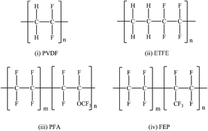
Energetic heavy ions passing through polymer foils cause modifications of properties, first of all structural changes. In order to evaluate ion beam modification of fluoropolymers, a comparison of polyvinylidene fluoride (PVDF), ethylene-tetrafluoroethylene (ETFE), tetrafluoroethylene-per-fluoromethoxyethylene (PFA), and tetrafluoroethylene-hexa-fluoropropylene (FEP) was undertaken. Foils were irradiated with heavy ions (Sm, Au) of a kinetic energy of a few MeV u−1 and fluences up to 6 × 10¹² ions cm−2 at room temperature in vacuum. The ion induced changes in the molecular structure were studied with Fourier transform infrared spectroscopy (FT-IR) and on-line mass spectrometry (Residual Gas Analysis, RGA). The results in terms of macromolecular changes and formation of fragments were compared and related to the individual polymer structures. In general, the polymers lose various small molecular fragments by bond scission of polymeric backbone and side chains, typically fluorine and hydrogen fluoride, as well as trifluoromethyl, and form new bonds, mainly CC double bonds. From the results, the main degradation reactions are derived. The findings are of technical interest for space applications when devices are hit by highly energetic heavy ions from galactic cosmic rays on long-term missions.


1. Introduction

Fluoropolymers are high performance polymers with a carbon chain structure in which terminating hydrogen atoms are at least partly replaced by fluorine atoms. Usually they show a very high resistance to solvents, acids and alkalis. A combination of interesting chemical and physical properties such as high mechanical and electrical resistance, thermal stability and low friction coefficient make it usable in hi-tech and biological applications.1,2

PTFE (polytetrafluoroethylene) being derived from polyethylene (PE) where all the hydrogen atoms have been replaced by fluorine is certainly the most prominent fluoropolymer. Together with its base polymer polyethylene (PE) several studies on ion beam effects have been carried out beforehand.3–9 ETFE (ethylene-tetrafluoroethylene) with only half of the hydrogen atoms replaced by fluorine is another kind of fluoropolymer which is well known for its excellent thermal and chemical stability as well as mechanical properties. On ETFE, the number of ion beam studies is quite limited. Minamisawa et al.10,11 carried out experiments on chemical modifications of ETFE exposed to protons (with a kinetic energy of 1 MeV) and Si ions (with 100 keV). Ion beam irradiation caused scission of the polymer backbone chain and cross linking.

Polyvinylidene fluoride (PVDF) is another excellent fluoropolymer material which is easy to shape by melting due to its low melting point; also, it shows the lowest density of all fluoropolymers. These features are favourable for certain industrial applications. PVDF has been investigated in a few studies by Duraud et al.6 and by Balanzat and Betz et al.12–14 They studied the changes in its chemical properties by exposing it to electrons (with 1 MeV) and Kr-ions (with 5 MeV u−1). ETFE and PVDF have a similar structure, with the difference being that ETFE contains vicinal C–F and C–H bonds in pairs of two, whereas PVDF is based on a backbone containing alternating C–F and C–H bonds, see Fig. 1. So far, these two polymers have not been compared for any potential differences in their ion-induced chemical degradation behaviour.


image file: c4ra04635h-f1.tif
Fig. 1 Molecular formulae of (i) polyvinylidene fluoride (PVDF), (ii) ethylene-tetrafluoroethylene (ETFE), (iii) tetrafluoroethylene-per-fluoromethoxyethylene (PFA) and (iv) tetrafluoroethylene-hexa-fluoropropylene (FEP).

Another couple of fluoropolymers is tetrafluoroethylene-per-fluoromethoxyethylene (PFA) and tetrafluoroethylene-hexa-fluoropropylene (FEP). Both of them have useful properties like low coefficient of friction and non-reactivity, show a better creep behaviour than other fluoropolymers and they can be melt-processed. They are similar to PTFE, with the difference being additional side chains of –CF3 and –OCF3, i.e. while FEP has a trifluoromethyl group attached to the carbon chain by a C–C bond, PFA contains an ether bond (Fig. 1(iii)). Hence, the composition differs in the presence of an O atom in the side-chain. These two polymers have hardly been investigated with respect to damage by swift ions, particularly heavy ions. Parada et al. studied the effect of proton irradiation on PFA and FEP,15,16 Minamisawa et al. reported on PFA damage by gold ions.17

The present work is part of a research program studying the swift heavy ion (here Au and Sm) induced modification of polymers, including fluoropolymers with different side groups, namely the above mentioned PVDF, ETFE, PFA and FEP. As mentioned before, very few studies have dealt with high energy heavy ion induced degradation of these polymers, and an investigation with all four polymers, particularly for comparing the differences, if any, has not yet been carried out. This paper presents a spectroscopic study of the transformations upon irradiation in vacuum at ambient temperature up to particle fluences of 6 × 10¹² ions cm−2. The ion degradation of these polymers was followed by the analysis of the evolved volatile products with on-line mass spectrometry (Residual Gas Analysis, RGA) and the changes in the polymer molecular structure by post irradiation experiments with Fourier transform infrared spectroscopy (FT-IR). This study addresses basic research on heavy ion induced modification of fluoropolymers and the underlying molecular degradation mechanisms.

2. Experimental

The fluoropolymers with thickness of 25 μm used for the present study were commercially available PVDF (1.7 g cm−3), ETFE (1.7 g cm−3), FEP (2.1 g cm−3) and PFA (2.1 g cm−3) supplied by DuPont de Nemours.

The samples were irradiated at the X0-Branch and M-Branch18 beam lines of the Universal Linear Accelerator (UNILAC) at GSI Helmholtz Centre of Heavy Ion Research (Darmstadt, Germany) with 11.1 MeV u−1 Samarium (atomic mass 150 u, total kinetic energy 1.76 GeV) and 4.8 MeV u−1 Gold (atomic mass 197 u, total kinetic energy 0.96 GeV) ions using a pulsed beam of 5 Hz repetition rate and 3 ms pulse length. The applied fluences for X0-Branch and for M3-Branch were 3 × 10¹⁰ to 6 × 10¹² ions cm−2 and 2 × 10¹¹ ions cm−2. A set of samples (PVDF, ETFE, PFA and FEP) were irradiated with Sm ions at M3-Branch with different fluences and were analyzed with on-line mass spectrometry (RGA). PVDF foils were irradiated with Au ions at X0-beamline. The particle flux was kept low (108 ions cm−2 s−1) in order to rule out thermal effects. Concerning the heavy ions, there is no particular reason why Sm and Au have been used. This was based on the availability at the large scale facility. However, from earlier studies on other polymers it is known that the basic mechanisms of ion induced modifications of polymers by heavy ions are essentially the same for different types of ions.19,20

FT-IR spectral measurements were performed using a NICOLET 6700 FT-IR spectrometer of Thermo Fisher Scientific in the range 400–4000 cm−1, with a resolution of 2 cm−1. Software OMNIC 7.0 was used for data processing. Quadrupole mass spectrometry (Residual Gas Analysis) was used to identify the outgassing degradation products. It was performed during irradiation at M3-Branch using a Microvision Plus quadrupole mass spectrometer from MKS in analogue mode with a resolution of 0.03 in a mass-to-charge (m/z) range from 1 to 200. The acquisition time of each mass spectrum was approximately 35 s and the spectra were acquired continuously throughout the irradiation experiment.

3. Results and discussion

3.1 Fourier transform infrared spectroscopy and mass spectrometry

As mentioned above, the ionizing radiation affects the molecular structure of the polymers. IR-spectroscopy and mass spectrometry are efficient and complementary tools for analysing structural changes. In the following, the individual behaviour of PVDF, ETFE, FEP and PFA as analysed by FT-IR and mass spectrometry is discussed in detail. Eventually, upon comparing, a general conclusion is drawn. In this study, we focus on chain scission and fragmentation, since they can be most easily detected with the employed techniques. Especially at high radical concentrations, cross linking of different polymer strands displays alternative reaction pathways. Therefore, further studies should use alternative techniques such as solid state NMR (to detect tertiary carbon atoms formed by cross linking) to evaluate the degree of cross linking.8
3.1.1 PVDF. Fig. 2 presents the FT-IR absorption spectra of pristine and irradiated PVDF foil with Au ions at different fluences up to 2 × 10¹² ions cm−2. At first glance, there are three general observations. The spectra show a decrease of the main bands, an increase in the background, and the appearance of new bands. Well visible on the background are the two absorption bands at 3024 and 2981 cm−1 which are identified as symmetric and asymmetric vibration modes of the aliphatic –CH2-groups which constitute half of the molecular structure of PVDF. Ion irradiation within the explored fluence regime leads to a decrease in intensity of these bands and to the development of two small shoulders at 2921 and 2851 cm−1 assigned to the asymmetric and symmetric stretching vibrations of other CH-groups, i.e. ones with a different molecular environment. The decrease of band intensity and appearance of two new shoulders reflect the molecular modification of the polymer. The observed –CH2 band degradation of PVDF by ion irradiation is known from the literature.12–14 In addition, in the spectrum of the PVDF, irradiated with 3 × 10¹¹ ions cm−2, three new peaks appear at 1752, 1711 and 1622 cm−1. These signals are explained by the stretching vibrations of developing unsaturated C-bonds: –CF[double bond, length as m-dash]CH–, and terminal –CH[double bond, length as m-dash]CF2– and –CF[double bond, length as m-dash]CH2. This finding is corroborated by the mass spectrometric observation of high amounts of outgassing H and F atoms, mostly found as their reaction products H2 and HF (Fig. 3). The carbon chain compensates the loss of H and F bonding partners by the formation of unsaturated C-bonds. As can be seen in Fig. 2, while the C–H bonds (around 3000 cm−1) and the C–F bonds (1140–1280 cm−1)21 are reduced in number, the number of C[double bond, length as m-dash]C bonds (1600–1800 cm−1) is rapidly increasing with ion fluence. The low wavenumber region between 1500 and 500 cm−1, the fingerprint region, contains a complex series of absorption bands. These may be ascribed to wagging, twisting and rocking vibration of the C–H and C–F groups. During irradiation all band intensities in this region are decreasing with increasing ion fluence. Besides, a broad band is observed in the high wavenumber region of 3100–3700 cm−1 (red curve in Fig. 2) which can be assigned to the vibration of –C[triple bond, length as m-dash]C– bonds. These triple bonds are formed when neighbouring carbon atoms completely lose their binding partners H or F.
image file: c4ra04635h-f2.tif
Fig. 2 FT-IR spectra of PVDF: Pristine (black), irradiated with Au ions of 4.5 MeV u−1 with fluences of 3 × 1011 cm−2 (blue) and 2 × 10¹² ions cm−2 (red).

image file: c4ra04635h-f3.tif
Fig. 3 (a) Outgassing spectra of PVDF online recorded without irradiation and during irradiation with 11.1 MeV u−1 Sm ions, (b) difference spectrum, indicating the outgassing species caused by the heavy ion irradiation.

In accordance with the FT-IR results, mass spectra show that not only the side elements bonded to the carbon backbone are cut off but also the PVDF backbone structure itself is broken up upon ion irradiation, yielding several C-containing fragments such as C, CH2F or CHF2. Prior to irradiation, a gas inventory composed of hydrogen, water, oxygen, carbon dioxide, and hydrocarbons has been found in the vacuum chamber, coming from wall desorption, leakage, and pump residues. The volatile fragments of the polymer add to these components. Since the ion beam transfers enough energy to cut all types of bonds easily, a complex fragmentation scheme with various species can be expected. However, the main outgassing fragments are the above-mentioned small volatile species hydrogen (H, m/z = 2) and hydrogen fluoride (HF, m/z = 20). From the viewpoint of risk evaluation, it therefore must be considered that the irradiation of PVDF with heavy ions releases substantial quantities of these gases, particularly of the aggressive HF. Interestingly, no F2 molecule ions were detected with mass spectrometry. This is probably related to the extreme reactivity of the F radical and the high bond strength within the HF molecule, leading to the nearly complete reaction of the outgassing F to HF, rather than to F2. This observation is in accord with literature describing the selective and fast abstraction of H atoms by fluorine radicals in the case of CHF3 molecules which contain both H and F substituents,22 and with the well-established mechanism of radicalic substitution of hydrocarbons by halogens, resulting in a consecutive replacement of hydrogen atoms.23

The results suggest the reactions shown in Fig. 4 as the main degradation mechanism. The degradation process can be initiated by the (i) homolytic C–C bond breaking, (ii) release of an H atom, or (iii) an F atom, creating either a terminal difluoro-ethylene radical or a dihydro-ethylene radical which later form carbon–carbon double bonds by elimination of hydrogen-fluoride (HF). The intensity decrease of hydrocarbon groups found by FT-IR spectroscopy is correlated with mass spectrometric results which show a main mass signal at m/z = 20 (HF). Hence, the proposed mechanism is supported by both analysis methods.


image file: c4ra04635h-f4.tif
Fig. 4 Schematic presentation of the main heavy ion induced degradation reactions of PVDF.

In this schematic the backbone scission, forming C1- and C2-based species, is not shown, as well as the cross linking reaction between C-atoms of different chains. Of course, these reactions take place, however, to a lesser extent, as the mass spectrum shows for the scissions.

3.1.2 ETFE. The vibrational-spectroscopic features observed for pristine and Sm-irradiated ETFE are presented in Fig. 5. Pristine ETFE exhibits two absorbance bands at 2975 and 2881 cm−1 in the wavenumber range 3000–2800 cm−1. They correspond to the symmetric and asymmetric stretching vibration of –CH2 groups. At the low wavenumber region (1500–500 cm−1), several absorbance bands can be assigned to the presence of hydrocarbon and C–F bonds.
image file: c4ra04635h-f5.tif
Fig. 5 FT-IR spectra of ETFE: Pristine (black), irradiated with Sm ions with a fluence of 3 × 10¹¹ ions cm−2 (blue) and 3 × 10¹² ions cm−2 (red).

A significant change of all characteristic vibration bands of irradiated ETFE foils has been observed with increasing ion fluence. During irradiation at fluences above 3 × 10¹² ions cm−2, the hydrocarbon bands at 2975 and 2881 cm−1 decrease and a broad shoulder appears which is an indication for a mixture of alkyne-containing degradation products. This is the same situation as with PVDF. No characteristic absorption bands were found in the wavenumber range 1800–1600 cm−1 for pristine ETFE foil, but the sample irradiated with 3 × 10¹¹ ions cm−2 exhibits two new absorption bands at 1722 and 1624 cm−1 corresponding to the carbon–carbon double bond of –CF[double bond, length as m-dash]CH– and –CH[double bond, length as m-dash]CH– groups. The overall decreasing tendency of absorption bands at low wavenumber region between 1500 and 500 cm−1 is basically the same as in the case of PVDF.

Hence, ion induced degradation shows a reduction in the number of aliphatic C–H groups at 2975 and 2881 cm−1, and an increase in carbon–carbon double bonds at 1722 and 1624 cm−1 due to the scission of H and F, under release of HF.

Fig. 6 shows the typical mass spectra of pristine and irradiated ETFE foils indicating outgassing fragments of hydrogen (H2, m/z = 2), fluorine (F, m/z = 19), hydrogen fluoride (HF, m/z = 20), CF (m/z = 31) and CHF2 (m/z = 51). A possible degradation mechanism of ETFE is depicted in Fig. 7 indicating the unsaturation of carbon bonds found by FT-IR spectroscopy and the presence of outgassing fragments by mass spectrometry. Hence, the degradation process for both polymers (PVDF and ETFE) is very similar and mainly based on the homolytic bond breaking within the backbone and the elimination of hydrogen and fluorine as free radicals, forming either hydrogen molecules or HF. This causes the formation of C[double bond, length as m-dash]C double bonds. As a consequence of the reaction, the hydrocarbon bands are decreased at 2975 and 2881 cm−1 and the C[double bond, length as m-dash]C double bond signal in the wavenumber range of 1800–1600 cm−1 is increased in intensity as observed by FT-IR spectroscopy. This mechanism is consistent with the mass spectrometric observation of ETFE.


image file: c4ra04635h-f6.tif
Fig. 6 (a) Outgassing spectra of ETFE online recorded during irradiation with 11.1 MeV u−1 Sm ions, (b) difference spectrum, indicating the outgassing species caused by the heavy ion irradiation.

image file: c4ra04635h-f7.tif
Fig. 7 Scheme representing the degradation mechanism of ETFE polymer.
3.1.3 PFA. The FT-IR spectra of PFA foil irradiated with Sm ions of two fluences in comparison with the non-irradiated foil are presented in Fig. 8. The major absorption bands of pristine PFA are found in the low wavenumber region. The bands at 994 and 775 cm−1 as well as at 741 and 720 cm−1 are assigned to the –CF3 vibration bands of the side chain. Other absorption bands above the fingerprint region (550–500 cm−1) indicate the presence of C–H and C–F bending vibrations.
image file: c4ra04635h-f8.tif
Fig. 8 FT-IR spectra of PFA: Pristine (black), irradiated with Sm ions with 3 × 1011 cm−2 (blue) and 6 × 10¹² ions cm−2 (red).

As shown in Fig. 8, a new broad IR absorption band at 1717 cm−1 appears with the increase of ion fluence. According to the literature,24 the new broad band at about 1717 cm−1 can be assigned to the stretching vibration of the –CF[double bond, length as m-dash]CF– group. Once the foil was irradiated with a fluence beyond 3 × 10¹¹ ions cm−2, the intensities of characteristic absorption bands at 738, 724 and 704 cm−1, as well as the bands at 550–500 cm−1 decreased, which is consistent with the results of the before mentioned other fluoropolymers (PVDF and ETFE).

Fig. 9 shows the mass spectrum of pristine and irradiated PFA. The main characteristic outgassing fragments are carbon (C, m/z = 12), fluorine (F, m/z = 19), hydrogen fluoride (HF, m/z = 20), carbon monoxide (CO; m/z = 28), CF (m/z = 31), CO2 (m/z = 44), CFO (m/z = 47), CF3 (m/z = 69) and C2F3 (m/z = 81). It is important to note that PFA has a fully fluorinated carbon chain structure with a small amount of oxygen atoms in the side chain so that the amount of oxygen-bearing fragments is rather small, while there are several carbon-based fragment molecules with different numbers of F atoms. It turns out that the mass signal m/z = 69 of CF3 is particularly large, considerably larger than CF and more than an order of magnitude above the value of CF2. A possible mechanism may be the scission of CF2 radicals from the polymeric backbone followed by recombination with adjacent fluorine atoms to form CF3 molecules.25 Since the C–F bonds are stronger than the C–C bonds, it can be assumed that the latter are more easily broken. With 490 kJ mol−1, the polar C–F bond is, indeed, the strongest single bond in organic chemistry, while the C–C bond has a dissociation energy of only 350 kJ mol−1.26,27 Consequently, the scission of the backbone occurs easier than the disintegration of the trifluoromethyl-group.


image file: c4ra04635h-f9.tif
Fig. 9 (a) Outgassing spectra of PFA polymer online recorded during irradiation with 11.1 MeV u−1 Sm ions, (b) difference spectrum, indicating the outgassing species caused by the heavy ion irradiation.

In PFA, double bond formation becomes visible at fluences of 1 × 10¹² ions cm−2. These are either isolated double bonds and dienes, but also a certain fraction of longer polyene sequences are found. This has also been observed for aliphatic polymers.28

Backbone scission and elimination of the side chain –OCF3 are believed to be free radical reactions, see Fig. 10. It starts at a low fluence irradiation and gives double bond formation within the macromolecular backbone. With increasing fluence, chain scission occurs to some extent, as a result of which more –OCF3 groups are eliminated and end-chain unsaturation appears. This mechanism explains both the appearance of CF3, CF and CFO in the mass spectrum, as well as the observed double bond formation in the IR spectrum.


image file: c4ra04635h-f10.tif
Fig. 10 Schematic presentation the degradation mechanism of PFA.
3.1.4 FEP. The FT-IR spectra of FEP observed after Sm irradiations at 11.1 MeV u−1 are presented in Fig. 11. The characteristic absorption bands of pristine FEP in the low wavenumber region are mainly 982, 779, 749 and 704 cm−1, which are assigned to the –CF3 side chain and –CF backbone vibration bands. This is similar to the case of PFA, since the structures of PFA and FEP are mostly identical, with PFA bearing only an additional ether bridge to the side chain.
image file: c4ra04635h-f11.tif
Fig. 11 FT-IR spectra of FEP: Pristine (black), irradiated with Sm ions with 3 × 10¹¹ ions cm−2 (blue) and 3 × 10¹² ions cm−2 (red).

During irradiation, all absorption bands of FEP polymer are affected in their shapes and intensities. Two new absorption bands appear at 1707 and 1672 cm−1 with increasing beam fluence. These are assigned to stretching vibrations of –CF[double bond, length as m-dash]CF2 and –CF[double bond, length as m-dash]C– groups, in accordance with the other three polymers.

A typical mass spectrum of pristine and irradiated FEP is presented in Fig. 12. Again, before irradiation, the usual gas inventory of the vacuum chamber has been found. During irradiation, the gas in the vacuum chamber shows an intensity increase of species like carbon (C, m/z = 12), fluorine (F, m/z = 19), hydrogen fluoride (HF, m/z = 20), carbon monoxide (CO; m/z = 28), CF (m/z = 31), CF2 (m/z = 50), CF3 (m/z = 69) and C2F3 (m/z = 81), with CFx (x = 1, 2, 3) causing the main part of partial gas pressure detected during irradiation. These fragments arise from the covalent bond scission of the fluorine-bearing polymeric backbone (–CF2–). Reaction with an adjacent fluorine atom creates CF3 (m/z = 69), in analogy to the case of PFA. The difference to the latter is that the oxygen-containing fragments (such as CFO) are not present, since the side chain has no ether bridge.


image file: c4ra04635h-f12.tif
Fig. 12 Outgassing spectra of FEP online recorded during irradiation with 11.1 MeV u−1 Sm ions, (b) difference spectrum, indicating the outgassing species caused by the heavy ion irradiation.

The main degradation mechanism of FEP is presented in Fig. 13. It is governed by homolytic C–C bond scission of the polymer backbone and between the backbone and the side chain.


image file: c4ra04635h-f13.tif
Fig. 13 Schematic presentation of the degradation mechanism of FEP polymer.

Elimination of fluorine radical from the backbone was detected above a fluence of 1 × 10¹¹ ions cm−2. The F radical reacts both with hydrogen from residual gas (H from H2O, H2) and with the highly reactive CF2 radical (m/z = 50), creating HF and CF3. These molecular species have also been found in an earlier investigation on ion irradiation with 1 MeV protons.16 CF3 as a volatile product can also directly be produced by homolytic bond breaking between –CF3 side chain and R–CF2 backbone.

In all cases, backbone scissions takes place, leading to all kinds of small fluorocarbons (CFx, C2Fx), as observed in the mass spectrum.

3.2 Comparison of the four polymers

In general, the absorption bands of all four polymers show an overall tendency to decrease with increasing ion fluence. This is an indication that the polymers are decomposed by the energetic ions. Carbon–carbon bond scission takes place at any position of the polymer backbone, also fluorine and hydrogen (for PVDF and ETFE) as well as the side chains are cut off. Upon bond scission and fragmentation, radicals and dangling bonds are formed which form new bonds. Since the volatile side elements fluorine, hydrogen and the side chains are depleted, the newly formed bonds will essentially be between carbon atoms of the backbone, leading to double bonds.

For comparison, the new bands appearing in the FT-IR spectra of the four investigated polymers are listed in Table 1. All show new bands in the 1600–1750 cm−1 region assigned to carbon double bonds (PVDF: 1752, 1711 and 1622 cm−1, ETFE: 1722 and 1624 cm−1, PFA: 1717 cm−1 and FEP: 1707 and 1672 cm−1). PVDF shows the development of two small shoulders at 2921 and 2851 cm−1 assigned to the asymmetric and symmetric stretching vibrations of the CH-groups. In case of hydrogen containing polymers PVDF and ETFE, additionally carbon triple bonds are found in the wavenumber region of 3100–3700 cm−1.

Table 1 Interpretation of the appearing new absorption bands in polymers (PVDF, ETFE, PFA and FEP)
Polymers Wavenumber cm−1 Assignment
PDVF 2921, 2851 Symmetric and asymmetric vibration of –CH groups
1752, 1711 and 1622 Carbon double bonds
3100–3700 Stretching vibration of (–C[triple bond, length as m-dash]C–)
ETFE 1722, 1624 Carbon double bonds
3100–3700 Stretching vibration of (–C[triple bond, length as m-dash]C–)
PFA 1717 Carbon double bonds
FEP 1707, 1672 Carbon double bonds


In Table 2, the RGA results for all polymers are listed. Each observed m/z value was attributed to an outgassing fragment and normalized to the value of the polymer with the highest intensity in %. All four polymers show more or less the same outgassing fragments. But the percentages of intensities of these fragments are strongly differing. Even polymers without hydrogen (PFA) and oxygen (PVDF, ETFE, FEP) in their molecular structure show hydrogen- and oxygen-containing fragments due to the omnipresence of hydrogen and oxygen in the vacuum chamber.

Table 2 Outgassing fragments of fluoropolymers (PVDF, ETFE, PFA and FEP) for Sm (11.1 MeV u−1) ion irradiation. Signal intensities from mass spectrometric measurements are calculated in percent (%) for each m/z value, normalized to the highest value which was set to 100%
PVDF intensity in % ETFE intensity in % PFA intensity in % FEP intensity in % Possible compounds Fragments/m/z
100 66.1 2.1 3.7 H2 2
100 79.8 13.6 16.2 F 19
100 70.4 0.6 0.9 HF 20
43.9 22.4 100 54.5 CO 28
9.0 16.4 96.8 100 CF 31
100 8.9 59.8 32.5 CH[double bond, length as m-dash]CF/CO2 44
3.3 1.5 100 4.1 CFO 47
8.0 5.7 96.0 100 CF2 50
76.7 100 23.6 31.3 CHF2 51
2.3 0 100 71.0 CF3 69
0 0 53.1 100 C2F3 81


H2, HF and F have been observed as dominant outgassing fragments (66.1% to 100% relative intensity) of PVDF and ETFE due to their large hydrogen content. The results showed that the ion induced process of these polymers yielded less heavy fragments than that of PFA and FEP. This is due to the scission of the CF3 side chain with its comparatively high mass. It is also interesting to note that the mass signal m/z = 19 of the F atom was detected only with a small intensity in the mass spectra of PFA and FEP. Despite the fact that they are fully fluorinated, a significantly reduced amount of F fragments is observed in contrast to the other two polymers (PVDF and ETFE), which possess only 50% F in the backbone (13.6 and 16.2% relative intensity for PFA and FEP, compared to 100 and 79.8% for PVDF and ETFE). This may be related to the F radical which reacts with the carbon backbone in the absence of H as an efficient F scavenger. This reaction consumes the large amount of F, leading to the observed small F mass signal, and at the same time to the large CF3 signal. For the same reason, also the CF2 and the C2F3 signal are much larger in case of PFA and FEP (53.1% to 100% relative intensity versus 0% to 8.0% relative intensity in the case of PVDF and ETFE). That the mass signal m/z = 51 of CHF2 is highest for ETFE is unexpected and is apparently due to the adjacent CH2–CH2 and CF2–CF2 building blocks in the polymer backbone.

The mass signals m/z = 28 and 47 of CO and CFO exhibit the highest intensity for PFA in comparison with other polymers (PVDF; ETFE and FEP). This is quite conceivable because of its O containing molecular structure. Furthermore, the mass signal m/z = 44 showed the highest intensity for PVDF, less for PFA (59.8%), and lowest for FEP (32.5%) and ETFE (8.9%). According to the molecular structure of the four polymers, this may be an indication that the mass consists of two different molecular species, molecules CH[double bond, length as m-dash]CF and CO2. Therefore, the mass signal m/z = 44 can most likely be assigned to CH[double bond, length as m-dash]CF for PVDF and to CO2 for PFA. In the case of PVDF, the especially high intensity of the CH[double bond, length as m-dash]CF fragment is also probably related to the specific backbone structure with alternating C–F and C–H bonds. Because each unit of neighbouring carbon atoms is terminated by both H and F atoms, upon heavy ion irradiation, the formation of mixed, F- and H-containing C2 fragments is favoured.

In general, concerning the two independent analysis methods FT-IR and mass spectrometry, complementary results were obtained.

In Fig. 14, five characteristic fragments of the four polymers are compared in bar plots. They significantly differ from each other. Each polymer gives a characteristic degradation product fingerprint. It is directly related to its molecular structure, particularly the side chains. In turn, one would be able to identify a polymer by this fingerprint under ion irradiation. Taking the very similar structural units of the polymers into account, these large differences are intriguing.


image file: c4ra04635h-f14.tif
Fig. 14 Outgassing fragments (m/z = 20, 31, 47, 51 and 69) of fluoropolymers (PVDF, ETFE, PFA and FEP). The intensities are calculated in percent (%) for each m/z value.

4. Conclusions

In the present study, the degradation of fluorine-containing polymers by highly energetic heavy ions was investigated. FT-IR spectroscopy for structural analysis of the transformation on one hand and mass spectrometry of the volatile degradation products on the other gave a reasonable picture of the processes on a molecular level. The results of both methods are complementary to each other. To our best knowledge, this is the first comprehensive analysis of the degradation mechanisms of multiple, heavy-ion irradiated fluoropolymers. All four polymers showed a general degradation of their structure in the IR spectra, by scission of the single bonds, particularly between C atoms, but also the appearance of new absorption bands indicating the formation of C[double bond, length as m-dash]C double bonds. Mass spectrometric results indicate that several small molecules are ejected from the polymers into vacuum. While for PVDF and ETFE hydrogen fluoride is the dominant species, it is rather trifluoromethyl in case of PFA and FEP. This is a direct consequence of the different molecular structures of the polymers. For instance, the presence of C–H bonds significantly reduced the loss of carbon-containing species from the polymer scaffold by transforming the highly reactive F radicals to HF. Also, the presence of side chains and the arrangement of the H and F substituents along the carbon backbone had strong impact on the degradation behaviour.

When looking at the findings with the eye of classical chemistry, one has to consider that the situation energetically is far away from any equilibrium chemistry. When the highly energetic heavy ion travels through the polymer, it creates immense electron excitation and internal ionization. The energy transferred into the material is in the order of keV per nanometer ion trajectory in the polymers.28 Since the chemical bonds are in the order of a few eV, each and every bond in the vicinity of the ion track can be broken. This situation is extremely hyperthermal. Regular organic chemistry comes into play in a certain distance from the ion track and when the ion is gone and the released electrons thermalize. Then, the generated molecular fragments and radicals behave regularly, either still in the bulk of the polymer or when they diffuse to the surface.

The results are certainly interesting from an academic point of view since this high energy chemistry is different from equilibrium chemistry. On the other hand, they do also have a practical impact. Extremely highly energetic ions are not only an artificial laboratory situation, but they occur naturally, not in our atmosphere on earth, but in space. Galactic cosmic rays (GCR) are composed of all kinds of energetic radiation, both electromagnetic and particles. While the latter are mainly nucleons and light ions, they do also contain a certain amount of heavy ions, particularly of the first row of transition metals, such as Fe.29 They have such a high energy that they penetrate space devices and vehicles. There, they will exactly exert the effects measured in the present study. It is obvious that one would not like to disintegrate polymers on board, leading to loss of e.g. mechanical stability and their electrical insulation character; also one would definitely not like to have highly aggressive and toxic F2 or HF in the vessel.

While the flux of such energetic heavy ions in space is very low, one has to consider that their damage adds to the one created by protons, electrons and gamma-rays, and, particularly, that their damage creation locally is orders of magnitude higher than the one of any other species. For long-term missions in space, these effects have to be kept in mind.

Acknowledgements

The authors acknowledge the financial support granted by the Bundesministerium für Bildung und Forschung (BMBF, PT-DESY), Germany, under Project no. 05P09RDRB0. The authors thank Prof. C. Trautmann and Dr T. Seidl, D. Severin and M. Bender for their valuable support during experimental work at the GSI Helmholtz Centre of Heavy Ion Research and the accelerator team of GSI for providing excellently stable beam conditions during the irradiation experiments.

Notes and references

  1. M. G. Dhara and S. Banerjee, Prog. Polym. Sci., 2010, 35, 1022–1077 CrossRef CAS PubMed .
  2. N. M. Hansen, K. Jankova and S. Hvilsted, Eur. Polym. J., 2007, 43, 255–293 CrossRef CAS PubMed .
  3. L. Calcagno, G. Compagnini and G. Foti, Nucl. Instrum. Methods Phys. Res., Sect. B, 1992, 65, 413–422 CrossRef .
  4. V. Picq, J. Ramillon and E. Balanzat, Nucl. Instrum. Methods Phys. Res., Sect. B, 1998, 146, 496–503 CrossRef CAS .
  5. V. Picq and E. Balanzat, Nucl. Instrum. Methods Phys. Res., Sect. B, 1999, 151, 76–83 CrossRef CAS .
  6. J. Duraud, A. L. Moël and C. L. Gressus, Radiat. Eff., 1986, 98, 151–157 CrossRef CAS .
  7. C. Wetteland, G. Gosnell, M. Hollander, J. Tesmer, R. Mooday and Y. Wang, Nucl. Instrum. Methods Phys. Res., Sect. B, 2006, 250, 396–401 CrossRef CAS PubMed .
  8. A. Oshima, K. Murata, T. Oka, N. Miyoshi, A. Matsuura, H. Kudo, T. Murakami, E. Katoh, M. Washio and Y. Hama, Nucl. Instrum. Methods Phys. Res., Sect. B, 2007, 265, 314–319 CrossRef CAS PubMed .
  9. R. K. Dhillo, S. Singh and R. Kumar, Nucl. Instrum. Methods Phys. Res., Sect. B, 2010, 268, 2189–2192 CrossRef CAS PubMed .
  10. R. A. Minamisawa, A. D. Almeida, S. Budak, V. Abidzina and D. Ila, Nucl. Instrum. Methods Phys. Res., Sect. B, 2007, 261, 1159–1161 CrossRef CAS PubMed .
  11. R. A. Minamisawa, A. De Almeida, V. Abidzina, M. A. Parada, I. Muntele and D. Ila, Nucl. Instrum. Methods Phys. Res., Sect. B, 2007, 257, 568–571 CrossRef CAS PubMed .
  12. E. Balanzat, S. Bouffard, A. Le Moël and N. Betz, Nucl. Instrum. Methods Phys. Res., Sect. B, 1994, 91, 140–145 CrossRef CAS .
  13. E. Balanzat, N. Betz and S. Bouffard, Nucl. Instrum. Methods Phys. Res., Sect. B, 1995, 105, 46–54 CrossRef CAS .
  14. N. Betz, A. Le Moël, E. Balanzat, J. M. Ramillon, J. Lamotte, J. P. Gallas and G. Jaskierowicz, J. Polym. Sci., Part B: Polym. Phys., 1994, 32, 1493–1502 CrossRef CAS .
  15. M. A. Parada, R. A. Minamisawa, A. D. Almeida, C. Muntele, R. L. Zimmerman, I. Muntele and D. Ila, Braz. J. Phys., 2004, 34, 948–950 CrossRef CAS PubMed .
  16. M. Parada, A. De Almeida, C. Muntele, I. Muntele and D. Ila, Surf. Coat. Technol., 2005, 196, 378–382 CrossRef CAS PubMed .
  17. R. A. Minamisawa, R. L. Zimmerman, S. Budak and D. Ila, Nucl. Instrum. Methods Phys. Res., Sect. B, 2008, 266, 1269–1272 CrossRef CAS PubMed .
  18. O. Baake, T. Seidl, U. H. Hossain, A. O. Delgado, M. Bender, D. Severin and W. Ensinger, Rev. Sci. Instrum., 2011, 82, 045103 CrossRef PubMed .
  19. U. H. Hossain, V. Lima, O. Baake, D. Severin, M. Bender and W. Ensinger, Nucl. Instrum. Methods Phys. Res., Sect. B, 2014, 326, 135–139 CrossRef CAS PubMed .
  20. T. Seidl, O. Baake, U. H. Hossain, M. Bender, D. Severin, C. Trautmann and W. Ensinger, Nucl. Instrum. Methods Phys. Res., Sect. B, 2012, 272, 400–404 CrossRef CAS PubMed .
  21. S. S. Madaeni, S. Zinadini and V. Vatanpour, J. Membr. Sci., 2011, 380, 155–162 CrossRef CAS PubMed .
  22. M. A. Clyne, D. J. McKenney and R. F. Walker, Can. J. Chem., 1973, 51, 3596–3604 CrossRef CAS .
  23. S. R. Jensen, W. A. Brown, E. Heath and D. G. Cooper, Biodegradation, 2007, 18, 703–717 CrossRef CAS PubMed .
  24. M. Hesse, H. Meier and B. Zeeh, Spektroskopische Methoden in der organischen Chemie, Georg Thieme Verlag, Zürich, 2013 Search PubMed .
  25. M. Parada, R. Minamisawa, M. Moreira, A. de Almeida, I. Muntele and D. Ila, Surf. Coat. Technol., 2007, 201, 8246–8249 CrossRef CAS PubMed .
  26. D. O'Hagan, Chem. Soc. Rev., 2008, 37, 308–319 RSC .
  27. D. M. Lemal, J. Org. Chem., 2004, 69, 1–11 CrossRef CAS PubMed .
  28. U. H. Hossain, T. Seidl and W. Ensinger, Polym. Chem., 2014, 5, 1001–1012 RSC .
  29. T. K. Gaisser, Cosmic rays and particle physics, Cambridge University Press, 1990 Search PubMed .

This journal is © The Royal Society of Chemistry 2014