PEG–lipid telechelics incorporating fatty acids from canola oil: synthesis, characterization and solution self-assembly

Muhammad Arshada, Sumayya Saiedb and Aman Ullah*a
aDepartment of Agricultural, Food and Nutritional Science, University of Alberta, T6G 2P5, Canada. E-mail: amanullah@ualberta.ca; Fax: +1 780 492 4265; Tel: +1 780 492 4845
bDepartment of Chemistry, University of Karachi, Karachi, 75270, Pakistan

Received 19th April 2014 , Accepted 3rd June 2014

First published on 6th June 2014


Abstract

The synthesis of amphiphilic, ABA type, novel PEG–lipid telechelics and their solution self-assembly have been reported. Natural fatty acids were functionalized with a propargyl group. Poly(ethylene glycol) (PEG) and glycerol ethoxylate (GE) were functionalized with terminal azides. The functionalized PEG, GE and fatty acids were then conjugated using click chemistry. Characterization of these conjugates has been carried out with the help of 1HNMR, FTIR, and GPC and further evaluated for their solution self-assembly. The particle size and morphology of nanoparticles were observed with transmission electron microscopy (TEM) and dynamic light scattering (DLS) measurements. The results prove the high conjugation efficiency and self-assembly of telechelics into nanoparticles of different average sizes in solution. This study highlights that these novel telechelic nanoparticles may serve as a potential drug carriers. The strategy presented herein can potentially be extended to the preparation of natural fatty acid functionalized assemblies with other hydrophilic polymers for therapeutic applications.

Introduction

Novel polymeric materials have attracted much attention in advanced material science, due to the growing requirements of emerging technologies in slow drug release,1–3 biosensing,4 or tissue engineering.5–8 Copolymers and/or polymeric conjugates containing hydrophilic and hydrophobic segments can form a micellar structure with a hydrophobic inner core and a hydrophilic outer shell in aqueous media as a result of their amphiphilic nature. Because hydrophobic interactions are utilized effectively for the formation of micellar structures, drug carrier systems for hydrophobic drugs may be constructed using polymeric micelles. Polymeric nanoparticles are particles with diameters generally between 10–200 nm and have shown great advantages compared with the conventional therapeutics.9,10 Several polymeric micelles11–16 and hybrid nanoparticles17–23 have been designed for drug delivery applications. The most widely used hydrophilic shell in the polymeric micelles is made of poly(ethylene glycol) (PEG), which is highly water-soluble, biocompatible, non-immunogenic, non-antigenic, and is approved by the FDA for use in drugs, foods, and cosmetics.24–27

In contrast to the pervasive use of PEG as a hydrophilic shell, numerous hydrophobic blocks, which include polyesters such as poly(lactic acid) (PLA), poly(e-caprolactone) (PCL), and poly(glycolic acid) (PGA),28 have been studied for pharmaceutical applications. However, among the array of hydrophobic cores being currently investigated by pharmaceutical scientists, lipid core containing nanoparticles have attracted much attention because of various advantages including their ability to control and target drug release. Also most of the lipids used are biodegradable, have excellent biocompatibility, are non-toxic, non-allergenic, non-irritating, easy to scale-up and easy to validate.29 PEG–lipid, such as PEG–phosphatidylethanolamine (PEG–PE),30–32 nanoparticles have been used for micellar drug delivery. The structure of these conjugates is similar to that of amphiphilic co-polymers of A–B type. The hydrophobic part, however, is represented by a lipid instead of hydrophobic polymer block. Mahato et al., reported that such lipid conjugated micelles have lower critical micelle concentration than conventional surfactants suggesting that these micelles are more stable than those formed by conventional surfactants.33 Although we could conjugate PEG with different lipids to make different A–B type PEG–lipid conjugates, the flexibility in the design of hydrophobic core is still limited.

Using azide–alkyne cycloaddition, we report a strategy to prepare ABA type conjugated macromolecules constituted by poly(ethylene glycol) (PEG) as a hydrophilic skeleton that bears unsaturated hydrophobic natural fatty acids, from canola oil, fixed at extremities (telechelic).34 Another advantage of incorporating unsaturated fatty acids into conjugates is that it provides flexibility to prepare highly stable core–shell type spherical micelle by cross-linking or polymerization of unsaturations in the core either chemically and photochemically. Telechelic macromolecules are those associating “sticker” groups only at the chain ends and are analogous to the triblock copolymers.35 The general difference is in the shortness of the hydrophobic “tail” group compared to the block size of typical triblocks. Therefore, a telechelic macromolecule containing surfactant-sized hydrophobic end-groups attached to a polymer-sized hydrophilic chain possesses features of both surfactants and block copolymers.36 In addition, Copper-catalyzed azide–alkyne cycloaddition commonly termed as “click chemistry” coined by Sharpless et al.,37 is a highly versatile approach which results in very specific and efficient preparation of triazole products under moderate reaction conditions.38,39 The click reaction is tolerant to aqueous or organic media, and little or no side reactions are observed. The copper-catalyzed azide–alkyne cycloaddition40–43 is considered as a versatile tool for the preparation or modification of tailor-made polymers,44–47 and an ideal approach for preparing telechelic macromolecules.48–50

Experimental

Materials and methods

Poly(ethylene glycol) (PEG; Mn = 200 & 400), glycerol ethoxylate (GE; Mn = 1000), canola oil, bromoacetyl bromide (98%), potassium hydroxide (85+%), triethyl amine (99%), sodium azide (99.5%) and thin layer chromatography plates (TLC, silica gel matrix) were purchased from Sigma Aldrich. N,N-Diisopropylethylamine (DIPEA, Acros, 99.5%), propargyl alcohol (Acros, 99%), copper bromide (Acros, 98%), toluene (Caledon, 99.5%), silica gel for chromatography (Whatman, 60 Å, 70–230 mesh), potassium carbonate (Fisher, 99%), methanol (Fisher, 99.9%), acetonitrile (Fisher, 99.9%), tetrahydrofuran (Fisher, 99.9%) and all other chemicals were used as received.

Transesterification of canola oil into canola methyl esters (CME)

A general methodology for transesterification of canola oil into CME and CPE is presented in Scheme 1A. Canola oil was first transesterified into methyl esters using previously reported method51 with some modifications. In a typical process, canola oil (30 g) was taken in 250 mL dry round bottom flask and solution of KOH (250 mg in 8 mL methanol) was added. The reaction mixture was stirred vigorously for 30 minutes at room temperature. Then the reaction was stopped and lower layer of glycerol formed was separated out with the help of separating funnel, while the upper layer of methyl ester was treated again with methanol and KOH by using the same method to ensure complete transesterification of canola oil. After that, the upper layer of methyl esters was separated out and washed with distilled water (100 mL × 4) to remove the KOH and methanol. Finally the methyl ester layer was dried over anhydrous Na2SO4 to get the desired product. The crude product was passed through a silica gel column by using a solvents mixture of 5% ethyl acetate in hexane as an eluent to remove the other contents present in the canola oil. The required product was obtained as a viscous liquid with 87% yield.
image file: c4ra03583f-s1.tif
Scheme 1 Synthesis of (A) Canola Propargyl Ester (CPE), (B) PEG-N3 (2a, 2b), (C) CPE–PEG triazole (3a, 3b).

Synthesis of canola propargyl ester (CPE)

For the conversion of CME into CPE, CME (14.1 mmol) and propargyl alcohol (56.4 mmol) were stirred at 100 °C in the presence of K2CO3 (7.0 mmol) in an open vessel for 12 hours. The methanol formed during the reaction was removed continuously by evaporation. After completion of the reaction, as confirmed by thin layer chromatography (TLC), the reaction mixture was washed with distilled water to remove potassium carbonate and then extracted with diethyl ether (25 mL × 3). The emulsion formed during the extraction process was removed by adding dil. HCl (15 mL). The combined organic layers were dried over anhydrous sodium sulphate. The crude product was purified by silica gel column chromatography by using a solvent mixture of 5% ethyl acetate in hexane as an eluent. The product as viscous liquid was obtained with 95% yield.

General procedure for one pot synthesis of azide teminated poly(ethylene glycol) (PEG-N3) and glycerol ethoxylate (GE-N3)

For the synthesis of PEG-N3 (Scheme 1B) and GE-N3 (Scheme 2), the desired amount of PEG and GE was separately dissolved in 150 mL of dry toluene in a round bottom flask equipped with a magnetic stirrer. The trace amount of water in PEG and GE was removed from toluene using azeotropic distillation. After removal of 40–50 mL of toluene from the reaction mixture by azeotropic distillation, the mixture was cooled to 0 °C and then triethyl amine (1.5 eq. with respect to one hydroxyl group) was added. Subsequently, bromoacetyl bromide (1.5 eq. with respect to each hydroxyl group) was dropped in over 45 min, under inert environment at same temperature, and white precipitates of triethylammonium bromide were formed immediately. The reaction mixture was warmed to room temperature over approximately 2 hours and then stirred for an additional 20 hours. The triethylammonium bromide salt formed was filtered off and solvent dried under vacuum. The procedure was repeated twice to ensure the complete coupling of the end groups. Then the toluene in the filtrate was removed on a Rotavapor by evaporation. The crude product was dissolved in acetonitrile and further treated with sodium azide (1.5 eq. with respect to each hydroxyl group) at 60 °C for 12 hours. The reaction mixture was then diluted with water and extracted with ethyl acetate (100 mL × 3). The combined organic layers were dried over anhydrous sodium sulphate. The crude product was purified by silica gel column chromatography (eluent: ethyl acetate–methanol = 7/3) to obtain the intended product as viscous liquid (yields: 2a = 75%, 2b = 68%, 4 = 60%).
image file: c4ra03583f-s2.tif
Scheme 2 General route for the synthesis of GE-N3 (4) and CPE–GE triazole (5).

General procedure for the click reaction of PEG-N3 (2a & 2b) and GE-N3 (4) with CPE

The coupling of CPE with PEG-N3 and GE-N3 to get CPE–PEG (Scheme 1C) and CPE–GE triazoles (Scheme 2) was carried out using alkyne–azide click reaction. The 1 mmol solutions of the azido terminated PEG and GE in 15 mL tetrahydrofuran (THF) containing CuBr (0.3 mmol) and DIPEA (1 mmol) were separately treated with 2.2 mmol and 3.3 mmol of acetylene terminated CPE respectively. The solution was purged with nitrogen for 15 minutes and let it react at the room temperature under continuous stirring in an inert atmosphere. The reaction progress was monitored continuously by using TLC. After 24 hours no starting material was left behind in the reaction mixture indicating complete conversion of reactants into the product. The reaction was then stopped and the mixture was treated with 1 N HCl and washed with water. The product was extracted with ethyl acetate and further washed with water two times and dried over anhydrous Na2SO4.

Purification method of synthesized CPE–PEG (3a, 3b), CPE–GE (5) triazoles

After the completion of reaction and work up as mentioned above, for compound 3a the solvent was evaporated on Rotavapor. The viscous crude product was solidified after few hours and washed with diethyl ether to remove unreacted canola propargyl ester (CPE). The pure product was obtained as an off white solid with 90% yield. While the compounds 3b and 5 were obtained in purified form with 70 & 60% yields respectively after passing through a silica gel column using ethyl acetate and methanol as a solvent system with the ratio of 9[thin space (1/6-em)]:[thin space (1/6-em)]1.

Self-assembly of CPE–PEG (3a, 3b) and CPE–GE (5) in solutions

The CPE–PEG and CPE–GE triazoles were proved to be insoluble in water. Depending on the solubility of amphiphilic CPE–PEG and CPE–GE, these were first dissolved in a small volume of THF, followed by the slow addition of a known amount of water for the micelles formation. In a typical process, CPE–PEG and CPE–GE (2.5 mg) were individually dissolved in 0.8 mL of THF, and the solution was diluted with 10 mL of deionized water. The mixture was shaken and then left in an open atmosphere for ∼72 hour at room temperature until the complete evaporation of THF due to its volatile nature. All the concentrations of micelle solutions were 0.25 mg mL−1.

Characterization

Nuclear magnetic resonance (NMR)

1H-NMR spectra were recorded in a Varian INOVA (400 MHz) instrument at 27 °C. The samples were dissolved in deuterated chloroform for the measurements.

Fourier transform infrared spectroscopy (FTIR)

FTIR measurements were conducted on a Thermo Nicolet 8700 instrument. The samples were prepared either by casting a thin film on silicon wafer or by preparing a KBr pellet for solid samples. The spectra were collected within the frequency range 4000–650 cm−1. All sample spectra were recorded at 128 scans and 4 cm−1 resolution. The infrared spectra were acquired using Bruker OPUS software (version 5.5) and analyzed by using Thermo Scientific OMNIC software package (version 7.1).

Gel permeation chromatography (GPC)

The determination of average molecular weight (Mn) and polydispersity index (D) values were carried out using gel permeation chromatography (GPC) system equipped with Styragel HR1 GPC column and detector (ELSD 2000). The eluent was THF at a flow rate of 0.5 mL min−1. The injected volume of sample was 10 μL with 0.5 mg mL−1 concentration. A series of polystyrene standards were used for GPC calibration.

Dynamic light scattering (DLS)

Dynamic light scattering (DLS) was used to measure the particle size of the polymeric nanoparticles. The measurement was carried out on Malvern Zetasizer Nano-ZS instrument equipped with 4.0 mW helium–neon laser with wavelength 633 nm at 25 °C and the scattering angle was 90°. The particle size measurement was repeated three times and the data were reported as the mean.

Transmission electron microscopy (TEM)

TEM images were taken on FEI Morgagni 268 equipped with Gatan Orius CCD Camera operating at 80 kV to observe the morphology as well as the size of nanoparticles. A drop of micelles in solution form (0.25 mg mL−1) was directly loaded on a copper grid followed by drying at room temperature.

Results and discussion

While there have been several reports on the preparation of PEG-based amphiphilic nanoparticles incorporating petroleum derived synthetic hydrophobic cores,33,52 there has been significantly less attention paid on incorporation of hydrophobic moieties from natural resources and to the best of our knowledge there are no reports on incorporation of natural unsaturated fatty acids from renewable lipid resource such as canola oil. Our preliminary studies focused on acetylene functionalization of unsaturated fatty acids from canola oil and azide termination of hydroxyl groups of PEG and GE having different molecular masses. The resulting acetylene terminated natural fatty acids and azide terminated PEG and GE were clicked together to prepare PEG–fatty acid amphiphilic conjugates. The effect of different volume fraction of the hydrophilic block and variation of number of hydrophobic moieties on solution self-assembly was also investigated.

The preparation of azide terminated PEG and GE as a hydrophilic moiety and hydrophobic part obtained from canola oil after its modification is depicted in Schemes 1 and 2. The synthesis of hydrophobic part named as canola propargyl ester (CPE) was carried out in two steps starting from canola oil as shown in Scheme 1A. The triglycerides of canola oil were first converted into its methyl esters. The transesterification of canola oil into its methyl esters (CME) was carried out according to the previously reported method51 with some modifications as described in Experimental section. The completion of esterification was confirmed by disappearance of glycerol part of triglycerides on TLC and further confirmed with the help of 1H-NMR (Fig. 1). In the transesterified product, the signals associated with glycerol part of canola oil triglycerides appearing at 4.12–4.31 ppm and 5.26 ppm (–O–CH2–CH–CH2–O–) labelled as 1 and 2 have disappeared, while a new peak representing methyl protons (–OCH3) of methyl ester, labelled as 11, appeared at 3.66 ppm. Canola methyl esters (CME) thus obtained from canola oil also contained some other saturated and unsaturated fatty esters. This crude mixture was passed through silica gel column to obtain only unsaturated fatty esters. After passing through silica gel column, a mixture of methyl ester of oleic acid (76%), linoleic acid (21%) and linolenic acid (3%) was obtained as calculated with the help of 1H-NMR (ESI page 3). Purified canola methyl esters (CME) thus obtained were further treated with propargyl alcohol in the presence of K2CO3 to obtain acetylene terminated product. The reaction was very efficient as no reactant was left behind as checked by TLC and 1H-NMR further confirmed complete conversion of starting material into product. The signal of methoxy group (3.66 ppm) of CME labelled as 11 was completely disappeared, while the two peaks g and h appeared at 4.68 ppm (d, –O–CH2–C–CH) and 2.46 ppm (t, –O–CH2–C–CH) respectively due to addition of propargyl alcohol into canola methyl ester (Fig. 1). The acetylene termination of CME was further confirmed by FTIR. The appearance of acetylene peak at 3310 cm−1 (ESI page S2) generally assigned to C–H stretching vibrations of alkynes,53 also confirmed the presence of acetylene group in CPE.


image file: c4ra03583f-f1.tif
Fig. 1 1H-NMR spectra of canola oil, CME, and CPE.

To achieve simple and efficient synthesis of terminated azides 2a, 2b, and 4 in a one pot, we first functionalised the hydroxyl groups of poly(ethylene glycol) (PEG; average Mn = 200 & 400) and glycerol ethoxylate (GE; average Mn = 1000) into bromo terminated product and then converted to azide by treating with sodium azide as mentioned in Experimental section. The pure product obtained after silica gel column chromatography was characterized with the help of 1H-NMR and FTIR. As can be seen from FTIR spectra of PEG-N3 (Fig. S1, ESI), the characteristic absorption peaks of the carbonyl stretching vibration and terminal azido group appearing at 1745 cm−1 and 2107 cm−1 respectively confirm functionalization of OH groups of PEG with azide group. Fig. 2A shows the FTIR spectra of azide terminated GE. The absorbance band at 1748 cm−1 and 2110 cm−1 corresponding to stretching vibrations of carbonyl group and terminal azides confirms successful termination of GE hydroxyl groups with azide. The absence of characteristic peak of hydroxyl groups and the presence of absorbance bands due to azide and carbonyl functional groups confirmed the complete functionalization of terminal hydroxyl groups of PEG (Fig. S1, ESI) and GE (Fig. 2). In 1H-NMR, the peak b at 3.91 ppm (Fig. S2, ESI) (4H, N3–CH2–CO) and 3.73 ppm (Fig. 3) (6H, N3–CH2–CO) was assigned to methylene in the α-position of the azide group for PEG and GE azide respectively.


image file: c4ra03583f-f2.tif
Fig. 2 FTIR spectra of GE-N3 (A), CPE (B), & CPE–GE (C).

image file: c4ra03583f-f3.tif
Fig. 3 1H-NMR spectra of GE-N3 (X), CPE (Y), & CPE–GE triazole (Z).

The “click reactions” of azide terminated 2a, 2b & 4 with acetylene part of CPE were performed in tetrahydrofuran at room temperature as described in Experimental section. The FTIR and 1H-NMR confirmed the formation of CPE–PEG and CPE–GE triazoles. FTIR spectra of CPE–PEG (Fig. S1C, ESI) and CPE–GE (Fig. 2C) show that the bands due to azide and acetylene functional groups have completely disappeared. The formation of triazole rings was confirmed further by the presence of peak near 3135 cm−1 in both IR spectra which is a characteristic peak assigned to [double bond, length as m-dash]C–H stretching vibration of 1,2,3-triazole rings.54 1H-NMR analysis of click products, CPE–PEG (Fig. S2, ESI) and CPE–GE (Fig. 3) shows the triazole proton at 7.77 ppm labelled as e for CPE–PEG and i for CPE–GE. The spectrum also shows the shift of signal for methylene protons in the α-position of the azide group from 3.91 to 5.19 ppm and from 3.73 to 5.17 ppm (labelled as b) for CPE–PEG and CPE–GE triazoles respectively. In addition a small triplet (labelled as e) due to acetylenic proton of CPE has shifted form 2.46 ppm to 7.77 ppm which appeared as a singlet of triazole proton. These clearly confirm the linkage between terminated azide and acetylene into CPE–PEG and CPE–GE triazoles.

The conjugation was further confirmed by Gel Permeation Chromatography (GPC). GPC traces of CPE, PEG-N3 and PEG–CPE click are shown in Fig. 4. The analysis of the obtained bioconjugates by GPC (Fig. 4), shows a clear shift towards higher molecular weight after click coupling of CPE with PEG-N3. These chromatograms of the conjugates confirm that click reaction occurred efficiently, with a characteristic shift towards the high molar mass region after CPE with PEG-N3 coupling. Similarly successful conjugation of GE-N3 with CPE was also confirmed by GPC Fig. S3 (ESI).


image file: c4ra03583f-f4.tif
Fig. 4 Gel permeation chromatography (GPC) traces of CPE (Mn = 210 g mol−1), PEG-N3 (Mn = 730 g mol−1), PEG200–CPE (Mn = 1404 g mol−1), and PEG400–CPE (Mn = 2075 g mol−1).

Size and self-assembly behaviour of amphiphilic conjugates

The amphiphilic nature of polymers/conjugates results in the formation of micelles (nanocarriers) with a hydrophobic core and a hydrophilic shell in an aqueous solution.55–57 The morphology of the nanoparticles depends upon the ratio of hydrophobic and hydrophilic part in the conjugate backbone. In this study, we have synthesized amphiphilic systems with varying hydrophilic backbone and hydrophobic chains to evaluate the self-assembly behaviour. Transmission electron microscopy (TEM) and dynamic light scattering (DLS) were used to determine the shape and size of amphiphilic CPE–PEG and CPE–GE conjugates (Fig. 5). The samples were prepared and directly loaded in solution form on the copper grid without additional staining. TEM images of CPE–PEG and CPE–GE (3a, 3b & 5) indicate that the micelles with short PEG backbone particularly PEG200–CPE (3a) are rather ellipsoids instead of spherical due to difficulty in self-assembling of hydrophobic parts probably because of the short PEG backbone. While micelles formed by PEG400–CPE (3b) have approximately spherical morphology but as can be seen from Fig. 5B, the contrast density within the sphere is inhomogeneous, namely with more dark domains dispersed in some of the nanoparticles, suggesting that all the hydrophobic chains were unable to self-assemble in the micelle core. Interestingly, micelles prepared from CPE–GE (5) with longer hydrophilic backbone and more lipid contents, compared to CPE–PEG (3a & 3b) have more spherical shape and smooth surface (Fig. 5C).
image file: c4ra03583f-f5.tif
Fig. 5 DLS data (Z) and TEM images of CPE–PEG and CPE–GE triazoles in aqueous solutions as shown in (A) 3a, (B) 3b, (C) 5.

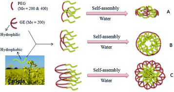
Dynamic light scattering (DLS) results revealed that the hydrodynamic radii of conjugates are in the range of 90–350 nm for 3a, 80–300 nm for 3b, and 110–370 nm for 5 respectively, which correlates with the TEM results (Fig. 5). It has also been observed that the variation of PEG contents and number of lipid chains in the amphiphilic lipid–PEG conjugate has significant effect on the shape and diameter of micelles. The graphical illustrations are given in Fig. 6 for the formation of micelles in which the hydrophobic part as a core shielded with hydrophilic shell.


image file: c4ra03583f-f6.tif
Fig. 6 Graphical illustration of micelles formation for compounds (A) 3a, (B) 3b, and (C) 5.

Conclusion

In summary, we have successfully synthesized amphiphilic lipid–PEG conjugates by “click reaction”, i.e., the Cu(I)-catalyzed Huisgen 1,3-dipolar cycloaddition between azide terminated PEG and alkyne terminated fatty acids from canola oil. These macromolecules were characterized with the help of 1H-NMR, FTIR and GPC. These amphiphilic lipid–PEG conjugates were evaluated for the first time for their self-assembled micellar structures. The formation of micelles results in an aqueous media due to the amphiphilic nature of CPE–PEG and CPE–GE consisting of hydrophobic core and a hydrophilic shell. From these results, It has been concluded that the lipid–PEG conjugate nanoparticles have different average sizes (159, 145 & 200 nm) due to different ratio of fatty acids and PEG contents in the bioconjugate backbone. The higher number of fatty acids and increased PEG contents in polymeric backbone of 5 result in the formation of more regular spherical micelles with smooth surface as compared to those having less ratio of PEG contents and shorter PEG backbone, particularly in case of PEG-200. The particle size and structure could be altered by changing the ratio of PEG and lipid contents. This study highlights that these nanocarriers may serve as a potential stealth drug carrier due to their amphiphilic nature and therefore ability to self-assemble in aqueous medium. These carrier systems are being further studied to explore their drug encapsulation efficiency and controlled release.

Acknowledgements

We gratefully acknowledge the financial support for current work by ALES, AFNS, and the Canadian Bureau for International Education (CBIE).

Notes and references

  1. N. A. Peppas, J. Z. Hilt, A. Khademhosseini and R. Langer, Adv. Mater., 2006, 18, 1345–1360 CrossRef CAS.
  2. H. Otsuka, Y. Nagasaki and K. Kataoka, Adv. Drug Delivery Rev., 2003, 55, 403–419 CrossRef CAS.
  3. Y. Wang, X. Li, G. Wu, J. Chen, Y. Wang, H. Gao and J. Ma, RSC Adv., 2013, 3, 13859–13868 RSC.
  4. R. J. Russell, M. V. Pishko, C. C. Gefrides, M. J. McShane and G. L. Coté, Anal. Chem., 1999, 71, 3126–3132 CrossRef CAS.
  5. J. A. Hubbell, Nat. Biotechnol., 1995, 13, 565–576 CrossRef CAS PubMed.
  6. J. S. Mohammed and W. L. Murphy, Adv. Mater., 2009, 21, 2361–2374 CrossRef CAS.
  7. A. M. Kloxin, A. M. Kasko, C. N. Salinas and K. S. Anseth, Science, 2009, 324, 59–63 CrossRef CAS PubMed.
  8. R. Ng, R. Zang, K. K. Yang, N. Liu and S.-T. Yang, RSC Adv., 2012, 2, 10110–10124 RSC.
  9. G. S. Kwon and K. Kataoka, Adv. Drug Delivery Rev., 1995, 16, 295–309 CrossRef CAS.
  10. M.-C. Jones and J.-C. Leroux, Eur. J. Pharm. Biopharm., 1999, 48, 101–111 CrossRef CAS.
  11. K. Miyata, R. J. Christie and K. Kataoka, React. Funct. Polym., 2011, 71, 227–234 CrossRef CAS PubMed.
  12. J. Rios-Doria, A. Carie, T. Costich, B. Burke, H. Skaff, R. Panicucci and K. Sill, J. Drug Delivery, 2012 DOI:10.1155/2012/951741.
  13. G. Li, J. Liu, Y. Pang, R. Wang, L. Mao, D. Yan, X. Zhu and J. Sun, Biomacromolecules, 2011, 12, 2016–2026 CrossRef CAS PubMed.
  14. J. Cheng, B. A. Teply, I. Sherifi, J. Sung, G. Luther, F. X. Gu, E. Levy-Nissenbaum, A. F. Radovic-Moreno, R. Langer and O. C. Farokhzad, Biomaterials, 2007, 28, 869–876 CrossRef CAS PubMed.
  15. P. De, S. R. Gondi and B. S. Sumerlin, Biomacromolecules, 2008, 9, 1064–1070 CrossRef CAS PubMed.
  16. Y.-L. Lo, G.-J. Chen, T.-H. Feng, M.-H. Li and L.-F. Wang, RSC Adv., 2014, 4, 11089–11098 RSC.
  17. J. M. Chan, L. Zhang, R. Tong, D. Ghosh, W. Gao, G. Liao, K. P. Yuet, D. Gray, J.-W. Rhee, J. Cheng, G. Golomb, P. Libby, R. Langer and O. C. Farokhzad, Proc. Natl. Acad. Sci. U. S. A., 2010, 107, 2213–2218 CrossRef CAS PubMed.
  18. J. M. Chan, L. Zhang, K. P. Yuet, G. Liao, J.-W. Rhee, R. Langer and O. C. Farokhzad, Biomaterials, 2009, 30, 1627–1634 CrossRef CAS PubMed.
  19. I. De Miguel, L. Imbertie, V. Rieumajou, M. Major, R. Kravtzoff and D. Betbeder, Pharm. Res., 2000, 17, 817–824 CrossRef CAS.
  20. Y. Liu, K. Li, J. Pan, B. Liu and S.-S. Feng, Biomaterials, 2010, 31, 330–338 CrossRef CAS PubMed.
  21. S. Sengupta, D. Eavarone, I. Capila, G. Zhao, N. Watson, T. Kiziltepe and R. Sasisekharan, Nature, 2005, 436, 568–572 CrossRef CAS PubMed.
  22. J. Thevenot, A.-L. Troutier, L. David, T. Delair and C. Ladavière, Biomacromolecules, 2007, 8, 3651–3660 CrossRef CAS PubMed.
  23. L. Zhang, J. M. Chan, F. X. Gu, J.-W. Rhee, A. Z. Wang, A. F. Radovic-Moreno, F. Alexis, R. Langer and O. C. Farokhzad, ACS Nano, 2008, 2, 1696–1702 CrossRef CAS PubMed.
  24. A. Lavasanifar, J. Samuel and G. S. Kwon, Adv. Drug Delivery Rev., 2002, 54, 169–190 CrossRef CAS.
  25. K. Knop, R. Hoogenboom, D. Fischer and U. S. Schubert, Angew. Chem., Int. Ed., 2010, 49, 6288–6308 CrossRef CAS PubMed.
  26. F. M. Veronese and G. Pasut, Drug Discovery Today, 2005, 10, 1451–1458 CrossRef CAS.
  27. K. D. Hinds and S. W. Kim, Adv. Drug Delivery Rev., 2002, 54, 505–530 CrossRef CAS.
  28. D. Attwood, C. Booth, S. G. Yeates, C. Chaibundit and N. M. P. S. Ricardo, Int. J. Pharm., 2007, 345, 35–41 CrossRef CAS PubMed.
  29. A. A. Attama, M. A. Momoh and P. F. Builders, in Recent Advances in Novel Drug Carrier Systems, ed. A. D. Sezer, InTech, Rijeka, Croatia, 2012, pp. 107–140 Search PubMed.
  30. V. P. Torchilin, Curr. Drug Delivery, 2005, 2, 319–327 CrossRef CAS.
  31. A. N. Lukyanov and V. P. Torchilin, Adv. Drug Delivery Rev., 2004, 56, 1273–1289 CrossRef CAS PubMed.
  32. R. Sawant and V. Torchilin, in Cancer Nanotechnology, ed. S. R. Grobmyer and B. M. Moudgil, Humana Press, 2010, vol. 624, ch. 9, pp. 131–149 Search PubMed.
  33. F. Li, M. Danquah and R. I. Mahato, Biomacromolecules, 2010, 11, 2610–2620 CrossRef CAS PubMed.
  34. S. Nomula, S. Ma and L. Cooper Stuart, in Associative Polymers in Aqueous Media, American Chemical Society, 2000, vol. 765, ch. 8, pp. 127–142 Search PubMed.
  35. O. Vorobyova and A. Winnik Mitchell, in Associative Polymers in Aqueous Media, American Chemical Society, 2000, vol. 765, ch. 9, pp. 143–162 Search PubMed.
  36. B.-S. Kim and P. T. Mather, Macromolecules, 2002, 35, 8378–8384 CrossRef CAS.
  37. H. C. Kolb, M. G. Finn and K. B. Sharpless, Angew. Chem., Int. Ed., 2001, 40, 2004–2021 CrossRef CAS.
  38. C. W. Tornøe, C. Christensen and M. Meldal, J. Org. Chem., 2002, 67, 3057–3064 CrossRef PubMed.
  39. V. V. Rostovtsev, L. G. Green, V. V. Fokin and K. B. Sharpless, Angew. Chem., Int. Ed., 2002, 41, 2596–2599 CrossRef CAS.
  40. M. J. Joralemon, R. K. O'Reilly, C. J. Hawker and K. L. Wooley, J. Am. Chem. Soc., 2005, 127, 16892–16899 CrossRef CAS PubMed.
  41. N. V. Tsarevsky, B. S. Sumerlin and K. Matyjaszewski, Macromolecules, 2005, 38, 3558–3561 CrossRef CAS.
  42. B. S. Sumerlin, N. V. Tsarevsky, G. Louche, R. Y. Lee and K. Matyjaszewski, Macromolecules, 2005, 38, 7540–7545 CrossRef CAS.
  43. R. A. Evans, Aust. J. Chem., 2007, 60, 384–395 CrossRef CAS.
  44. J.-F. Lutz, Angew. Chem., Int. Ed., 2007, 46, 1018–1025 CrossRef CAS PubMed.
  45. P. L. Golas, N. V. Tsarevsky, B. S. Sumerlin, L. M. Walker and K. Matyjaszewski, Aust. J. Chem., 2007, 60, 400–404 CrossRef CAS.
  46. D. J. Coady and C. W. Bielawski, Macromolecules, 2006, 39, 8895–8897 CrossRef CAS.
  47. R. K. O'Reilly, M. J. Joralemon, C. J. Hawker and K. L. Wooley, J. Polym. Sci., Part A: Polym. Chem., 2006, 44, 5203–5217 CrossRef.
  48. A. P. Vogt and B. S. Sumerlin, Macromolecules, 2006, 39, 5286–5292 CrossRef CAS.
  49. J.-F. Lutz, H. G. Börner and K. Weichenhan, Macromol. Rapid Commun., 2005, 26, 514–518 CrossRef CAS.
  50. K. L. Heredia, Z. P. Tolstyka and H. D. Maynard, Macromolecules, 2007, 40, 4772–4779 CrossRef CAS.
  51. X. Lang, A. K. Dalai, N. N. Bakhshi, M. J. Reaney and P. B. Hertz, Bioresour. Technol., 2001, 80, 53–62 CrossRef CAS.
  52. J. Wang, K. Yao, C. Wang, C. Tang and X. Jiang, J. Mater. Chem. B, 2013, 1, 2324–2332 RSC.
  53. J. Coates, in Encyclopedia of Analytical Chemistry, ed. R. A. Meyers, John Wiley & Sons Ltd, Chichester, 2000 Search PubMed.
  54. R.-X. Yao, L. Kong, Z.-S. Yin and F.-L. Qing, J. Fluorine Chem., 2008, 129, 1003–1010 CrossRef CAS PubMed.
  55. G. S. Kwon and T. Okano, Adv. Drug Delivery Rev., 1996, 21, 107–116 CrossRef CAS.
  56. S. Yusa, K. Fukuda, T. Yamamoto, K. Ishihara and Y. Morishima, Biomacromolecules, 2005, 6, 663–670 CrossRef CAS PubMed.
  57. K. Bury, F. Du Prez and D. Neugebauer, Macromol. Biosci., 2013, 13, 1520–1530 CrossRef CAS PubMed.

Footnote

Electronic supplementary information (ESI) available. See DOI: 10.1039/c4ra03583f

This journal is © The Royal Society of Chemistry 2014