Effect of ligands on the characteristics of (CdSe)13 quantum dots

Yang Gao a, Bo Zhoub, Seung-gu Kangc, Minsi Xina, Ping Yangd, Xing Daia, Zhigang Wang*ae and Ruhong Zhou*acf
aInstitute of Atomic and Molecular Physics, Jilin University, Changchun, 130012, China. E-mail: wangzg@jlu.edu.cn; ruhongz@us.ibm.com
bBio-X Lab, Department of Physics, Zhejiang University, Hangzhou, 310027, China
cComputational Biology Center, IBM Thomas J. Watson Research Center, Yorktown Heights, NY 10598, USA
dEnvironmental Molecular Sciences Laboratory, Pacific Northwest National Laboratory, Richland, WA 99352, USA
eState Key Laboratory of Theoretical and Computational Chemistry, Institute of Theoretical Chemistry Jilin University, Changchun, 130023, China
fDepartment of Chemistry, Columbia University, New York, NY 10027, USA

Received 9th April 2014 , Accepted 9th June 2014

First published on 10th June 2014


Abstract

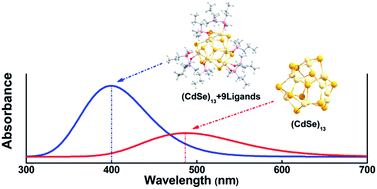
The widespread applications of quantum dots (QDs) have spurred an increasing interest in the study of their coating ligands, which can not only protect the electronic structures of the central QDs, but also control their permeability through biological membranes with both size and shape. In this work, we have used density functional theory (DFT) to systematically investigate the electronic structures of (CdSe)13 passivated by OPMe2(CH2)nMe ligands with different lengths and various numbers of branches (Me = methyl group, n = 0, 1–3) as well as different number of ligands ((CdSe)13 + [OPMe2(CH2)2Me]m (m = 0, 1, 9, 10)). Our results show that the absorption peak in the ultraviolet-visible (UV-vis) spectra displays a clear blue-shift, on the scale of ∼100 nm, upon the binding of ligands. Once the total number of ligands bound with (CdSe)13 reached a saturated number (9 or 10), no more blue-shift occurred in the absorption peak in the UV-vis spectra. On the other hand, the aliphatic chain length of ligands has a negligible effect on the optical properties of the QD core. Analyses of the bonding characteristics confirm that optical transitions are dominantly governed by the central QD core rather than the organic passivation. These findings might provide insights on the material design for the passivation of quantum dots for biomedical applications.


Introduction

The unique optical properties of quantum dots (QDs) have been widely applied to the design of functional materials, including photoenergy converters and nano-sized sensors.1–9 Their versatilities are largely originated from QDs' easy adaptability on various physicochemical factors such as particle sizes, atomic compositions, and surface modifications.10–17 More interestingly, inorganic QDs with comparable sizes to many biomolecules are recently proposed as potential platform for nanodrug carriers and/or diagnostic agents.18–29 These advances have raised serious requirements on a solid premise on the biosafety of QDs in a highly degradable biological environment.

Biocompatibility is usually achieved by the surface passivation on QDs with biocompatible organic molecules, so as to secure the toxic QDs in the core as well as to avoid their direct contacts with biomolecules.26,30 The passivation is also inevitable in stabilizing and dispersing QDs in an aggregation-prone aqueous medium, where the organic layer reduces the highly reactive surface potential,31–33 an intrinsic property of colloidal semiconductor nanoparticles due to high curvature.20,21 As such, the surface modification is accompanied by its critical influence to the chemical and electronic structures of QDs.11,16 Recent theoretical studies show that the passivating ligands can form stable coordinations on QD surface, sustaining the chemical structure of the QD.31−34 On the other hand, experimental study (e.g. PbSe–QD) revealed a complex behaviour between the PbSe-QD core and passivating organic layer in controlling the optical property.35 Thus, it is crucial to understand electrochemical effect of the passivating ligands on QDs in developing biocompatible and stable QDs, while simultaneously maintaining their desired electro-optical functions.36

Here, we systematically investigate the ligand effect on the electronic structure of a typical (CdSe)13 quantum dot.37–39 A bare CdSe-QD is toxic which can cause massive cell deaths in vivo tests,26,30 despite its excellent optical property and promising potential for biomedical applications. Thus, an effective passivation would be a critical step for avoiding any detrimental effect on the host biosystem as well as stabilizing the chemical and structural integrity of the QD.40,41 A spherical structure of (CdSe)13 and a large surface-to-volume ratio facilitate a high density coating with various organic ligands.25–28 Among many available ligands coupled to CdSe-QDs,42–47 we selected trimethyl phosphine oxide (TMPO) and its derivatives (i.e., OPMe2(CH2)nMe, n = 0, 1–3) due to their high ligand exchange capacity on the CdSe surface.48 Despite several studies on this important ligand coating,17,34,48 there are still quite a few interesting questions remaining, such as what kind of length effect the ligand might have, whether the number of branches matter, and how sensitive the coating is to the number of ligands. In this study, we used the first-principal density functional theory (DFT) to systematically study these important effects, which has been proven as a useful methodology for similar systems of molecular adsorption.17,31–33,42–52 We investigated the optical property changes of the (CdSe)13 core, including electronic spectra, electron transfer and density of state, binding energy with varying surface capping density and alkyl chain length of the passivation ligands.

Computational methods

The structure of the bare (CdSe)13 QD was revealed from mass spectrometry, which could exist as a stable cage structure.37 Cadmium ions were determined as the primary cause of cytotoxicity in their applications in vivo.30 The passivated structures have thus been generated with TMPO on top of the toxic Cd atoms.30 In order to study the capping density effect, we prepared three different systems depending on numbers of TMPOs (i.e., 1, 9 and 10; ESI Part S1). The ligands were firstly set to coordinate the unsaturated Cd atoms for cases of 1 and 9 TMPOs. The last ligand with 10 TMPOs was placed on the saturated Cd atom (i.e., Cd (2) in Fig. 1), which was located on the axis of the 3-fold symmetry of the (CdSe)13-cage. For the effect of alkyl chain lengths of the passivated ligands, we also varied the chain length of one of the methyl groups of TMPO from methyl to butyl (i.e., OPMe2-(CH2)nMe, n = 0, 1–3).
image file: c4ra03202k-f1.tif
Fig. 1 Left: the optimized bare (CdSe)13-cage structure with C3 symmetry; right: a single ligand structure: OPMe2(CH2)2Me; middle: the optimized (CdSe)13 + 9OPMe2(CH2)2Me complex structure with C3 symmetry. The dotted lines indicate the ligand passivated sites (Cd: faint yellow, Se: deep yellow, O: red, P: pink, C: gray, and H: white. similarly hereinafter).

Previous study have shown that the hybrid exchange correlation functional B3LYP53–55 of DFT with LANL2DZ basis set is appropriate for describing electronic properties of CdSe QDs, whether it is pure theory or the combination of the theoretical and experimental work.48,52,56–60 And the organic passivation undergoes with a weakly-coupled interaction which is based on electrostatic interaction formation,34 rather than the weak van der Waals interaction between molecules. Thus the results of the DFT-B3LYP method without the empirical dispersion term have been gived in the text, and a compare with the results including the empirical dispersion contribution (using the B3LYP-D3 method) was performed in the ESI Part S2. Accordingly, the B3LYP method was employed to optimize all geometric structures.17 Considering that the molecular passivation may create atypical bonds, we added d polarization functions for all the C, O, and P atoms, and also added p polarization functions for all the H atoms by using double-ξ basis set61 for the ligands. We also examined the symmetry effect on the molecular geometry and electronic structures by imposing the systems with C3 symmetry.37 The vibrational frequencies were calculated at the same level of theory to verify the minima of the corresponding structures (ESI Part S3). In addition, the time-dependent density functional theory62 (TD-DFT) was also used to study UV-vis absorption spectra. Previous research has shown the solvent effect is not significant on UV-vis spectra of passivated CdSe QD system,33 therefore it will not be introduced in the calculation. All the calculations were performed with the Gaussian 09 program package.63

Results and discussion

UV-vis absorption spectra of QD

It is well established that CdSe-QD has unique optical properties. In this work, we first focus on the significant absorption peak with the maximum excitation wavelength, which is contribution by valence orbital transitions, and the calculated results is comparable with previous reports (ESI Part S4). The UV-vis absorption spectra are dramatically shifted upon passivation, where the peak is blue-shifted by ∼95 nm from 487 nm of the bare QD core (Fig. 2a). However, it seems converged to ∼395 nm after 9 ligands, whereafter the peak moves only 3 nm to the blue (i.e., 392 nm) with 10 ligands. On the contrary, the UV-vis spectra are not sensitive to the allipatic chain length of the coordinated ligands (Fig. 2b). Our result clearly indicates that the organic passivation has a significant effect on electronic absorption bands of the CdSe core. Similar blue-shift effect is consistent with recent study on UV-vis spectra of (CdSe)13 passivated by methylamine ligands.56 And then, we also considered the effect of dispersion interaction between QD and ligands on the UV-vis spectra using the B3LYP-D3 method and found that the max absorption peak has only a little blue-shift (about 10 nm) compared with the result above (ESI Part S2).
image file: c4ra03202k-f2.tif
Fig. 2 The UV-vis absorption spectra of (CdSe)13 + [OPMe2-(CH2)2Me]m (m = 0, 1, 9, 10). Different colors represent different QD complex structures. Dash-dot lines represent the corresponding structure with C3 symmetry. The numbers indicate the wavelengths of electronic absorption peaks.

In order to further validate our current theoretical approach, we compared our findings with available data from previous studies. There are several previous studies with both theoretical and experimental methods on the UV-vis spectra of CdSe QDs.64–66 Table S1 (ESI Part S4) summarizes UV-vis spectra observed for various sizes of QDs saturated in TOPO solvent. It is clear that the maximum excitation wavelength decreases from 550 nm to 415 nm as with decrease of particle size 3.1 nm to 1.2 nm. Since the experimental results are all for QD nanostructures of larger than our system of ∼0.9 nm, it is difficult to directly compare.67 Nonetheless, the trend seems reasonable to be extrapolated to our calculated excitation wavelength 400 nm. In other words, with the decrease of the size of the center quantum particles, the excitation wavelength displays a clear blue-shift. In this work, the diameter of the bare (CdSe)13-cage is 0.9 nm, so it can be inferred from Table S1 that the maximum excitation wavelength should be less than 415 nm, which agrees well with our result of about 400 nm.

On the other hand, Table S2 (ESI Part S4) shows the peak positions of the 0.9 nm (CdSe)13-cage depending on degree of passivation. They ranges from 487 nm in bare (CdSe)13 to 400 nm in (CdSe)13 + 9OPMe2(CH2)0,1–3Me-symmetry passivation. As shown above, passivation obviously induced significant blue-shift in the excitation wavelength, consistent with previous findings.71 These comparisons confirmed that our current approach is reliable.

We then computed the detailed excitation transition energies, oscillator strengths, and related molecular orbital composition, to further understand the origin of the electronic absorption peak (Table 1 and ESI Part S5). Electronic transitions mostly occurred in four highly occupied molecular orbitals (i.e., HOMO − 3 to HOMO). For example, (CdSe)13 + 9OPMe2(CH2)2Me shows the electronic transition from the first four front occupied orbitals to LUMO, where the symmetry constraint only causes a few nanometer shift of the absorption peaks. It is noteworthy that all the electronic transitions are only originated from the central QDs, independent of ligands. The orbital localization is depicted in Table 2 and S4 (ESI Part S6), where HOMO and LUMO are dominantly created in the CdSe–core regardless of the ligands. The vibration spectra clearly show that there is no collaborative vibration between central (CdSe)13 and ligands (ESI Part S2).

Table 1 The excitation energies (E in eV), excitation wavelength (λ in nm), oscillator strengths (f > 0), and excited-state composition with contribution percentages [similar statistical methods have been reported]68−70 of the two kinds of asymmetrical structure
structure E λ F Transition
(CdSe)13 2.52 493 0.032 49% HOMO − 2 [Cd 5p 5%, Se 4p 95%] → LUMO [Cd 5s 70%, Se 4p 30%]
2.56 485 0.030 1% HOMO − 1 [Cd 5p 8%, Se 4p 92%] → LUMO
47% HOMO [Cd 5p 8%, Se 4p 92%] → LUMO
2.56 485 0.030 47% HOMO − 1 → LUMO
1% HOMO → LUMO
(CdSe)13 + 9OPMe2(CH2)2Me 3.02 411 0.085 49% HOMO [Cd 5p 6%, Se 4p 94%] → LUMO [Cd 5s 68%, Se 4p 32%]
3.13 396 0.096 48% HOMO − 1 [Cd 5p 7%, Se 4p 93%] → LUMO
3.23 384 0.125 2% HOMO − 3 [Cd 5p 9%, Se 4p 91%] → LUMO
46% HOMO − 2 [Cd 5p 8%, Se 4p 92%] → LUMO


Table 2 The highest occupied molecular orbital (HOMO) and the lowest unoccupied molecular orbital (LUMO) of bare (CdSe)13 cage asymmetrical structure, (CdSe)13 + 9OPMe2(CH2)2Me asymmetrical structure and (CdSe)13 + 10OPMe2(CH2)2Me structure
MO (CdSe)13 (CdSe)13 + 9OPMe2(CH2)2Me (CdSe)13 + 10OPMe2(CH2)2Me
LOMO image file: c4ra03202k-u1.tif image file: c4ra03202k-u2.tif image file: c4ra03202k-u3.tif
HOMO image file: c4ra03202k-u4.tif image file: c4ra03202k-u5.tif image file: c4ra03202k-u6.tif


Mulliken charge populations for CdSe

In addition, the charge delocalization can be assessed by the extent of charge transfer, which is found to be relatively small overall. Using the case of (CdSe)13 + 9OPMe2(CH2)nMe (n = 0, 1–3) as an example, Mulliken charge populations are varied by less than 0.1 e for the representative core atoms (i.e., Se(1), Cd(2), Cd(4), Cd(5), Cd(6)) from the various passivation. Also, compared to the bare system (Cd13Se13), QDs with surface passivation are slightly easier to lose and obtain electrons for Cd and Se atoms, respectively. Nevertheless, the net charge transfer in (CdSe)13 + 9OPMe2(CH2)3Me (from the ligands to the core) is only about 0.156 e per ligand.

The chain length has a negligible effect on the charge distribution, which is particularly obvious for atoms Cd(2) and Cd(3) due to no dangling bonds for passivating ligands. It is also non-sensitive with the symmetry constraint (see red and blue curves in Fig. 3). This indicates that there is no obvious chemical bonding between QD and ligands (ESI Part S7), but only partial charge polarization. Our results support that the CdSe-QD is kept stable even with TMPO saturated on the surface, which would be important for QD to properly serve as an optical probe in the highly degradable biological media, although the optical properties are tuned by the degree of passivation.


image file: c4ra03202k-f3.tif
Fig. 3 Mulliken partial charge populations for surface Se and Cd atoms. The positions of these atoms are shown in Fig. 1 (left) in which the numbers are corresponding to the position of Se and Cd. The abscissa I, II, III, and IV represents bare (CdSe)13-cage symmetrical structures and (CdSe)13 + 9OPMe2(CH2)nMe (n = 0, 1–3) symmetrical structures, respectively.

DOSs of the system

We calculated the density of states (DOSs) of the system. The DOS varies sensitively depending on the degree of passivation (Fig. 4a). The Fermi level is shifted up to ∼2.5 eV. The HOMO–LUMO gap increased from 3.0 eV to 3.6 eV for the bare and the passivated systems (ESI Part S4), respectively, further confirming the significant blue-shifts in aforementioned UV-vis spectra. This is also qualitatively consistent with recent experiment with (CdSe)6 passivated by OPH3.52 Especially, DOS are highly fluctuating between −10.0 and −16.0 eV with the increase of passivation. These fluctuations are mainly contributed by ligands. From orbital component analysis we found that these ligand molecular orbitals (MOs) are mostly localized on the 2p orbitals of C atoms, 2p orbitals of O atoms, and 3p orbitals of P atoms. On the other hand, the fluctuations in DOS from −6.5 and −10 eV are from MOs of Se and Cd atoms, which are strongly localized on the 4p orbitals and 5s orbitals for Se and Cd respectively. There are also some hybrid orbitals between QD and ligands. DOSs becomes stable once ligands are saturated (after nine ligands). On the contrary, DOSs are relatively less sensitive to the symmetry constraints and the chain length variation (Fig. 4b). The shift is only about 10−2 eV for Fermi levels of QDs with different chain lengths. Interestingly, the density of states (DOS) share similar characteristics as vibrational spectra (ESI Part S3), even though there is no coordination vibration mode between the ligands and the central QD. Therefore, we conclude that the increase of the chain length of ligands would not significantly affect the energy level position of the QD.
image file: c4ra03202k-f4.tif
Fig. 4 The different QD structures of DOSs. The dash-dot and solid lines represent symmetry and asymmetry, respectively. The arrows represent the position of HOMOs and LUMOs in different structures and the vertical dotted lines indicate the Fermi level.

The stability of the ligand coating on the (CdSe)13 surface is further studied. We calculated the average binding energy Eb per each ligand molecule between the ligand and the (CdSe)13 cluster. Eb is defined as

image file: c4ra03202k-t1.tif
where E(CdSe)13+ligands is the total energy of the ligands coating on the (CdSe)13 surface, while image file: c4ra03202k-t2.tif and image file: c4ra03202k-t3.tif represent the total energies of (CdSe)13 and ligands in its relaxed geometry, respectively, with both the (CdSe)13 and ligands being in the same atomic configurations as in the relaxed (CdSe)13 + ligands system.

Our calculations show the binding energy Eb is −1.15 eV for each OPMe2(CH2)2Me when absorbing onto the (CdSe)13 surface, which is comparable to a recent theoretical study, −1.08 eV per ligand for (CdSe)6 passivated by OPMe3.57 We also calculated the average binding energy Eb for (CdSe)13 + 9OPMe2(CH2)nMe (n = 0, 1–3) structures, and the resulting binding energies are 0.88 eV, 0.89 eV, 0.89 eV and 0.89 eV respectively for n = 0, 1–3 (ESI Part S8), which suggests the chain length has only minor effect on the binding energy, consistent with above electronic structure analyses. These binding energies also suggest the (CdSe)13 clusters passivated by various OPMe2(CH2)nMe ligands are quite stable. Furthermore, the IR vibrational spectra data also support that the QD is stable with only relatively weak coupling to the passivation organic molecules (ESI Part S3).

Conclusions

In this work, we systematically studied the electronic structures of (CdSe)13 nanocluster passivated TMPO ligands, with different chain lengths, different number of branches, and different number of ligands. Our calculations indicate that the coating ligands can significantly blue-shift the position of the absorption peaks of UV-vis spectra (on the scale of 95 nm). Once the passivation is saturated on the surface of (CdSe)13 (i.e., 9 ligands in this case), the absorption peaks in the UV-vis spectra tend to be stabilized. The analyses of valence MOs of passivated QD structures indicate that the active orbital regions are localized in the center of (CdSe)13 QD. Moreover, from the UV-vis spectra of (CdSe)13 coated with 9-ligands, we find that (CdSe)13 QD is insensitive to the branch length of ligands. Similar conclusion is also reflected in the binding energy, DOS spectra and vibration spectra.

Since CdSe QDs are widely used in many applications including biomedical ones, it is critical to fully understand their electronic structures as well as effects from coating ligands. Our current extensive study clearly shows that the optical characteristics of QD will not change much even in the saturated passivation as well as the length variation of ligand aliphatic chains. Even though our current findings are based on (CdSe)13, we believe these conclusions can be applied to other QDs of different sizes, and provide new insight on biomedical applications of QDs.

Acknowledgements

The work described in this paper is supported by IBM Blue Gene Science Program, and grants from the National Science Foundation of China under grant no. 11374004. Z. W. acknowledges the High Performance Computing Center (HPCC) of Jilin University.

Notes and references

  1. M. Bruchez, M. Moronne, P. Gin, S. Weiss and A. P. Alivisatos, Science., 1998, 281, 2013–2016 CrossRef CAS.
  2. N. Tessler, V. Medvedev, M. Kazes, S. Kan and U. Banin, Science., 2002, 295, 1506–1508 CrossRef PubMed.
  3. J. Gorman, D. G. Hasko and D. A. Williams, Phys. Rev. Lett., 2005, 95, 090502 CrossRef CAS.
  4. S. Csonka, I. Weymann and G. Zarand, Nanoscale, 2012, 4, 3635–3639 RSC.
  5. J. Y. Lek, L. Xi, B. E. Kardynal, L. H. Wong and Y. M. Lam, ACS Appl. Mater. Interfaces, 2011, 3, 287–292 CAS.
  6. P. V. Kamat, J. Phys. Chem. C, 2008, 112, 18737–18753 CAS.
  7. I. Lokteva, N. Radychev, F. Witt, H. Borchert, J. Parisi and J. Kolny-Olesiak, J. Phys. Chem. C, 2010, 114, 12784–12791 CAS.
  8. D. Gerion, F. Pinaud, S. C. Williams, W. J. Parak, D. Zanchet, S. Weiss and A. P. Alivisatos, J. Phys. Chem. B, 2001, 105, 8861–8871 CrossRef CAS.
  9. D. A. Tryk, A. Fujishima and K. Honda, Electrochim. Acta, 2000, 45, 2363–2376 CrossRef CAS.
  10. A. P. Alivisatos, Science, 1996, 271, 933–937 CAS.
  11. A. Pandey and P. Guyot-Sionnest, Science, 2008, 322, 929–932 CrossRef CAS PubMed.
  12. C. B. Murray, D. J. Norris and M. G. Bawendi, J. Am. Chem. Soc., 1993, 115, 8706–8715 CrossRef CAS.
  13. A. Shiohara, S. Prabakar, A. Faramus, C. Y. Hsu, P. S. Lai, P. T. Northcote and R. D. Tilley, Nanoscale, 2011, 3, 3364–3370 RSC.
  14. L. H. Qu and X. G. Peng, J. Am. Chem. Soc., 2002, 124, 2049–2055 CrossRef CAS PubMed.
  15. G. D. Scholes and G. Rumbles, Nat. Mater., 2006, 5, 683–696 CrossRef CAS PubMed.
  16. M. Beard, A. Midgett, M. Law, R. Ellingson and A. Nozik, Nano Lett., 2009, 9, 1217–1222 CrossRef PubMed.
  17. S. Kilina, S. Ivanov and S. Tretiak, J. Am. Chem. Soc., 2009, 131, 7717–7726 CrossRef CAS PubMed.
  18. S. G. Kang, G. Q. Zhou, P. Yang, Y. Liu, B. Y. Sun, T. Huynh, H. Meng, L. Zhao, G. M. Xing, C. Y. Chen, Y. L. Zhao and R. H. Zhou, Proc. Natl. Acad. Sci. U. S. A., 2012, 109, 15431–15436 CrossRef CAS PubMed.
  19. S. G. Kang, T. Huynh and R. H. Zhou, Sci. Rep., 2012, 2, 00957 Search PubMed.
  20. W. J. Parak, D. Gerion, T. Pellegrino, D. Zanchet, C. Micheel, S. C. Williams, R. Boudreau, M. A. L. Gros, C. A. Larabell and A. P. Alivisatos, Nanotechnology., 2003, 14, R15–R27 CrossRef CAS.
  21. X. Michalet, F. Pinaud, T. D. Lacoste, M. Dahan, M. P. Bruchez, A. P. Alivi-satos and S. Weiss, Single Mol., 2001, 2, 261–276 CrossRef CAS.
  22. X. Michalet, F. F. Pinaud, L. A. Bentolila, J. M. Tsay, S. Doose, J. J. Li, G. Sun-daresan, A. M. Wu, S. S. Gambhir and S. Weiss, Science, 2005, 307, 538–544 CrossRef CAS PubMed.
  23. I. L. Medintz, H. T. Uyeda, E. R. Goldman and H. Mattoussi, Nat. Mater., 2005, 4, 435–446 CrossRef CAS PubMed.
  24. R. Gill, M. Zayats and I. Willner, Angew. Chem., Int. Ed., 2008, 47, 7602–7625 CrossRef CAS PubMed.
  25. C. S. S. R. Kumar, Nanomaterials for Medical Applications. Kirk-Othmer Encyclopedia of Chemical Technology, Wiley, 2007, http://mrw.interscience.wiley.com/emrw/9780471238966/kirk/article/nanokuma.a01/current/abstract, accessed February 11 2008 Search PubMed.
  26. J. Drbohlavova, V. Adam, R. Kizek and J. Hubalek, J. Mol. Sci., 2009, 10, 656–673 CrossRef CAS PubMed.
  27. M. S. Andrew and S. M. Nie, Nat. Biotechnol., 2009, 27, 732–733 CrossRef PubMed.
  28. M. S. Andrew and S. M. Nie, J. Am. Chem. Soc., 2008, 130, 11278–11279 CrossRef PubMed.
  29. Y. Wang, Y. H. Liu, Y. Zhang, F. Wang, P. J. Kowalski, H. W. Rohrs, R. A. Loomis, M. L. Gross and W. E. Buhro, Angew. Chem., Int. Ed., 2012, 51, 6154–6157 CrossRef CAS PubMed.
  30. M. A. Walling, J. A. Novak and J. R. E. Shepard, J. Mol. Sci., 2009, 10, 441–491 CrossRef CAS PubMed.
  31. S. Y. Chung, S. Lee, C. Liu and D. Neuhauser, J. Phys. Chem. B, 2009, 113, 292–301 CrossRef CAS PubMed.
  32. H. Kim, S. W. Jang, S. Y. Chung and S. Lee, J. Phys. Chem. B, 2010, 114, 471–479 CrossRef CAS PubMed.
  33. V. V. Albert, S. A. Ivanov, S. Tretiak and S. V. Kilina, J. Phys. Chem. C, 2011, 115, 15793–15800 CAS.
  34. J. M. Azpiroz, J. M. Matxain, I. Infante, X. Lopez and J. M. Ugalde, Phys. Chem. Chem. Phys., 2013, 15, 10996–11005 RSC.
  35. C. M. Evans, L. Guo, J. J. Peterson, Z. S. Maccagnano and T. D. Krauss, Nano Lett., 2008, 8, 2896–2899 CrossRef CAS PubMed.
  36. A. Nag, A. Hazarika, K. V. Shanavas, S. M. Sharma, I. Dasgupta and D. D. Sarma, J. Phys. Chem. Lett., 2011, 2, 706–712 CrossRef CAS.
  37. A. Kasuya, R. Sivamohan, Yu. A. Barnakov, I. M. Dmitruk, T. Nirasawa, V. R. Romanyuk, V. Kumar, S. V. Mamykin, K. Tohji, B. Jeyadevan, K. Shinoda, T. Kudo, O. Terasaki, Z. Liu, R. V. Beloslu-dov, V. Sundararajan and Y. Kawazoe, Nat. Mater., 2004, 3, 99–102 CrossRef CAS PubMed.
  38. J. Pedersen, S. Bjornholm, J. Borggreen, K. Hansen, T. P. Martin and H. D. Ra-smussen, Nature, 1991, 353, 733–735 CrossRef CAS.
  39. Y. Wang, Y. H. Liu, Y. Zhang, P. J. Kowalski, H. W. Rohrs and W. E. Buhro, Inorg. Chem., 2013, 52, 2933–2938 CrossRef CAS PubMed.
  40. M. A. Schreuder, J. R. McBride, A. D. Dukes III, J. A. Sammons and S. J. Ro-senthal, J. Phys. Chem. C, 2009, 113, 8169–8176 CAS.
  41. K. Knowles, D. B. Tice, E. A. McArthur, G. C. SolOMOn and E. A. Weiss, J. Am. Chem. Soc., 2009, 132, 1041–1050 CrossRef PubMed.
  42. K. Eichkorn and R. Ahlrichs, Chem. Phys. Lett., 1998, 288, 235–242 CrossRef CAS.
  43. P. Deglmann, R. Ahlrichs and K. Tsereteli, J. Chem. Phys., 2002, 116, 1585–1597 CrossRef CAS PubMed.
  44. K. Leung and K. B. Whaley, J. Chem. Phys., 1999, 110, 11012–11022 CrossRef CAS PubMed.
  45. M. C. Troparevsky and J. R. Chelikowsky, J. Chem. Phys., 2001, 114, 943–946 CrossRef CAS PubMed.
  46. M. C. Troparevsky, L. Kronik and J. R. Chelikowsky, J. Chem. Phys., 2003, 119, 2284–2287 CrossRef CAS PubMed.
  47. A. Puzder, A. J. Williamson, F. Gygi and G. Galli, Phys. Rev. Lett., 2004, 92, 217401 CrossRef.
  48. P. Yang, S. Tretiak, A. Masunov and S. Ivanov, J. Chem. Phys., 2008, 129, 074709 CrossRef PubMed.
  49. C. J. Tian, P. Xiu, Y. Meng, W. Y. Zhao, Z. G. Wang and R. H. Zhou, Chem.–Eur. J., 2012, 18, 14305–14313 CrossRef CAS PubMed.
  50. Z. G. Wang, M. G. Yao, S. F. Pan, M. X. Jin, B. B. Liu and H. X. Zhang, J. Phys. Chem. C, 2007, 111, 4473–4476 CAS.
  51. X. Dai, C. Cheng, W. Zhang, M. S. Xin, P. Huai, R. Q. Zhang and Z. G. Wang, Sci. Rep., 2013, 3, 1341 CAS.
  52. A. E. Kuznetsov, D. Balamurugan, S. S. Skourtis and D. N. Beratan, J. Phys. Chem. C, 2012, 116, 6817–6830 CAS.
  53. A. D. Becke, J. Chem. Phys., 1993, 98, 5648–5652 CrossRef CAS PubMed.
  54. C. Lee, W. Yang and R. G. Parr, Phys. Rev. B: Condens. Matter Mater. Phys., 1988, 37, 785–789 CrossRef CAS.
  55. B. Miehlich, A. Savin, H. Stoll and H. Preuss, Chem. Phys. Lett., 1989, 157, 200–206 CrossRef CAS.
  56. R. Nadler and J. F. Sanz, Theor. Chem. Acc., 2013, 132, 1–9 CrossRef CAS.
  57. P. Yang, S. Tretiak and S. Ivanov, J. Cluster Sci., 2011, 22, 405–431 CrossRef CAS.
  58. X. Q. Wang, Q. Zeng, J. Shi, G. Jiang, M. L. Yang, X. Y. Liu, G. Enrightb and K. Yu, Chem. Phys. Lett., 2013, 568, 125–129 CrossRef PubMed.
  59. B. P. Bloom, L. B. Zhao, Y. Wang and D. Waldeck, J. Phys. Chem. B, 2013, 117, 22401–22411 CAS.
  60. S. K. Muzakir, N. Alias, M. M. Yusoff and R. Jose, Phys. Chem. Chem. Phys., 2013, 15, 16275–16285 RSC.
  61. R. Ditchfield, W. J. Hehre and J. A. Pople, J. Chem. Phys., 1971, 54, 724–728 CrossRef CAS PubMed.
  62. M. A. L. Marques and E. K. U. Gross, Annu. Rev. Phys. Chem., 2004, 55, 427–455 CrossRef CAS PubMed.
  63. M. J. Frisch, G. W. Trucks, H. B. Schlegel, G. E. Scuseria, M. A. Robb, J. R. Cheeseman, G. Scalmani, V. Barone, B. Mennucci, G. A. Petersson, H. Nakatsuji, M. Caricato, X. Li, H. P. Hratchian, A. F. Izmaylov, J. Bloino, G. Zheng, J. L. Sonnenberg, M. Hada, M. Ehara, K. Toyota, R. Fukuda, J. Hasegawa, M. Ishida, T. Nakajima, Y. Honda, O. Kitao, H. Nakai, T. Vreven, J. A. Montgomery, Jr, J. E. Peralta, F. Ogliaro, M. Bearpark, J. J. Heyd, E. Brothers, K. N. Kudin, V. N. Staroverov, R. Kobayashi, J. Normand, K. Raghavachari, A. Rendell, J. C. Burant, S. S. Iyengar, J. Tomasi, M. Cossi, N. Rega, J. M. Millam, M. Klene, J. E. Knox, J. B. Cross, V. Bakken, C. Adamo, J. Jaramillo, R. Gomperts, R. E. Stratmann, O. Yazyev, A. J. Austin, R. Cammi, C. Pomelli, J. W. Ochterski, R. L. Martin, K. Morokuma, V. G. Zakrzewski, G. A. Voth, P. Salvador, J. J. Dannenberg, S. Dapprich, A. D. Daniels, O. Farkas, J. B. Foresman and J. V. Ortiz, Gaussian 09 Revision D.01, Wallingford CT, 2013 Search PubMed.
  64. C. Landes, C. Burda and M. A. El-Sayed, Nano Lett., 2001, 1, 667–670 CrossRef CAS.
  65. G. Kalyuzhny and R. W. Murray, J. Phys. Chem. B, 2005, 109, 7012–7021 CrossRef CAS PubMed.
  66. A. Peng, J. Wickham and A. P. Alivisatos, J. Am. Chem. Soc., 1998, 120, 5343–5344 CrossRef.
  67. A. H. Margaret and G. S. Philippe, J. Phys. Chem., 1996, 100, 468–471 CrossRef.
  68. R. A. Vogt, T. G. Gray and C. E. Crespo-Hernández, J. Am. Chem. Soc., 2012, 134, 14808–14817 CrossRef CAS PubMed.
  69. H. Braunschweig, R. D. Dewhurst, K. Hammond, J. K. Mies, K. Radacki and A. Vargas, Science, 2012, 336, 1420–1422 CrossRef CAS PubMed.
  70. M. Del Ben, R. W. A. Havenith, R. Broer and M. Stener, J. Phys. Chem. C, 2011, 115, 16782–16796 CAS.
  71. K. A. Nguyen, P. N. Day and R. Pachter, J. Phys. Chem. C, 2010, 114, 16197–16209 CAS.

Footnotes

Electronic supplementary information (ESI) available: The bare (CdSe)13-cage structure. The dispersion correction of UV UV-vis absorption spectra. The vibration modes of central QD and the vibration spectra with different number/length of ligands. Comparison of our calculated results with previous reports. The excitation energy, oscillator strengths and corresponding molecular orbital compositions of QDs. Molecular valence orbitals of (CdSe)13 with ligands. Contour maps of charge density deformation in the local area of Cd and O atoms. The average binding energy Eb for per ligand molecule between the ligands and the (CdSe)13 clusters. See DOI: 10.1039/c4ra03202k
These authors contributed equally to this work.

This journal is © The Royal Society of Chemistry 2014