Lysis dynamics and membrane oligomerization pathways for Cytolysin A (ClyA) pore-forming toxin

M. S. Vaidyanathan a, Pradeep Sathyanarayanabc, Prabal K. Maitid, Sandhya S. Visweswariahbc and K. G. Ayappa*ab
aDepartment of Chemical Engineering, Indian Institute of Science, Bangalore, India. E-mail: ayappa@chemeng.iisc.ernet.in; Fax: +91 80 23608121; Tel: +91 80 22932769
bBioengineering Programme, Indian Institute of Science, Bangalore, India. E-mail: pradeep@mrdg.iisc.ernet.in; Fax: +91 80 23600999; Tel: +91 80 22932659
cDepartment of Molecular Reproduction, Development and Genetics, Indian Institute of Science, Bangalore, India. E-mail: sandhya@mrdg.iisc.ernet.in; Fax: +91 80 23600999; Tel: +91 80 22932659
dDepartment of Physics and Center for Condensed Matter Theory, Indian Institute of Science, Bangalore, India. E-mail: maiti@physics.iisc.ernet.in; Fax: +91 80 22932602; Tel: +91 80 22932315

Received 16th September 2013 , Accepted 22nd October 2013

First published on 23rd October 2013


Abstract

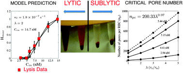
Pore-forming toxins are known for their ability to efficiently form transmembrane pores which eventually leads to cell lysis. The dynamics of lysis and underlying self-assembly or oligomerization pathways leading to pore formation are incompletely understood. In this manuscript the pore-forming kinetics and lysis dynamics of Cytolysin-A (ClyA) toxins on red blood cells (RBCs) are quantified and compared with experimental lysis data. Lysis experiments are carried out on a fixed mass of RBCs, under isotonic conditions in phosphate-buffered saline, for different initial toxin concentrations ranging from 2.94–14.7 nM. Kinetic models which account for monomer binding, conformation and oligomerization to form the dodecameric ClyA pore complex are developed and lysis is assumed to occur when the number of pores per RBC (np) exceeds a critical number, npc. By analysing the model in a sublytic regime (np < npc) the number of pores per RBC to initiate lysis is found to lie between 392 and 768 for the sequential oligomerization mechanism and between 5300 and 6300 for the non-sequential mechanism. Rupture rates which are first order in the number of RBCs are seen to provide the best agreement with the lysis experiments. The time constants for pore formation are estimated to lie between 1 and 20 s and monomer conformation time scales were found to be 2–4 times greater than the oligomerization times. Cell rupture takes places in 100s of seconds, and occurs predominantly with a steady number of pores ranging from 515 to 11[thin space (1/6-em)]000 on the RBC surface for the sequential mechanism. Both the sequential irreversible and non-sequential kinetics provide similar predictions of the hemoglobin release dynamics, however the hemoglobin released as a function of the toxin concentration was accurately captured only with the sequential model. Each mechanism develops a distinct distribution of mers on the surface, providing a unique experimentally observable fingerprint to identify the underlying oligomerization pathways. Our study offers a method to quantify the extent and dynamics of lysis which is an important aspect of developing novel drug and gene delivery strategies based on pore-forming toxins.


1. Introduction

Pore-forming toxins (PFTs) are a class of proteins produced by a wide variety of organisms including bacteria1 and humans.2 They have the unique property of generating pores in the membranes of target cells.3–5 PFTs are classified on the basis of the secondary structure (α or β) of the pore-forming region, and usually undergo a monomer to oligomer transition that is a pre-requisite for pore formation.6,7 Cytolysin A (ClyA, HlyE or SheA) from E. coli is a well characterized α-PFT3,8 and crystal structures are available for both the water-soluble monomeric9 and membrane-associated, oligomeric forms.10 The monomer of ClyA possesses 5 α helices and a hydrophobic β sheet, the “β-tongue”, that is buried within the helices. More recent crystal structure data10 indicates that the toxin oligomerizes as a dodecamer. A pathway for the transition from the monomer to the membrane-bound protomer, and finally to the dodecameric pore complex has been proposed based on a comparison of the monomeric and oligomeric crystal structures.10 Large conformational changes in the N-terminus and the β sheet regions need to occur during the monomer to oligomer transition, that appear to involve initial flipping out of the β-tongue to bind to the lipid membrane, followed by larger translocation of the N-terminus into the lipid membrane. ClyA forms cation-selective pores which have an internal diameter of 4–7 nm.10

A variety of experimental techniques such as lytic experiments, gel electrophoresis, site-directed mutagenesis and cryo-electron microscopy have been used to unravel the mechanisms of pore formation in ClyA11–14 and in other widely studied PFTs such as cholestrol-dependent toxins (CDCs)15 and S. aureus α-Hemolysin.1,16 Despite the interest in unravelling structural and mechanistic pathways for the action of PFTs,10 the kinetics of membrane oligomerization, rates of pore formation, and the dynamics of ensuing lysis, have not been the subject of much quantitative investigation.17 Pore formation kinetics and dynamics of the release of self-quenching dye molecules from liposomes have been quantified by Schwarz and co-workers,18,19 where the marker release dynamics is fitted to either a single or double exponential function with suitably motivated kinetic models for pore formation. Models which quantify the permeation rates across bacterial membranes due to pore-forming protegrin peptides20 and lysenin-induced permeation in giant unilamellar vesicles21 have appeared in the recent literature. Recently an investigation of permeation rates due to α-Hemolysin on liposomes using optical contrast microscopy and micropipette experiments,22 reveal pore densities of about 100 pores μm−2.

Quantifying the phenomenon of pore formation and lysis is crucial for developing PFT-based drug/gene delivery therapies and controlling pore formation in vesicle-based bioreactors23 during the development of artificial cells. In this study, the lytic activity of ClyA is modeled based on experiments carried out on red blood cells. We formulate an adsorption-kinetic model which incorporates monomer binding, conformational changes and sequential as well as non-sequential oligomerization pathways to determine the rate of hemoglobin released as a function of time and ClyA concentration. A first order rupture model is used to quantify the lysis dynamics. Our model captures the experimental hemoglobin data as a function of toxin concentration. From this observation, we extract the critical number of pores per RBC above which cell lysis occurs.

Experimental procedure

1.1. Expression and purification of His-tagged Cytolysin A (ClyA)

pGS1111 plasmid containing the ClyA gene as a fusion with glutathione S-transferase was obtained from Dr J. Green, University of Sheffield, UK. The ClyA gene was subcloned from pGS1111 into pPRO Ex-HTb using EcoRI and SalI to obtain pPROb ClyA containing an N-terminal hexahistidine tag. E. coli BL21 endo cells transformed with pPROb ClyA were grown in terrific broth. ClyA full length (ClyA FL) proteins were expressed on induction with 500 μM isopropyl thiogalactopyranoside. Cells were lysed by sonication in buffer containing 100 mM Tris–HCl (pH 8.0), 5 mM β-mercaptoethanol, 100 mM NaCl, 1 mM benzamidine, 2 mM phenylmethylsulfonyl fluoride and 10% glycerol. Centrifugation was carried out at 30[thin space (1/6-em)]000 g and the cell-free extract was interacted with nickel–nitrilotriacetic acid beads. Beads were washed with buffer containing 100 mM Tris–HCl (pH 8.0), 5 mM β-mercaptoethanol, 500 mM NaCl, 20 mM imidazole to remove nonspecific proteins on the beads. His6 ClyA was eluted in buffer containing 100 mM Tris–HCl (pH 8.0), 5 mM β-mercaptoethanol, 100 mM NaCl, 300 mM imidazole, 10% glycerol. Proteins were desalted in buffer (100 mM Tris–HCl (pH 8.0), 5 mM β-mercaptoethanol, 100 mM NaCl and 10% glycerol).

His6 ClyA was treated with TEV protease to obtain tagless protein. 1 part of purified hexahistidine TEV protease was taken per 30 parts by mass of ClyA and incubated overnight at 4 °C. TEV was separated by interacting further with Ni-NTA beads. Protein quantity was estimated by the Bradford method.24

1.2. Hemolysis assay

The hemolysis assay was carried out as described previously.12 Rabbit erythrocytes were washed and diluted 1[thin space (1/6-em)]:[thin space (1/6-em)]100 v/v in PBS (phosphate-buffered saline pH 7.4). Aliquots of RBC suspension were transferred to microcentrifuge tubes. ClyA was added to suitable aliquots of RBCs and incubated at 37 °C in a shaking incubator for 1 hour. Lysis experiments were carried out for ClyA concentrations ranging from 2.94–14.7 nM. These correspond to 100–500 ng ml−1 respectively, since ClyA is a 34 kDa monomer. Unlysed cells and debris were sedimented by centrifugation at 5000 rpm for 1 min. Released hemoglobin in the supernatant was quantified by spectrometric detection at 540 nm. The numbers of cells remaining after lysis were counted in a hemocytometer.

1.3. Turbidity assay

A suspension of rabbit erythrocytes (1% v/v in phosphate-buffered saline; 1 ml) was treated with varying amounts of Cytolysin A as indicated. To assess turbidity, 200 μl of the cell suspension was transferred to a clear-bottomed 96-well plate and light scattering was measured at 620 nm. The cells in the plate were centrifuged at 3000 rpm for 2 min, and the extent of haemolysis was estimated by measuring the absorbance of the supernatant at 570 nm. Optical density measurements were carried out on a Tecan Infinite F50 microplate reader.

2. Modeling

2.1. Membrane binding and bulk toxin concentration

We develop a model to predict the hemoglobin release kinetics of the RBCs as a function of initial toxin concentration. The series of steps that lead to pore formation are illustrated in Fig. 1. The model is developed in the mean field framework, wherein all cells are assumed to be identical. Diffusion is assumed to be fast relative to membrane binding and oligomerization. Assuming a protein diffusion coefficient of 10−13 m2 s−1, the diffusion time on the membrane is of the order of 15–50 ms for toxin concentrations ranging from 2.94–14.7 nM. Membrane binding is assumed to be irreversible and of similar time scale to that of oligomerization. The amount of hemoglobin released due to cell lysis is significantly larger (∼108 times) than that released from the pores of unlysed cells, both due to the size of the hemoglobin molecule25 (5–6 nm) as well as the small effective pore diameter available for transport. The inner pore diameter exposed to the cytosol is 4 nm in the crystal structure and in a fully solvated environment, the effective diameter is expected to decrease further. Further, osmotic protection assays of ClyA conclude that the effective pore sizes range from 2.0–3.5 nm.26 The solution is assumed to remain isotonic as lysis proceeds, since lysis did not occur in the absence of toxin, when RBCs were incubated in buffer solution made up of fully lysed (sonicating 1% RBC (v/v)) RBCs. We have assumed that the conformational change follows first order irreversible kinetics since the conformational step involves a transition from a water-soluble monomer to a membrane-inserted protomer via a series of conformational changes in the regions around the β-tongue region of the monomer and the N-terminus.10 This is succeeded by a fast oligomerization step to form the pore complex.
image file: c3ra45159c-f1.tif
Fig. 1 Schematic indicating the various steps leading to pore formation. The water-soluble monomer adsorbs onto the cell membrane and undergoes a conformational change to form the membrane-bound protomer. This is followed by an oligomerization step to form the dodecameric pore complex.

The rate equation for the membrane-bound monomer, whose surface molar concentration is denoted as m, is

 
image file: c3ra45159c-t1.tif(1)
where ka is the adsorption rate constant, kd is the desorption rate constant, ms is the saturated surface molar concentration and the last term represents the rate at which the membrane-bound monomer (m) undergoes a conformational change to the membrane-bound protomer (p1) with a rate constant kc and pl is the surface molar concentration of the oligomer containing l-mers. If the bulk concentration of the toxin monomer is constant, eqn (1) is similar in form to the Langmuir–Hinshelwood equation, traditionally used to describe the concentration of surface species undergoing both adsorption and reaction.

Since the initial toxin concentration (Cin) in the aliquots is in the range of 2.94–14.7 nM, an additional balance is used to describe the concentration change of toxins in solution. This yields

 
image file: c3ra45159c-t2.tif(2)
where Vsol denotes the volume of solution in the aliquot, ARBC is the area of a single RBC and NRBC is the number of erythrocytes present in Vsol at any instant.

2.2. Oligomerization kinetics

Oligomerization involves the formation of dimers, trimers and higher mers from the protomer, until an n-mer complex (pore) is formed. Data obtained from scanning transmission electron microscopy (STEM) and single-wavelength anomalous diffraction (SAD) indicate that the Cytolysin A (ClyA) pore complex consists of n = 12 and 13 mers respectively.10,14 Oligomerization can occur in a number of distinct kinetic pathways. In Fig. 2, the two main mechanisms are illustrated. In the sequential mechanism, the nth mer is formed by the addition of a 1 mer to a (n − 1) mer complex. In the non-sequential mechanism, the nth mer can be formed by allowed integer combinations of the smaller mers. As an example, a 4 mer can be formed by a combination of 2 + 2 mers as well as a 3 + 1 mers as illustrated in Fig. 2.
image file: c3ra45159c-f2.tif
Fig. 2 Two possible modes of oligomerization, (top) sequential oligomerization and (bottom) non-sequential oligomerization are shown. In sequential oligomerization a protomer is necessary for the formation of a higher oligomer whereas in the non-sequential mechanism, a higher oligomer can be formed from an allowed combination of lower oligomers.

If oligomerization occurs sequentially and irreversibly, the reaction mechanism is,

 
image file: c3ra45159c-t3.tif(3)
where kl is the reaction rate constant for the lth oligomerization step. If the lth oligomer is formed in an irreversible non-sequential process, the reaction mechanism is,
image file: c3ra45159c-t4.tif

image file: c3ra45159c-t5.tif
for the formation of the lth oligomer. In the above non-sequential mechanism for oligomerization the number of distinct reaction rate constants for the formation of a 12-mer pore complex is 66. In what follows, we develop the model for the irreversible sequential mechanism. We are unaware of any experiments which shed light on either of these mechanisms. Results for the reversible sequential mechanism and non-sequential irreversible kinetics are presented later in the text.

In the sequential mechanism there are 11 rate constants. Molecular simulation of hydrophobic association of small solutes in water27 reveal that a sequential aggregation procedure is favored during cluster formation. The sequential aggregation mechanism is also used for modelling micellar aggregation.28 We assume that all the rate constants for the sequential mechanism are identical. This assumption is widely used in sequential polymerization reactions. With the assumption that all rate constants for oligomerization (kl) are identical, a balance on the protomer yields,

 
image file: c3ra45159c-t6.tif(4)
where, the first term on the right hand side represents the formation of the protomer from the monomer and the other terms represent sequential oligomerization steps, wherein, the protomer binds with the other ‘mers’ to form the higher ‘mers’ with a rate constant kl. From rate considerations, for dimer formation, (l = 2), a prefactor of 1/2 appears in the term which corresponds to the formation of the dimer. This is a necessary condition for satisfying the species mass balance. The governing equation for the dimer (l = 2) is given by,
 
image file: c3ra45159c-t7.tif(5)
where p2 represents the concentration of dimer and k2 represents the rate constant for the reaction. The equation for the formation of the lth oligomer (l > 2) is
 
image file: c3ra45159c-t8.tif(6)
where δl,12 represents the Kronecker delta function. The corresponding number of pores per RBC is obtained using
 
np = p12NavARBC, (7)
where Nav is the Avogadro number.

2.3. Cell lysis

Every dodecamer corresponds to a stable pore in the membrane. If the rate at which lysis occurs is directly proportional to the number of cells that are present at any instant of time then cell lysis follows a first order process. On physical grounds, we further assume that cell lysis occurs only when the number of pores exceeds a critical number of pores in each cell. Since our experiments are carried out under isotonic conditions, lysis is associated with rupture. Lysis can be described using the following first order process,
 
image file: c3ra45159c-t9.tif(8)
where
image file: c3ra45159c-t10.tif
x represents the fraction of unlysed cells at any instant of time, np is number of pores per RBC at any instant and npc is the critical number of pores per RBC above which cell lysis occurs. The constant κl represents the decay rate constant for cell lysis. In eqn (8), the ramp function [Fraktur R](npnpc) incorporates the increased lysis as a function of the excess pores, npnpc. We also investigate other functional forms for [Fraktur R] such as a unit step function and a higher power dependence on npnpc. The influence of these on the model predictions are discussed later in the text. As cells lyse, the number of RBCs, NRBC, at any instant is
 
NRBC = NinRBCx, (9)
NinRBC is the initial number of RBCs.

The rate at which hemoglobin is released from the RBCs into solution is

 
image file: c3ra45159c-t11.tif(10)
where the first term represents the contribution due to lysis (rupture) and the second term is the diffusive flux contribution from the pores of unlysed cells. In the above equation, Vh, is the volume of hemoglobin present in a single RBC, ρh is the density of hemoglobin, Dh is the diffusivity of hemoglobin, lp is the diffusion length along the pore, Ap is the average area of a pore, msh, Hout and ρh represents the saturated hemoglobin mass in one RBC, amount of hemoglobin present in the solution at any instant of time and the density of hemoglobin respectively.

3. Solution procedure

While analyzing the problem, it is useful to recast the equations in suitable dimensionless forms. If [t with combining macron] = t/τ, [C with combining macron]m = Cm/Cin, [m with combining macron] = m/ms and [p with combining macron]l = pl/ms then eqn (1), (2) and (4)–(6) in dimensionless forms are,
 
image file: c3ra45159c-t12.tif(11a)
 
image file: c3ra45159c-t13.tif(11b)
 
image file: c3ra45159c-t14.tif(11c)
 
image file: c3ra45159c-t15.tif(11d)
 
image file: c3ra45159c-t16.tif(11e)

From eqn (11a)–(11e), we can extract the following set of time constants.

 
image file: c3ra45159c-t17.tif(12)

3.1. Model parameters

Since we do not have experimental data to independently determine the various time constants, it is more convenient to define a ratio between time constants. We define the ratio between conformational and adsorption times as,
 
image file: c3ra45159c-t18.tif(13)

It has been observed that the conformational times, τc are larger than the time for membrane binding and oligomerization10,14 suggesting that λ > 1. We can also rewrite the adsorption time constant and conformational time constant in terms of λ, provided we have an estimate of the time required for initiation of pore formation. Since the processes leading to pore formation occur in series, the total time constant, τnet for pore formation is the sum of the time constants for the individual steps.

 
τnet = τc + τa + τtl + τd (14)

Using eqn (12) and (13) and with the added assumption that desorption rate is negligible and the adsorption and reaction time constants are similar (τaτtl = (n − 1)τl), the constants ka and kc can be expressed in terms of λ. Hence

 
image file: c3ra45159c-t19.tif(15a)
and
 
image file: c3ra45159c-t20.tif(15b)

Under these assumptions, for a fixed initial concentration of toxins and cell mass, specifying λ, τnet and Cin is sufficient to make predictions for the rate at which pores are formed in the sublytic regime. With these assumptions, eqn (11) can be expressed solely in terms of the constant λ. The values of various system properties used in the simulation are given in Table 1 and the values of different parameters are given in Table 2. For τnet, the model predictions were tested for a range of values as indicated. The parameters related to the pore geometry, radius of the pore, rp and length of the pore, lp are obtained from the crystal structure of the ClyA pore.10 The initial number of RBCs are counted using the hemocytometric technique. Typical liquid diffusivities are used for hemoglobin. Since the diffusivity only influences the hemoglobin release in the sublytic regime, obtaining a precise value of the diffusivity is not of special consequence.

Table 1 Various system properties and parameters used in the simulation. In some cases only the range of parameters that were tested are given
Area of RBC29 (ARBC) 136 μm2
Volume of RBC29 (Vh) 90 fL
Radius of pore10 (rp) 3.5 nm
Length of pore10 (lp) 13 nm
Diffusivity of hemoglobin (Dh) 10−9 m2 s−1
Initial number of RBC (NinRBC) 3.2 × 10⁷ cells per ml
Volume of lysis assay (Vsol) 1 ml
Saturated surface concentration (ms) 10−9 mol m−2
Net reaction time constant (τnet) 1–25 s


Table 2 Values of constants obtained from sequential and non-sequential oligomerization
Parameter Sequential oligomerization Non-sequential oligomerization
λ 2–4 2–4
npc 392–768 pores 5300–6300 pores
Decay rate constant (κl) 1.5–1.8 × 10⁻⁷ s−1 1.25–1.35 × 10⁻⁷ s−1


3.2. Simulation details

We used an explicit Euler scheme for discretizing the governing ordinary differential equations and the equations were solved with a reduced time step of 0.005 (0.016 s, λ = 4). Calculations performed with a reduced time step of 0.001 did not alter the reported results. We developed an in-house program using Matlab 7.0 to solve the discretized equations. Calculations were checked with a mass balance on the monomers.

4. Results and discussions

4.1. Lysis experiments

The OD data from lysis experiments at 60 minutes are illustrated in Fig. 3a as a function of the bulk monomer toxin concentration, Cin. The data represents an average over 5 independent experiments. Based on the time evolution (Fig. 3b) data, no further lysis was observed above 30 minutes for all the toxin concentrations investigated in the study. Hence data at 30 minutes is expected to represent the steady state in the system. From the cell counts in the hemocytometer we find 98% lysis at 14.7 nM and about 10–15% lysis at 5.88 nM. The data clearly reveals that the RBC lysis occurs only above the critical toxin concentration (npc) which we estimate at 5.44 nM. Below this critical concentration lies the regime of low hemolytic activity where the absolute OD values are an order of magnitude below values obtained with lysis, indicating that leakage from pores is not significant. Although pore formation occurs in this regime, the concentration of pores is not sufficient to initiate lysis. To further support this hypothesis we carried out turbidity assay experiments (Fig. 4) for initial toxin concentrations ranging from 2.94–29.4 nM. The decrease in turbidity is seen to occur simultaneously with an increase in the OD supporting the view that the increase in OD is due to cell lysis. The turbidity decrease is also mirrored with the corresponding OD data. Since the cell mass used in the turbidity experiments is lower than that used in the lysis experiments, sublytic toxin concentrations lie below 2.94 nM.
image file: c3ra45159c-f3.tif
Fig. 3 (a) The normalized optical density values as a function of the initial toxin concentration, observed after 60 minutes during the lysis experiments. A distinct jump is observed above a toxin concentration of 5.44 nM. (b) Time evolution data of optical density values during RBC lysis. At 14.7 nM 98% lysis is observed.

image file: c3ra45159c-f4.tif
Fig. 4 Turbidity (left axis) and lysis data (right axis) show that the decrease in turbidity occurs simultaneously with an increase in the OD. The sublytic initial toxin concentration is less than 2.94 nM (100 ng ml−1).

4.2. Model predictions

In this section model predictions for the sequential irreversible kinetics are compared with the lysis data.

4.2.1. Sublytic regime

In the sublytic regime (np < npc) the evolution of np with time is obtained by solving eqn (11a)–(e). The number of pores np is obtained from eqn (7). From the simulations we observe that the number of pores per RBC for a given initial toxin concentration saturates within 10 s (Fig. 5a). This saturation in np is due to the limiting amount of toxin present in solution. In Fig. 5b, we plot the variation in np at saturation (20 s) with image file: c3ra45159c-t21.tif, for different initial toxin concentration assuming that np < npc. The curve corresponding to 5.44 nM is fitted to the form y = axb and the relation, npc = 200.33λ0.97 is obtained which can be used to fix the value of npc for a given value of λ. In order to simulate the lysis data, a value of τnet (eqn (14)), which is the time constant associated with the time required for formation of the first pore, has to be specified. Initial estimates are in the range of 1–25 s and we use a value of 1 s in all our simulations, unless specified. We show later that our results are relatively insensitive to the value of τnet in this range. The parameters λ, npc and τnet are obtained in the sublytic regime as discussed above.
image file: c3ra45159c-f5.tif
Fig. 5 Relation between λ and npc in the sublytic regime. (a) Number of pores per RBC as a function of time. The data plotted are for λ = 2 and τnet = 1 s. (b) A regression analysis of the curve at 5.44 nM (dash-dotted line) yields the relation, npc = 200.33λ0.97.

4.2.2. Lysis regime

In the lysis regime, in addition to the kinetic eqn (11a–e) we also solve the lysis and hemoglobin release equations, eqn (8) and eqn (10). Cell lysis occurs, once np > npc, and the OD increases with increasing toxin concentration as shown in Fig. 3a. Once values of λ, npcand τnet are fixed in the sub-lytic regime the only unknown parameter in the model is the value of κl (eqn (8)). In all cases κl is fixed by matching the maximum extent of lysis of 98% obtained at 14.7 nM. Subsequent simulations are run with different values of Cin to compare with the experimental data. Fig. 6 illustrates the comparison between the model prediction and the experimental OD data. Since the OD varies linearly with the amount of hemoglobin released during lysis,30 we scale both the experimental and predicted data by their respective maximum OD to facilitate a meaningful comparison. We also carried out independent lysis experiments to verify the linear relationship between the OD versus hemoglobin data. Hence we normalize the long time data and define Hmax as the ratio of the mass of the steady state hemoglobin released at a given Cin to the corresponding value at Cin = 14.7 nM which is the highest Cin considered in the study. This facilitates a comparison of Hmax predicted from the model directly with the normalized OD values. For λ = 2, the corresponding npc = 392 and the comparison of Hmax at a value of κl = 1.8 × 10⁻⁷ s−1 is illustrated in Fig. 6a. The comparison for λ = 4 (npc = 768) and κl = 1.5 × 10⁻⁷ s−1 is illustrated in Fig. 6c. Comparison of the hemoglobin release dynamics (Hout vs. time) for the corresponding set of parameters are illustrated in Fig. 6b and d respectively.
image file: c3ra45159c-f6.tif
Fig. 6 Comparison of model predictions (open circles) with experimental results (open squares). The amount of hemoglobin released (Hmax) at the end of 30 min for (a) npc = 392, (c) npc = 768. Dynamics of hemoglobin released, (Hout) for (b) npc = 392, (d) npc = 768. A value of λ between 2 and 4 is seen to accurately capture the Hmax versus Cin data [(a) and (c)]. The amount of hemoglobin released is scaled with the maximum amount to facilitate a comparison. Simulations corresponding to τnet = 24 s are shown in bold lines and τnet = 1 s in dashed lines.

We observe that in this range of λ (2 ≤ λ ≤ 4) values and κl ∼ 1.5–1.8 × 10⁻⁷ s−1 the OD vs. Cin data is captured quite accurately. Since λ is the ratio of the ClyA monomer conformation time to the monomer adsorption time, this range of λ value is consistent with experiments10 which indicates that conformation is preceded by fast adsorption followed by rapid oligomerization. The Hout dynamics predicted by the model is seen to capture the experimental data quite well (Fig. 6b and d). Upon increasing λ we find that a lower value of κl is required to match the OD vs. Cin data (Fig. 6d). A value of τnet = 1 s captures the early time release in the Hout data at 14.7 nM, quite accurately and increasing τnet to 24 s results in a short delay at early times. Since τnet represents the time taken to form the first pore, in situ monitoring of the hemoglobin release dynamics would be required to determine τnet more precisely. In our experiments the time evolution of the lysis data is carried out by intermittently arresting lysis at different times and quenching the aliquots in ice for a period of 3–5 minutes, while the OD is determined. Experiments carried out continuously for the different time points shown in Fig. 6b and d did not alter the data obtained from the intermittent experiments. Fluorescence permeation experiments by Yamazaki and co-workers21 by lysenin (33.4 kDa) induced pore formation on single giant unilamellar vesicles show that pore formation is complete within about 10 s for toxin concentrations of 200 ng ml−1 and 50 s at 40 ng ml−1. These are similar to the time scales deduced in our model.

At a toxin concentration of 14.7 nM, a steady distribution of mers is achieved on the time scale of 10–20 s (for a τnet = 1 s). Within this time interval, the pore density goes through a phase of rapid increase, to exceed the critical pore density (Fig. 7) during which very little lysis is observed (Fig. 6a and d). Lysis is predominantly observed after a steady number of pores have formed on the RBC surface. This steady number of pores ranges from 515 at 5.88 nM to 11[thin space (1/6-em)]657 at 14.7 nM. Given this situation it is instructive to define an effective lysis time constant, κl,eff = κl(npnpc) where np is the steady state value of the number of pores at a given value of Cin (Fig. 7). The value of κl,eff at 14.7 nM is 2.027 × 10⁻³ s−1 which results in an effective lysis time constant of 493 s.


image file: c3ra45159c-f7.tif
Fig. 7 The pore density is plotted as a function of time in the post-lysis regime. The number of pores (np) per RBC ranges from 515 at 5.88 nM to 11[thin space (1/6-em)]657 at 14.7 nM. λ = 2 and κl = 1.8 × 10⁻⁷ s−1, npc = 392.

4.2.3. Oligomer and pore concentration

In both the low (<5.44 nM) and high toxin (≥5.44 nM) regimes, the amount of toxin is found to be limiting. Even at the highest toxin concentration, Cin = 14.7 nM the toxin in bulk solution is depleted within 10 s. Selected oligomer concentrations as a function of time are plotted in Fig. 8a and b for both high and low toxin concentrations as predicted by the kinetic model, (eqn (4) and (6)). The pore density is illustrated in Fig. 7. The governing equations for the formation of an l-mer are given in eqn (6). Since pore formation occurs via a sequential oligomerization mechanism, a protomer (1-mer) is necessary for the formation of all other l mers. Hence a steady monomer concentration on the RBC is achieved once the 1-mer concentration reduces to zero and lag times are observed for the formation of the l + 1-mer. From the distribution of mers on the membrane we observe that a large fraction of protomers remain trapped as intermediate mers on the membrane. At 14.7 nM the number of monomers per ml is 8.854 × 10¹². The initial number, NRBC = 3.2 × 10⁷, the monomers per RBC is 2.76 × 10⁵. If all the monomers were converted to pores, each RBC would have 2.3 × 10⁵ pores. However the number of pores formed per RBC at 14.7 nM is 1.1657 × 10⁵ pores (Fig. 7) indicating that about 50% of the mers remain on the membrane surface as intermediate n-mers (n = 1–11). At a sublytic concentration of 5.44 nM only about 4.5% of the mers are converted to pores resulting in 375 pores per RBC (Fig. 5a).
image file: c3ra45159c-f8.tif
Fig. 8 Model predictions for oligomer concentration profiles (pl) for λ = 2 and κl = 1.8 × 10⁻⁷ s−1. (a) Initial bulk concentration = 5.44 nM. (b) Initial bulk concentration = 14.7 nM. Steady state distribution of oligomers for (c) Cin = 5.44 nM, (d) Cin = 14.7 nM.

The effect of λ is more prominent at the higher toxin concentration where both the life time and the maximum concentration for 1-mers is found to decrease as λ is increased from 2 to 4. The steady state concentrations of various n-mers as predicted by the model are illustrated in Fig. 8c and d. A change in the value of λ results in a shift in the distribution for a particular initial toxin concentration. An increase in λ implies an increase in the conformational time relative to the adsorption and reaction times. Hence as λ is increased, occurrence of the lower mers on the surface decreases due to the faster reaction time scales, relative to conformation. At low bulk toxin concentrations (Fig. 8c) the distribution of higher mers and consequently the number of pores (12 mers) is very low due to the limited supply of monomers in the system. However, at higher concentration (Fig. 8d), the number of monomers is no longer the limiting factor and the distribution shifts towards the higher mers, thereby increasing the number of pores on the surface.

4.2.4. Parameter sensitivity

We briefly summarize the results of simulations carried out to test the influence of the estimated parameters on the model predictions. In the absence of monomer membrane binding equilibria, the value of saturated surface concentration (ms) is unknown. In order to test the influence of ms on the model prediction, we carried out a few simulations for ms = 1 × 10⁻⁸ mol m−2 and ms = 1 × 10⁻¹⁰ mol m−2 for various Cin values. For ms = 1 × 10⁻⁸ mol m−2, a negligible number of pores were formed and the HmaxCin data (Fig. 3a) is underpredicted. At ms = 1 × 10⁻¹⁰ mol m−2, pore formation was extremely rapid and little variation in pore density between Cin = 8.82 nM and 14.7 nM was observed. As a consequence HmaxCin data (Fig. 3a) is grossly overpredicted. Hence a value of ms = 1 × 10⁻⁹ mol m−2 was used in the simulations (Fig. 6). We further note that the amount of saturated surface concentration, ms implicitly changes the reaction rate constant kl (eqn (12)). Increasing ms effectively decreases the reaction rate constant (eqn (12))

Once τnet is fixed, npc is related to λ through the relation npc = b with the constants a and b being fixed for a given initial toxin concentration Cin (Fig. 5). We have found that 2 ≤ λ ≤ 4 fits the hemoglobin release data very closely (Fig. 6) and although the hemoglobin released as a function of time is slightly underestimated by the model the agreement is reasonable. Upon increasing λ to 7, and keeping ms = 10−9 mol ms−1, the corresponding value of npc is 1349. With a value of κl = 1.3 × 10⁻⁷ s−1, although Hmax versus Cin data is accurately predicted, the hemoglobin versus time data is grossly underpredicted. Varying ms between 10−8 and 10−10 mol m−2 further deteriorated the prediction. Finally we point out that other functional forms of the dependence on npnpc in the cell lysis equation eqn (8) such as the unit step function or a quadratic dependence (npnpc)2 only overestimated the Hmax versus Cin data.

4.3. Sequential oligomerization with reversible kinetics

In the previous discussion we present the results for the oligomerization kinetics which was assumed to be irreversible. The sequential oligomerization model with reversible kinetics is,
 
image file: c3ra45159c-t22.tif(16)
where kf and kb represent the forward and backward reaction rate constants. The kinetic equations are,
 
image file: c3ra45159c-t23.tif(17a)
 
image file: c3ra45159c-t24.tif(17b)
 
image file: c3ra45159c-t25.tif(17c)

A reversible time constant can be defined from eqn (17) asimage file: c3ra45159c-t26.tif. The ratio, R is defined as the ratio of forward to backward time constants (R = τf/τb) to study the effect of reversibility. Upon examining the number of pores as a function of time we observe that the time taken to reach a steady number of pores is significantly larger than the time taken to reach steady state in the lysis experiments. In order to make comparisons with the irreversible mechanism we evaluated the number of pores at the threshold concentration of 5.44 nM. It is observed that the number of pores required for lysis initially increases and then decreases for increments in R values. The distribution of oligomers at steady state are shown for different R values in Fig. 9a and b for Cin = 14.7 nM. The steady state concentrations of ‘mers’ change from a predominantly 11-mer concentration to a predominant 1-mer concentration as R is varied between 0 and 1. For R < 1, we find that the concentration of pores (12 mers) are significantly higher than the intermediate ‘mer’ concentrations shown in Fig. 9b and range from 0.142 nmol m−2 (R = 0) to 0.28 nmol m−2 (R = 0.01). For R = 1, the concentration of 12 mers is 0.24 nmol m−2 and decreases with a further increase in R. The predictions using reversible sequential kinetics for the Hmax vs. Cin data (Fig. 9c) indicate greater deviation from the experimental data when compared with R = 0. These results indicate that the irreversible mechanism provides the best agreement with the experimental data. We point out that the critical number of pores, npc in the reversible framework is non-monotonic, ranging from 392 (R = 0) to 5128 (R = 1) for λ = 2.


image file: c3ra45159c-f9.tif
Fig. 9 Oligomer distribution as a function of the ratio of forward and backward time constants, R = τf/τb for Cin = 14.7 nM. (a) R varies between 0 and 1. (b) R varies between 0 and 0.1. (c) Hemoglobin release data as a function of toxin concentration is shown for various R. R = 0 has the closest agreement with the experimental data (open squares).

4.4. Non-sequential oligomerization

In contrast to the 392 pores obtained for the critical number of pores via the sequential mechanism, a substantially larger critical number of pores are observed via the non-sequential oligomerization (∼6000 pores per cell). The mass balance for the protomer (p1) concentration remains identical to that of the sequential oligomerization mechanism (eqn (4)). The governing equations for pl (l < ln) oligomer undergoing non-sequential irreversible oligomerization are,
 
image file: c3ra45159c-t27.tif(18)
where n represents the number of monomers in a pore. The relation between the critical number of pores and npc for non-sequential oligomerization is npc = 5439λ0.15. The critical number of pores for λ = 2 is 6035 pores, which is about 20 times greater than that obtained from a sequential oligomerization mechanism. In this scheme, the hemoglobin release (Fig. 10a) data is overpredicted at intermediate toxin concentrations when compared with the sequential oligomerization. The predictions of the Hout vs. time data at 14.7 nM (Fig. 10b) are similar when compared with the sequential oligomerization (Fig. 6b and d). A comparison of the fitted parameters between the sequential and non-sequential oligomerization mechanisms are given in Table 2.

image file: c3ra45159c-f10.tif
Fig. 10 (a) Model predictions from the non-sequential mechanism. Simulated HmaxCin (open circles) curves grossly overpredicts the experimental data (open squares). (b) Simulated hemoglobin release compares well with the experimental data. Dashed line, τnet = 1 s, solid line, τnet = 24 s.

Oligomer distributions obtained from the non-sequential mechanism (Fig. 11b) show an entirely different trend when compared to that obtained from the sequential mechanism (Fig. 8). In the sequential mechanism, the higher ‘mers’ attained a steady state once the protomer was depleted. In the non-sequential mechanism, the contribution to the dodecamers (12 mer) can be obtained from a large number of combinations of the lower oligomers, leading to a larger dodecamer or pore concentration. The time scale required to attain the oligomer steady state concentration (Fig. 11a) is about 20 s for an initial concentration of 14.7 nM. Similar time scales are observed in the sequential mechanism as well.


image file: c3ra45159c-f11.tif
Fig. 11 Oligomer concentrations as a function of time are shown for (a) 5.44 nM and (b) 14.7 nM. Steady state oligomer concentrations are shown for (c) 5.44 nM and (d) 14.7 nM. At steady state, 1–4 mer concentrations are zero for both 5.44 and 14.7 nM. The 12-mer concentration at steady state is quite large compared to the other oligomer concentrations present in the system.

Further experiments are required to distinguish between the various mechanisms. Western Blot experiments conducted on Hemolysin E12 and Clostridium perfringens ε toxins31 showed the presence of intermediate oligomers. On the other hand, single-molecule fluorescence imaging of α-hemolysin on a droplet interface bilayer showed the presence of only monomers and heptamers (pores).32

7. Discussion and conclusions

Lysis experiments on RBCs with the ClyA pore-forming toxin show that a threshold initial toxin concentration is required to initiate lysis. From this observation we analyzed the problem in two regimes; a low toxin concentration regime where rupture of cells is absent and a high toxin concentration regime, where lysis occurs and hemoglobin is released. Kinetic models which accounts for monomer binding, conformation (membrane-bound monomer to protomer) and oligomerization to form the dodecameric pore complex are developed. Models which account for sequential and non-sequential oligomerization are tested. Cell rupture is assumed to be first order in the number of live cells and directly proportional to the pores in excess of the critical number of pores, npc. In the sublytic regime the number of pores is found to have a power law dependence on λ which is the ratio of conformational time to the reaction time. This leads to the construction of a “phase diagram” between the number of pores np and λ for different values of the initial toxin concentration. Comparing simulations with experimental data, the range of npc was 392–768 for the sequential mechanism and 5300–6300 pores for the non-sequential mechanism for 2 ≤ λ ≤ 4. The range of λ values is consistent with available experimental data on ClyA which indicates that the membrane-bound conformational step is slower than the preceding adsorption and subsequent oligomerization steps.10

From the model we are also able to comment on the time constants for the various processes. The time constant for pore formation is about 1 s, indicating that pore formation itself is a fast process relative to the time taken for the pore population on a single RBC to reach steady state which is about 20–30 s. Since rupture kinetics is dynamic and depends on the fraction of live cells as well as the number of pores on the cell, lysis occurs in the time scale of 10s of minutes. Due to this separation of time scales lysis is seen to occur once the number of pores has reached a steady state. This steady number of pores ranges from 515 to 11[thin space (1/6-em)]657 as the toxin concentration ranges from 5.88–14.7 nM. For the non-sequential mechanism, the critical number of pores required to initiate lysis is about 20 times higher when compared to that of sequential oligomerization. Comparison of three different kinetic models reveals that the irreversible sequential kinetics provides the closest match with the hemoglobin released as a function of the initial toxin concentration. Although we observe an overprediction of the hemoglobin release data with the non-sequential mechanism, the hemoglobin release kinetics are similar to that of the sequential mechanism. The distribution of lower oligomers is distinctly different in both cases with a negligible numbers of lower mers observed in the non-sequential oligomerization. These differences in the distribution of mers offer a fingerprint to identify the underlying mechanism for pore formation. Experiments which can determine the number of pores or the steady state ‘mer’ distributions on the membrane surface will shed light on the pathways for oligomerization and enable a more definite conclusion of the underlying kinetics. Further, the rupture kinetics model contains only one adjustable parameter. Lysis experiments conducted with Vibrio cholerae El Tor cytolysin33 and Monalysin34 show similar lysis times (in the order of 10s of minutes) as observed in our study, suggesting similarities in the underlying kinetic pathways that lead to pore formation and rupture. The model developed in this manuscript is generic and could be recast with some variation to study the dynamics of other PFTs.

We briefly discuss some of the limitations of the model in its present form. The model is based on the mean field approximation where all cells are assumed to be identical and for the purpose of binding and oligomerization this is an adequate starting point. The more complicated process is the mechanics of rupture with the correct functional dependence on the pore density. In general there could exist a distribution of cells with different densities of pores. Preliminary experiments by varying the number of RBCs at a fixed toxin concentration led to an increase in lysis, suggesting that cell heterogeneity could be playing a role. Although a population balance model35 could include these variations, this is at an added cost of complexity. A second aspect inherent to the model is the presence of lysis beyond the time at which steady state is observed in the experiments (30 minutes). Once the number of pores has reached a steady state, which occurs within 30 s (Fig. 7a) cell lysis continues to occur at a fixed number of pores, proportional to npnpc. Running the simulation to steady state would eventually lead to lysis of all the cells in the system, albeit at an exceedingly slow rate at the lower and intermediate toxin concentrations. We have also assumed complete and irreversible binding of the monomer to the membrane and hence the number of pores predicted represent an upper limit within the proposed kinetic framework. Despite the mean field approximation, the model proposed in this manuscript is able to capture the inherent time scales in the process of pore formation and rupture as well as predict the variation of hemoglobin release as a function of the initial toxin concentration observed in the experiments. We finally point out that the effect of temperature on the kinetics and lysis has not been investigated in this work. Preliminary experiments at 14.7 nM toxin concentration carried out to steady state indicated a marked drop in lytic activity for temperatures below 15 °C and lysis was not observed at 10 °C. Data on binding isotherms would be required to understand temperature effects in these systems.

Our work has implications in optimizing dosage and developing novel drug delivery strategies based on PFTs. The observation that lysis occurs in a given window of toxin concentration has implications while developing PFT-based drug and gene therapy protocols. In recent studies, E. coli used in conjunction with radiation therapy was found to retard the growth of cancer tumors when compared with only radiation therapy protocols.36 Quantifying the dynamics and extent of lysis is an important aspect of developing appropriate treatment protocols in these combination therapies. In other applications where pores are used for gene delivery and in the development of artificial cells, lysis must be prevented and pore formation restricted to concentrations below the lysis threshold.

Acknowledgements

We acknowledge funding from Department of Science and Technology (DST) India under the IRHPA grant. The authors thank Sanjeev Kumar Gupta and Jaydeep Basu for several useful discussions on the development of the model as well as the reviewers for their critical comments.

References

  1. S. Bhakdi and J. Tranum-Jensen, Microbiol. Rev., 1991, 55, 733–751 CAS.
  2. J. Thiery, D. Keefe, S. Boulant, E. Boucrot, M. Walch, D. Martinvalet, I. S. Goping, R. C. Bleackley, T. Kirchhausen and J. Lieberman, Nat. Immunol., 2011, 12, 770–777 CrossRef CAS PubMed.
  3. A. Ludwig, C. von Rhein, S. Bauer, C. Huttinger and W. Goebel, J. Bacteriol., 2004, 186, 5311–5320 CrossRef CAS PubMed.
  4. A. W. Bernheimer and L. S. Avigad, Proc. Natl. Acad. Sci. U. S. A., 1976, 73, 467–471 CrossRef CAS.
  5. M. V. Sousa, M. Richardson, W. Fontes and L. Morhy, J. Protein Chem., 1994, 13, 659–667 CrossRef CAS.
  6. G. Anderluh and P. Macek, Toxicon, 2002, 40, 111–124 CrossRef CAS.
  7. I. Iacovache, F. G. van der Goot and L. Pernot, Biochim. Biophys. Acta, Biomembr., 2008, 1778, 1611–1623 CrossRef CAS PubMed.
  8. N. R. Wyborn, A. Clark, R. E. Roberts, S. J. Jamieson, S. Tzokov, P. A. Bullough, T. J. Stillman, P. J. Artymiuk, J. E. Galen, L. Zhao, M. M. Levine and J. Green, Microbiology, 2004, 150, 1495–1505 CrossRef CAS PubMed.
  9. A. J. Wallace, T. J. Stillman, A. Atkins, S. J. Jamieson, P. A. Bullough, J. Green and P. J. Artymiuk, Cell, 2000, 100, 265–276 CrossRef CAS.
  10. M. Mueller, U. Grauschopf, T. Maier, R. Glockshuber and N. Ban, Nature, 2009, 459, 726–730 CrossRef CAS PubMed.
  11. J. Oscarsson, Y. Mizunoe, L. Li, X.-H. Lai, A. Wieslander and B. E. Uhlin, Mol. Microbiol., 1999, 32, 1226–1238 CrossRef CAS.
  12. A. Atkins, N. R. Wyborn, A. J. Wallace, T. J. Stillman, L. K. Black, A. B. Fielding, M. Hisakado, P. J. Artymiuk and J. Green, J. Biol. Chem., 2000, 275, 41150–41155 CrossRef CAS PubMed.
  13. A. Ludwig, G. Volkerink, C. von Rhein, S. Bauer, E. Maier, B. Bergmann, W. Goebel and R. Benz, J. Bacteriol., 2010, 192, 4001–4011 CrossRef CAS PubMed.
  14. N. Eifler, M. Vetsch, M. Gregorini, P. Ringler, M. Chami, A. Philippsen, A. Fritz, S. A. Muller, R. Glockshuber, A. Engel and U. Grauschopf, EMBO J., 2006, 25, 2652–2661 CrossRef CAS PubMed.
  15. L. A. Shepard, O. Shatursky, A. E. Johnson and R. K. Tweten, Biochemistry, 2000, 39, 10284–10293 CrossRef CAS PubMed.
  16. A. J. Ratner, K. R. Hippe, J. L. Aguilar, M. H. Bender, A. L. Nelson and J. N. Weiser, J. Biol. Chem., 2006, 281, 12994–12998 CrossRef CAS PubMed.
  17. I. Iacovache, M. Bischofberger and F. G. van der Goot, Curr. Opin. Struct. Biol., 2010, 20, 241–246 CrossRef CAS PubMed.
  18. G. Schwarz and C. H. Robert, Biophys. Chem., 1992, 42, 291–296 CrossRef CAS.
  19. G. Schwarz, R.-t. Zong and T. Popescu, Biochim. Biophys. Acta, Biomembr., 1992, 1110, 97–104 CrossRef CAS.
  20. D. Bolintineanu, E. Hazrati, H. T. Davis, R. I. Lehrer and Y. N. Kaznessis, Peptides, 2010, 31, 1–8 CrossRef CAS PubMed.
  21. J. M. Alam, T. Kobayashi and M. Yamazaki, Biochemistry, 2012, 51, 5160–5172 CrossRef CAS PubMed.
  22. J. Lemière, K. Guevorkian, C. Campillo, C. Sykes and T. Betz, Soft Matter, 2013, 9, 3181–3187 RSC.
  23. V. Noireaux and A. Libchaber, Proc. Natl. Acad. Sci. U. S. A., 2004, 101, 17669–17674 CrossRef CAS PubMed.
  24. M. M. Bradford, Anal. Biochem., 1976, 72, 248–254 CrossRef CAS.
  25. H. Xu, E. K. Bjerneld, M. Käll and L. Börjesson, Phys. Rev. Lett., 1999, 83, 4357–4360 CrossRef CAS.
  26. J. Oscarsson, Y. Mizunoe, L. Li, X. H. Lai, A. Wieslander and B. E. Uhlin, Mol. Microbiol., 1999, 32, 1226–1238 CrossRef CAS.
  27. T. M. Raschke, J. Tsai and M. Levitt, Proc. Natl. Acad. Sci. U. S. A., 2001, 98, 5965–5969 CrossRef CAS PubMed.
  28. L. A. Rodríguez-Guadarrama, S. K. Talsania, K. K. Mohanty and R. Rajagopalan, Langmuir, 1999, 15, 437–446 CrossRef.
  29. C. E. McLaren, G. M. Brittenham and V. Hasselblad, Am. J. Physiol., 1987, 252, 3181–3187 Search PubMed.
  30. J. G. Kim, M. Xia and H. Liu, IEEE Eng. Med. Biol. Mag., 2005, 24, 118–121 CrossRef CAS.
  31. M. Nagahama, H. Hara, M. Fernandez-Miyakawa, Y. Itohayashi and J. Sakurai, Biochemistry, 2006, 45, 296–302 CrossRef CAS PubMed.
  32. J. R. Thompson, B. Cronin, H. Bayley and M. I. Wallace, Biophys. J., 2011, 101, 2679–2683 CrossRef CAS PubMed.
  33. A. Zitzer, I. Walev, M. Palmer and S. Bhakdi, Med. Microbiol. Immunol., 1995, 184, 37–44 CrossRef CAS.
  34. O. Opota, I. Vallet-Gely, R. Vincentelli, C. Kellenberger, I. Iacovache, M. R. Gonzalez, A. Roussel, F. G. van der Goot and B. Lemaitre, PLoS Pathog., 2011, 7, e1002259 CAS.
  35. S. Kumar and D. Ramkrishna, Chem. Eng. Sci., 1996, 51, 1311–1332 CrossRef CAS.
  36. S. N. Jiang, T. X. Phan, T. K. Nam, V. H. Nguyen, H. S. Kim, H. S. Bom, H. E. Choy, Y. Hong and J. J. Min, Mol. Ther., 2010, 18, 635–642 CrossRef CAS PubMed.

Footnote

Present address: Department of Chemical Engineering, University of Texas at Austin, Texas, USA.

This journal is © The Royal Society of Chemistry 2014