Temporal evolution of cell focal adhesions: experimental observations and shear stress profiles
Abstract
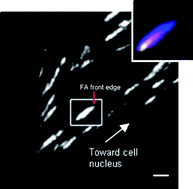
Live cells create adhering contacts with substrates viafocal adhesion (

Maintenance work is planned for Wednesday 1st May 2024 from 9:00am to 11:00am (BST).
During this time, the performance of our website may be affected - searches may run slowly and some pages may be temporarily unavailable. If this happens, please try refreshing your web browser or try waiting two to three minutes before trying again.
We apologise for any inconvenience this might cause and thank you for your patience.
* Corresponding authors
a
Department of Materials and Interfaces, Weizmann Institute of Science, Rehovot 76100, Israel
E-mail:
Daniel.wagner@weizmann.ac.il
b Department of Molecular Cell Biology, Weizmann Institute of Science, Rehovot 76100, Israel
Live cells create adhering contacts with substrates viafocal adhesion (

 Please wait while we load your content...
Something went wrong. Try again?
D. Raz-Ben Aroush, R. Zaidel-Bar, A. D. Bershadsky and H. D. Wagner, Soft Matter, 2008, 4, 2410 DOI: 10.1039/B804643N
To request permission to reproduce material from this article, please go to the Copyright Clearance Center request page.
If you are an author contributing to an RSC publication, you do not need to request permission provided correct acknowledgement is given.
If you are the author of this article, you do not need to request permission to reproduce figures and diagrams provided correct acknowledgement is given. If you want to reproduce the whole article in a third-party publication (excluding your thesis/dissertation for which permission is not required) please go to the Copyright Clearance Center request page.
Read more about how to correctly acknowledge RSC content.
 Fetching data from CrossRef.
This may take some time to load.
Loading related content
