
A novel diagnostic method of Raman spectroscopy for malignant pheochromocytoma/paraganglioma†
Abstract
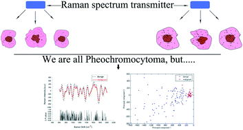
We evaluated the feasibility of Raman spectroscopy in the diagnosis of malignant pheochromocytoma/paraganglioma. Raman spectra were detected from pheochromocytoma/paraganglioma tissues using a Labram HR 800 RS, then principal component analysis and linear discriminant analysis were applied to prediction. There are 25 qualified patients, including 20 with benign pheochromocytoma/paraganglioma and 5 with malignant pheochromocytoma/paraganglioma. The spectra at 680, 733, 780, 847, 939, 993, 1049, 1121, 1170, 1222, 1268, 1314, 1370, 1433, 1487, 1615, 1757 cm−1 of benign and malignant pheochromocytoma/paraganglioma are significantly different, assigned to specific amino acids and DNA. Principal component analysis/linear discriminant analysis yielded a sensitivity of 80.0% and a specificity of 100.0% for diagnosing malignant pheochromocytoma/paraganglioma. Furthermore, we evaluated the malignant behavior of pheochromocytoma/paraganglioma by both Raman spectroscopy and PASS scale. By using a survival analysis, Raman spectroscopy became more efficient than PASS score. Finally, Raman spectroscopy has potential in the diagnosis of malignant pheochromocytoma/paraganglioma in clinical practice.
 Please wait while we load your content...

