
NMR analysis of the binding mode of two fungal endo-β-1,4-mannanases from GH5 and GH26 families†
Abstract
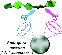
The enzymatic digestion of the main components of lignocellulosic biomass, including plant cell wall mannans, constitutes a fundamental step in the renewable biofuel production, with great potential benefit in the industrial field. Despite several reports of X-ray structures of glycoside hydrolases, how polysaccharides are specifically recognized and accommodated in the enzymes binding site still remains a pivotal matter of research. Within this frame, NMR spectroscopic techniques provide key binding information, complementing and/or enhancing the structural view by X-ray crystallography. Here we provide deep insights into the binding mode of two endo-β-1,4 mannanases from the coprophilous ascomycete Podospora anserina, PaMan26A and PaMan5A, involved in the hydrolysis of plant cell wall mannans and heteromannans. The investigation at a molecular level of the interaction between the wild-type enzymes and inactive mutants with manno-oligosaccharides, revealed a different mode of action among the two glycoside hydrolases most likely due to the presence of the additional and peculiar −4 subsite in the PaMan26A binding pocket.
 Please wait while we load your content...

