
Color resolution improvement of the dark-field microscopy imaging of single light scattering plasmonic nanoprobes for microRNA visual detection†
 ab
Cheng Zhi
Huang
*ac
ab
Cheng Zhi
Huang
*ac
Abstract
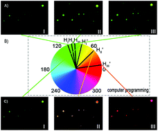
Imaging of light scattering plasmonic nanoparticles (PNPs) with the aid of the dark-field microscopy imaging (iDFM) technique has attracted wide attention owing to its high signal-to-noise ratio, but to improve the color resolution and contrast of dark-field microscopy (DFM) images of single light scattering PNPs in a small spectral variation environment is still a challenge. In this study, a new color analytical method for resolving the resolution and contrast in DFM images has been developed and further applied for colorimetric analysis using the digital image processing technique. The color of single light scattering PNP images is automatically coded at first with the hue values of the HSI color model, and then amplified using the MATLAB program even for marginal spectral changes, leading to significant improvement of the color resolution of DFM images and easy detection with the naked eye. As a proof of concept, this method is then applied to distinguish single PNPs with various sizes and to visually detect hepatocellular carcinoma-associated microRNA. As it greatly improved the color resolution of iDFM and its detection sensitivity, this method shows promise to serve as a better alternative for sensitive visual analysis and spectrometer-based spectral analysis.
 Please wait while we load your content...

