T1-Weighted MR imaging of liver tumor by gadolinium-encapsulated glycol chitosan nanoparticles without non-specific toxicity in normal tissues†
Abstract
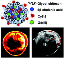
Herein, we have synthesized Gd(III)-encapsulated glycol chitosan nanoparticles (Gd(III)-CNPs) for tumor-targeted T1-weighted magnetic resonance (MR) imaging. The T1 contrast agent, Gd(III), was successfully encapsulated into 1,4,7,10-tetraazacyclododecane-1,4,7,10-tetraacetic acid (DOTA)-modified CNPs to form stable Gd(III)-encapsulated CNPs (Gd(III)-CNPs) with an average particle size of approximately 280 nm. The stable nanoparticle structure of Gd(III)-CNPs is beneficial for liver tumor accumulation by the enhanced permeation and retention (EPR) effect. Moreover, the amine groups on the surface of Gd(III)-CNPs could be protonated and could induce fast cellular uptake at acidic pH in tumor tissue. To assay the tumor-targeting ability of Cy5.5-labeled Gd(III)-CNPs, near-infrared fluorescence (NIRF) imaging and MR imaging were used in a liver tumor model as well as a subcutaneous tumor model. Cy5.5-labeled Gd(III)-CNPs generated highly intense fluorescence and T1 MR signals in tumor tissues after intravenous injection, while DOTAREM®, the commercialized control MR contrast agent, showed very low tumor-targeting efficiency on MR images. Furthermore, damaged tissues were found in the livers and kidneys of mice injected with DOTAREM®, but there were no obvious adverse effects with Gd(III)-CNPs. Taken together, these results demonstrate the superiority of Gd(III)-CNPs as a tumor-targeting T1 MR agent.


 Please wait while we load your content...