In vivo studies on angiogenic activity of two designer self-assembling peptide scaffold hydrogels in the chicken embryo chorioallantoic membrane
Abstract
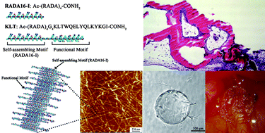
The rapid promotion of angiogenesis is critical for tissue engineering and regenerative medicine. The angiogenic activity of tissue-engineered scaffolds has already been the major criterion for choosing and designing ideal biological materials. We here report systematic in vivo studies on the angiogenic activity of two functionalized self-assembling peptides PRG (Ac-(RADA)4GPRGDSGYRGDS-CONH2) and KLT (Ac-(RADA)4G4KLTWQELYQLKYKGI-CONH2) using the chicken embryo chorioallantoic membrane (CAM) assay. 3D migration/sprouting bead assays showed that the two functional motifs PRGDSGYRGDS and KLTWQELYQLKYKGI improved the bioactivities of the self-assembling peptide RADA16-I (Ac-(RADA)4-CONH2) dramatically and provided ideal synthetic microenvironments for endothelial cell migration and cordlike structure sprout formation. A CAM assay was carried out to assess the efficiency of various peptide scaffolds in inducing capillary invasion in vivo. Among these three peptide scaffolds, the functionalized peptide scaffold RAD/KLT presented a significantly better angiogenic activity inducing CAM tissue invasion and new capillary vessel formation within the scaffolds in the absence of VEGF. With the addition of VEGF, more newly formed vessel lumen could be observed in all peptide scaffolds. Our results suggested that the functionalized peptide scaffolds had satisfactory angiogenic properties, and may also have wide potential applications in tissue regeneration.
 Please wait while we load your content...

