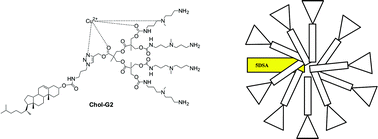
The self-assembly of dendritic molecules is an effective way of generating functionalised nanoscale architectures. This study reports a computer-aided analysis of the EPR spectra of 5 doxyl-stearic acid (5DSA) and Cu(II) probes interacting with self-assembling dendrons in water. The dendrons investigated have previously been reported as potential gene delivery vehicles, and possess amine surface groups, different dendritic architectures based on ether-amide or ester branching, and hydrophobic groups at the focal point which can encourage self-assembly in aqueous solution. The parameters extracted from computation provide information about both the structure and dynamics in solution and the interacting ability of the dendrons to be used in gene therapy. The hydrophobic 5DSA probe is able to effectively probe the hydrophobic core of self-assembled dendron nanostructures. It reports on the polarity of its local environment and is most affected by dendrons with two cholesterol units at the focal point, partly affected by dendrons with cholesterol groups at their focal point, but is unaffected by dendrons with a simple phenyl group at the focal point. This reflects the different modes of self-assembly observed for these dendrons. When Cu(II) is used as an EPR probe of the branched environment, it was found that at pH7, much of the Cu(II) was ‘external’ to the dendritic structure, presumably due to protonation of the peripheral amine groups. On gradually increasing the Cu(II) loading, and using computer-aided analysis, it was possible to quantify the levels of ‘internal’ (dendron-bound) and ‘external’ Cu(II) and it was found that this was strongly dependent on the structure of the dendritic branching and the ability of the dendron to self-assemble, with self-assembling ester dendrons being best able to bind the Cu(II). It was also possible to propose the nature of the copper binding sites associated with the ‘internal’ signal as either Cu–NO3 and Cu–N2O2 distorted square-planar coordination sites. The self-assembling ester based dendrons which also contain 1,2,3-triazole units, had higher levels of ‘internal’ Cu(II) and showed the latter form of coordination, while the other dendrons, with lower levels of Cu(II) uptake showed the former. In summary, this paper demonstrates that two complementary EPR probes can be used to provide information about different regions of a self-assembled dendritic architecture.

 Please wait while we load your content...

